1. www.postersession.com
Conclusions
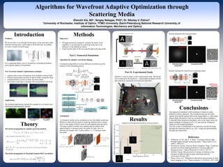
Algorithms for Wavefront Adaptive Optimization through
Scattering Media
Zhenzhi Xia, BS1; Sergey Nalegae, PhD2; Dr. Nikolay V. Petrov2
1University of Rochester, Institute of Optics; 2ITMO University (Saint-Petersburg National Research University of
Information Technologies, Mechanics and Optics)
Problems
We are not able to see through frosted glass, tissue and many other
materials because they scatter light so much that they lose ability
to create a clearly focused image
For a scattering object, wave is focused on a disordered medium,
and a speckle pattern is transmitted
New wavefront adaptive optimization technique
• captures and extracts information from multiple scattered light
• measure spatial phase profiles of optical fields exiting the tissue
• computationally recover an ideal wavefront shape
• micrometer-scale spot is focused with reconstructed wavefront
Applications
The potential applications include for example in-vivo deep tissue
and targeted photodynamic therapy.
Introduction
Theory
Wavefront propagation by angular spectrum method
Wavefront propagation by Rayleigh-Sommerfeld Convolution
Objectives
• Through numerical simulation, estimate the limits of the
capability to see through the scattering media due to the
influence of the diffraction effects
• Experimental study of the focusing through scattering media
Part I. Numerical Simulation
Algorithm for adaptive wavefront shaping
Construction algorithm of inverse diffusion wavefront uses the
linearity of the scattering process
Methods
Targeted image specified
CCD provides feedback to
SLM
Stepwise scanning and
cycle through 0 to 2pi for
every single segment to
determine optimal phase
Store the phase when the
target intensity is maximum
for each element
All
measurements
done Set the phase of the
segments to their stored
values
Global
Maximum
Feedback loop for achieving inverse diffusion
Results
NMSE: 0,0508
NMSE: 0,0455
NMSE: 0,0463
The numerical simulation of the technique is based on scalar
diffraction theory approach. The correlation between the input
optical field and the optical field on the target plane (i.e. after phase
plates) drops off quickly when we increase the phase retardance.
Concluded from numerical simulation, only in the region when the
phase retardance is among 0 to 0.9, we can treat the volumetric
media as a flat scattering diffuser
The new system is established in the laboratory and the focusing
through the scattering object (e.g. a fly’s wing) was demonstrated
through real experiment.
Reference
• Vellekoop, Ivo M., and A. P. Mosk. "Focusing coherent light
through opaque strongly scattering media." Optics letters 32.16
(2007): 2309-2311.
• Horstmeyer, Roarke, Haowen Ruan, Changhuei Yang. "Guidestar-
assisted wavefront-shaping methods for focusing light into
biological tissue." Nature Photonics 9.9 (2015): 563-571.
• Akbulut, Duygy. Measurements of strong correlations in the
transport of light through strongly scattering materials.
Universiteit Twente, 2013.
• Wang, Chen, et al. "Multiplexed aberration measurement for deep
tissue imaging in vivo." Nature methods 11.10 (2014): 1037-1040.
Assumption
A volumetric media can be considered as one flat highly-scattering
diffuser so that with any kind of scattering medium until a certain
limit a clear image could be retrieved with targeted area specified.
The paper authors consider only samples that are thicker than
approximately 6 transport mean free paths for light, here we
simulate the sample with 13 phase plates, i.e. 12 transport mean
free paths
Part II. Experimental Study
Wind fly is used as object in this experimental study. Wavefront
AO is realized by the hardware -- spatial light modulator (SLM)
and continuous sequential reconstruction algorithm.
Results obtained with Gaussian Beam
Results obtained for wind fly as a scattering object