This document provides information on hypertrophic cardiomyopathy (HCM), including its definition, history, etiology, pathophysiology, diagnosis, and clinical presentation. HCM is a genetic heart condition characterized by thickened heart muscle not caused by other conditions like high blood pressure. It affects around 1 in 500 people and can cause chest pain, dizziness, and heart failure. The pathophysiology involves left ventricular outflow tract obstruction, diastolic dysfunction, myocardial ischemia, and mitral regurgitation. Diagnosis involves imaging of the thickened heart muscle. Most patients are asymptomatic but may develop limiting symptoms like shortness of breath or chest pain in their 40s-60s as obstruction develops.
1. The document discusses different types of supraventricular tachycardia including AV nodal reentrant tachycardia (AVNRT), atrioventricular reentrant tachycardia (AVRT), and atrial tachycardia (AT).
2. It provides details on electrocardiogram patterns that can help differentiate the different types of SVT. Features like retrograde P waves, RP intervals, and the effect of ventricular pacing are discussed.
3. Ablation techniques for AVNRT and AVRT are covered, including approaches for mapping the circuits and terminating arrhythmias. The importance of making an accurate diagnosis is emphasized.
La Taquicardia Ventricular en pacientes sin cardiopatía puede ser idiopática, es decir, debida a una anomalía eléctrica exclusiva sin cardiopatía estructural subyacente. Estas arritmias idiopáticas suelen tener un pronóstico benigno. Sin embargo, en algunos casos puede ser difícil diagnosticar si existe una cardiopatía oculta o mínima. Identificar correctamente cada caso como una de las entidades clínicas conocidas es clave para el diagnóstico, pronóstico y tratamiento adecuados.
The document discusses challenges that can occur during coronary sinus lead implantation for cardiac resynchronization therapy, including anatomical abnormalities, difficulties accessing the coronary sinus or its branches, lead instability, and complications like phrenic nerve stimulation or high pacing thresholds. Specific challenges covered include stenosis, tortuosity, small vessel size, failure to cannulate, dissections, and potential solutions using devices, angiography, and stenting. The document also presents a case example of a patient who experienced left ventricular lead dislocation after initial successful cardiac resynchronization therapy device implantation.
This document describes equipment and settings used in electrophysiology (EP) studies. It lists various pieces of equipment including fluoroscopy units, recording systems, cardiac stimulators, ablation generators, 3D mapping systems, and intracardiac ultrasound units. It provides details on catheter types, positions, and settings for standard EP studies including studies of arrhythmias like supraventricular tachycardia. It also explains pacing protocols and techniques used in EP studies to evaluate conduction properties and induce arrhythmias.
The document provides criteria for diagnosing ST-elevation myocardial infarction (STEMI) on electrocardiogram (ECG). It lists cut-off values for ST elevation in different leads used to identify STEMI based on the patient's age, sex and lead location. It cautions that baseline ECG abnormalities like left bundle branch block (LBBB) could obscure interpretation and provides examples of STEMI in different heart locations identified by affected leads on ECG.
Case scenario of a patient with idiopathic ventricular Tachycardia (VT), followed by a topic review including diagnosis and management guidelines. It is defined as Monomorphic VT in patients without any structural heart disease or coronary disease”. Classified on the basis of site of origin broadly into three different categories i.e Outflow Tract VT, Annular VT, Fascicular VT
This document discusses strategies to minimize right ventricular pacing, which can have deleterious effects. It summarizes several clinical trials that evaluated ventricular versus atrial or dual-chamber pacing. The trials generally found that atrial or dual-chamber pacing reduced atrial fibrillation compared to ventricular pacing, though effects on other outcomes like mortality were less clear. The document recommends that right ventricular pacing be avoided or minimized when possible, through use of AAI pacing, DDD pacing with long fixed AV delays, search AV hysteresis algorithms, or mode-switching algorithms that favor intrinsic conduction.
This document provides guidance on pre-procedural preparation and tips for CRT implantation. It discusses evaluating patients before implantation through imaging, labs, and clinical assessments. Key steps in implantation include accessing veins, placing the RV lead first usually in the septum, cannulating the coronary sinus, performing venograms to select target veins, and placing the LV lead aiming for the lateral or posterior wall. Positioning the leads to maximize electrical delay and hemodynamics is important.
This document contains a quiz with multiple choice questions about ECG interpretations. Some key findings included in the questions are right bundle branch block with left posterior hemiblock, third degree atrioventricular block, left bundle branch block with Cabrera's sign indicating possible myocardial infarction, Wolff-Parkinson-White syndrome type A, ventricular tachycardia, atrial fibrillation, premature ventricular contractions, left ventricular hypertrophy, anterior myocardial infarction, hyperkalemia, and more. The document also includes explanations of ECG patterns and signs such as bundle branch blocks, ventricular tachycardia criteria, Wellens' phenomenon, hyperacute T waves, and more.
This document discusses cardiac axis determination from an electrocardiogram (ECG). It describes how to determine the QRS axis using the first method of inspecting leads I and aVF, and whether this indicates a normal, right, left, or bizarre axis. The second method involves finding the isoelectric limb lead and calculating the perpendicular axis. Common causes of left axis deviation, right axis deviation, and bizarre northwest axes are provided.
- Catheter ablation is an effective treatment for ventricular tachycardia (VT) in both structurally normal hearts and those with structural heart disease.
- For normal hearts, activation mapping and pace mapping are used to identify the origin of VT, with typical sites including the right ventricular outflow tract. Success rates are high with few complications.
- In structural heart disease, substrate mapping is used to identify scar tissue which hosts reentrant VT circuits. Entrainment mapping during VT can confirm circuits. Ablation targets abnormal potentials in scar or the scar border zone. Integration of cardiac imaging helps define substrate. Long-term outcomes after ablation are improved compared to antiarrhythmic drugs alone.
INOCA, or ischemia with non-obstructive coronary arteries, affects a large proportion of patients undergoing angiography who do not have obstructive coronary artery disease. INOCA can result from heterogeneous mechanisms like coronary vasospasm and microvascular dysfunction and is not benign, as it is associated with increased cardiovascular events and impaired quality of life. The diagnosis of MINOCA, a type of INOCA, requires meeting criteria for an acute myocardial infarction but having non-obstructive arteries on angiography and no other clear cause identified. Further evaluation is then needed to determine the underlying cause of MINOCA.
The document discusses congestive cardiac failure (heart failure) and its management. It provides details on:
- The high prevalence and mortality of heart failure.
- Current medical therapies including ACE inhibitors, beta-blockers, and aldosterone antagonists that have been shown to improve survival.
- Device therapies like cardiac resynchronization therapy and implantable cardioverter defibrillators that treat symptoms and reduce mortality.
- The benefits of multidisciplinary and integrated care approaches including telehealth monitoring in improving outcomes for heart failure patients.
This document provides information about electrophysiologic studies (EPS), including the purpose, requirements, procedures, and how to read EPS tracings. An EPS involves placing electrode catheters in the heart to record electrical activity and stimulate heart tissues to evaluate arrhythmias. Key aspects of an EPS include determining the sequence of impulse propagation in the atria, ventricles, and conduction system during normal sinus rhythm, pacing, and arrhythmias to diagnose arrhythmia type and location.
This document discusses various electrocardiogram (ECG) criteria that can be used to differentiate ventricular tachycardia (VT) from supraventricular tachycardia (SVT) with aberrant conduction. It reviews criteria proposed in several studies from 1965 to 2020, including Sandler's criteria on QRS morphology, Marriott's chest lead concordance, Wellens' criteria on QRS duration and morphology, and the Brugada algorithm - the first multistep algorithm for differentiation. More recent automated methods using computerized ECG interpretation may also help clinicians establish the likelihood of VT.
This document provides an overview of cardiac arrhythmias including their classification, mechanisms, clinical manifestations, diagnostic approaches and management strategies. It discusses various specific arrhythmias in detail such as atrial fibrillation, atrial flutter, supraventricular tachycardia, ventricular arrhythmias, sick sinus syndrome and heart block. Treatment options covered include pharmacological therapies using different classes of antiarrhythmic drugs, procedures like cardiac ablation and use of devices like pacemakers.
The document discusses assessment of mechanical dyssynchrony for cardiac resynchronization therapy. It defines electrical and mechanical dyssynchrony, and describes the deleterious hemodynamic effects of left ventricular dyssynchrony. It then summarizes various echocardiographic tools for assessing atrioventricular dyssynchrony, interventricular dyssynchrony, and intraventricular dyssynchrony, including M-mode, tissue Doppler imaging, and three-dimensional echocardiography. Measurement techniques for different dyssynchrony parameters such as septal-posterior wall motion delay, lateral wall postsystolic displacement, and time to peak systolic velocity are outlined.
1. This document provides an overview of a training course on complex supraventricular tachycardia (SVT) differentiation. It discusses various SVT etiologies and electrocardiogram patterns.
2. Mechanisms of SVT discussed include atrioventricular nodal reentrant tachycardia (AVNRT), atrioventricular reentrant tachycardia (AVRT), and atrial tachycardia (AT). The document also reviews electrophysiology study findings that help differentiate the mechanisms.
3. Case examples are presented to demonstrate electrophysiology study techniques for SVT diagnosis and ablation, including ventricular overdrive pacing, ventricular extrastimuli, and induction protocols.
This document discusses the management of various cardiac arrhythmias that present as emergencies, including tachyarrhythmias like ventricular tachycardia and fibrillation, as well as bradyarrhythmias. Algorithms are provided to guide clinicians in differentiating between ventricular tachycardia and supraventricular tachycardia based on electrocardiogram findings. Risk factors that increase the likelihood of ventricular tachycardia are outlined. Causes and management of other arrhythmias like atrial fibrillation, heart block, sinus bradycardia, and device malfunctions are also summarized. The goal is to aid clinicians in rapidly diagnosing serious arrhythmias and initiating the appropriate treatment in emergency settings
ECG LOCALISATION OF CULPRIT ARTERY IN STEMIPraveen Nagula
The document discusses coronary artery anatomy and ECG localization of the culprit vessel during acute myocardial infarction. It describes the typical blood supply and branches of the right coronary artery, left main coronary artery, left anterior descending artery, and left circumflex artery. It then covers how the ECG can be used to localize whether the right coronary artery, left circumflex artery, or other vessels are responsible for an acute MI based on the leads showing ST elevation and depression. Factors such as the ratio of ST changes in different leads help indicate whether proximal or distal vessels are involved.
This document provides an overview of hypertrophic cardiomyopathy (HCM). It begins with definitions of cardiomyopathy and HCM. It then discusses the historical perspective, genetic basis, morphology, pathophysiology, clinical features, diagnosis, and management of HCM. Some key points include:
- HCM is a genetic heart condition characterized by unexplained thickening of the heart muscle. It is the most common cause of sudden cardiac death in young people.
- The genetic basis involves mutations in genes encoding sarcomere proteins. This leads to impaired relaxation and increased calcium sensitivity of the heart muscle.
- Morphologically, HCM involves asymmetric left ventricular hypertrophy and abnormalities of the mitral valve apparatus. Hist
Este documento presenta información sobre la interpretación adecuada del electrocardiograma (ECG) en pediatría. Explica que el ECG es un registro gráfico de los cambios en la corriente eléctrica del corazón que no requiere de gran complejidad técnica. Luego, detalla los pasos para leer un ECG pediátrico, incluyendo el cálculo de la frecuencia cardiaca, análisis del ritmo, eje de la onda P, complejo QRS e intervalos. Finalmente, menciona algunas alteraciones no
- The document discusses the history and evolution of implantable cardioverter defibrillators (ICDs). Key points include:
- ICDs were first conceived in 1966 and the first human implant was in 1980.
- ICDs function by sensing cardiac rhythms, detecting arrhythmias like ventricular tachycardia or fibrillation, and delivering electrical therapies like anti-tachycardia pacing or shocks to treat them.
- Important aspects of ICD function include sensing, detection algorithms, therapy delivery, and troubleshooting problems. Studies show programming ICDs more conservatively with longer detection intervals and fewer unnecessary therapies can reduce inappropriate shocks and mortality.
Infective endocarditis remains a serious disease with high mortality and morbidity. While improvements have been made in diagnostics, antibiotics, and surgery, the incidence has remained unchanged over the past 20 years. Native valve infective endocarditis is now often caused by degenerative valve disease, congenital heart disease, or intravenous drug use rather than rheumatic heart disease. Prosthetic valve endocarditis has early and late onset forms and is commonly caused by Staphylococcus aureus and coagulase-negative staphylococci. New diagnostic criteria have been proposed along with treatments such as antibiotics and cardiac surgery. Vaccines and new antibiotics may help with prevention and treatment in the future.
EEN GROEIENDE BERG PILLEN….?
Themabijeenkomst medicijnen 21 oktober
Apotheek Monster / Poeldijk organiseert in samenwerking met Careyn en Stichting Welzijn Westland een themabijeenkomst medicijnen.
O documento descreve um curso de ECG dividido em quatro módulos. O primeiro módulo trata do ECG normal, incluindo anatomia, princípios de eletrofisiologia e formação do traçado. O segundo módulo aborda alterações no ECG como sobrecargas, hipertrofias e bloqueios. O terceiro módulo estuda mecanismos e tipos de arritmias. O quarto módulo trata de antiarrítmicos e testes de traçados.
This document outlines the content of a course on electrocardiography (ECG), covering normal ECG parameters and patterns, vectorial analysis of the ECG, basic ECG interpretation, and beginning recognition of rhythms such as sinus rhythm and heart block. The course is divided into modules that cover topics including normal ECG features, abnormalities, arrhythmias, antiarrhythmic drugs, and a test with 20 ECG strips.
O documento discute anestesia em neurocirurgia, focando no controle da pressão intracraniana e do fluxo sanguíneo cerebral. Aborda os efeitos dos anestésicos inalatórios, venosos e outros fármacos no sistema nervoso central, assim como fatores que regulam a perfusão cerebral, como a pressão de dióxido de carbono. Também discute hidratação e manejo de fluidos em pacientes neurocirúrgicos.
- O eletrocardiograma (ECG) registra os fenômenos elétricos originados durante a atividade cardíaca por meio de eletrodos posicionados no corpo.
- O ECG é dividido em derivações que fornecem diferentes ângulos de visualização do coração, incluindo derivações periféricas no plano frontal e derivações precordiais no plano horizontal.
- As principais áreas do traçado ECG analisadas incluem frequência cardíaca, ritmo, infarto e hipertrofia.
The document discusses several types of cardiac arrhythmias including:
1. Extra-systoles which can be supraventricular or ventricular.
2. Atrial flutter and fibrillation.
3. Bradycardias, blocks, and tachycardias which have specific classifications and characteristics.
The mechanisms of arrhythmias are described as being either due to reentry of electrical impulses, abnormal automaticity, or conduction abnormalities. Specific conditions must be met for the reentry mechanism, and abnormal automaticity can arise from reduced membrane potentials self-propagating electrical activity.
O documento discute arritmias cardíacas e fornece as seguintes informações essenciais:
1) Apresenta as bases eletrofisiológicas das arritmias cardíacas, incluindo o potencial de ação e de repouso das células cardíacas.
2) Discutem os mecanismos responsáveis pelas arritmias, incluindo anormalidades na geração e condução do impulso cardíaco.
3) Detalha métodos de diagnóstico de arritmias como ECG, er
O documento discute determinação do eixo elétrico cardíaco e hipertrofia ventricular esquerda e direita. Ele fornece detalhes sobre os critérios diagnósticos para sobrecarga atrial esquerda e direita usando índices como o de Macruz e Morris. Também descreve o critério de Romhilt e Estes para diagnóstico de hipertrofia ventricular esquerda.
Este documento descreve um curso de ECG dividido em 4 módulos. O Módulo I introduz o ECG normal e inclui aulas sobre a teoria do dipolo, derivações eletrocardiográficas e identificação do ritmo sinusal. O Módulo II aborda alterações do ECG. O Módulo III estuda arritmias. E o Módulo IV trata de antiarrítmicos e testes de ECG.
O documento discute o impacto da apneia do sono nas arritmias cardíacas, destacando que afeta até 50% dos pacientes com insuficiência cardíaca e mais de 30% dos pacientes com hipertensão. Também aborda formas de diagnóstico e tratamento da apneia do sono.
21. Systematische beoordeling
1. Ritme
2. Frequentie
3. Geleidingstijden
4. Hart-as
5. P top morfologie
6. QRS morfologie
7. ST morfologie
1. Vergelijking met oud ECG
2. Conclusie
22. 1 Ritme
Eigenschappen van
normaal sinusritme
•Op een P-top volgt
meestal een QRS complex
•Het ritme is regelmatig,
maar varieert licht met de
ademhaling
•De frequentie ligt tussen
de 60 en 100 / minuut.
•De p top is positief in II en
AVF, en bifasisch in V1
•De PQ tijd is tussen de
0,12 en 0,2 seconden
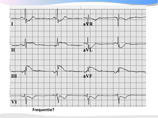
25. 2 Frequentie
3 methoden:
1. Aftelmethode
2. Berekenen: 1500 /
aantal kleine hokjes
tussen 2 hartslagen
3. Marker methode
De hartfrequentie wordt
beïnvloed door:
Het autonome
zenuwstelsel
De vulling van het hart
27. 3 Geleidingstijden
PQ tijd tussen 0.12 en 0.20
seconde
QRS duur <= 0.10-0.12
seconde
Te lang LBTB / RBTB
QTc tijd = repolarisatie
Mannen < 450ms
Vrouwen < 460ms
28. Check de QT tijd die
de computer
uitrekent!
Verlengde QTc tijd
geeft verhoogd risico
op plotse dood. Met
name > 480-500 ms.
Dan geen QTc
verlengende
medicatie:
•Sotalol QT
•Amiodarone QTc
•Erythromycine RR interval
(sec)
•Clarithromycine
•Haldol
Zie www.torsades.org Eyeballing: als T top eindigt voorbij het punt
halverwege RR is de QT meestal verlengd
29. 4 Hartas
Geeft de gemiddelde
electrische activiteit aan
Normaal is tussen -30 en
+90 graden.
Positief in I en AVF?
hartas = normaal
Kijk op het ECG! De
computer heeft het
meestal goed.
36. 5 P top morfologie
•De maximale hoogte van
de p top is 2,5 mm in II en /
of III
•De p top is positief in II en
AVF, en bifasisch in V1
•De breedte van de p top is
normaal korter dan 0.12
seconde
37. Linkeratriumdilatatie Rechteratriumdilatatie
Terminaal deel in V1 > 1mm2 P >2,5 mm in II en/of III en/of
en/of P >0,12 sec in I en/of II aVF
en/of P >1,5 mm in V1
49. 7+2 STAPPENPLAN
Stap 6: QRS morfologie
• R-top progressie?
– Overgangs complex
in V3, V4
• Normaal zit het
overgangs complex
(waar de R-top groter
wordt dan de S)
bij V3 tot V4
50. 7 ST morfologie
ST elevatie
Ischemie
Pericarditis
Aneurysma cordis
Normale variant
ST depressie
Reciproke bij ischemie
LVH
Digitalis
Hypokaliemie
Neurologisch
T top verandering
Ischemie
Pericarditis
Myocarditis
LVH / RVH
57. Vlak = < 0.5mm in I, II, V3-V6
Negatief = > 0.5mm in I, II, V3-V6
58. 7+1 Vergelijken met oud ECG
• Nieuwe LBTB?
• Asdraai?
• Nieuwe pathologische Q?
• Afname R top hoogte?
59. 7+2 Conclusie
Voorbeelden:
• "Sinustachycardie met ST elevatie over de voorwand,
passend bij een acuut voorwandinfarct"
• "Supraventriculaire tachycardie van 200/min op basis van
een AV nodale re-entry"
• "Oud onderwandinfarct met nu een acuut lateraal
myocard-infarct met QRS verbreding ten opzichte van het
ECG van 14 augustus vorig jaar"
• "Normaal ECG"