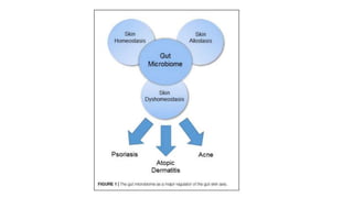
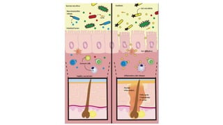
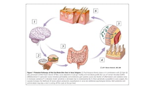
Intestinal Microbiome in Skin
disorders
13.11.2018
“AlldiseaseBeginsinthegut”
“Deathsitsinthebowels”
“Baddigestionistherootof
allevil”
The Hippocrates
What constitutes the intestinal microbiome ?
collective human gut microbiota is composed of over
35,000 bacterial species
Strict anaerobes >facultative anaerobes & Aerobes
1.Bacteroidetes
2.Firmicutes
1.Proteo bacteria
2.Verruco microbia
3.Actino bacteria
4.Fuso bacteria
5.Cyano bacteria
Baohong Wang, Mingfei Yao, Longxian Lv, Zongxin Ling, Lanjuan Li,The Human Microbiota in Health and Disease, Engineering,Volume 3, Issue 1, 2017, Pages 71-82.
Where are they & how they evolve ?
Baohong Wang, Mingfei Yao, Longxian Lv, Zongxin Ling, Lanjuan Li,The Human Microbiota in Health and Disease, Engineering,Volume 3, Issue 1, 2017, Pages 71-82.
Protection against Infection
Physical barrier to incoming pathogens
by competitive exclusion,
• Occupation of attachment sites,
• Consumption of nutrient sources,
• Production of antimicrobial substances.
Stimulations of various AMPs • defensins,
• cathelicidins,
• C-type lectins,
• Directs paneth cell to express AMP
Short-chain fatty acids (SCFAs) and
lithocholic acid
• Induce expression of LL-37 cathelicidin
• The induction involved the MEK/ERK pathway, AP-1 transcription factor,
and histone acetylation
Converts AMP to Active form Prodefensin to defensin by B. thetaiotaomicron
The Gm+ Lactobacillus and
Bifidobacterium
Prevents Listeria infection
• elaboration of secreted compounds
• modulation of the epithelial cells’ immune response
Lactobacillus Decreases colonization by pathogenic E. coli
SFB on ileal mucosa physically exclude
S. enteritidis from its attachment sites
INTRODUCTION:
• Gut and skin- densely vascularised and richly innervated organs with
crucial immune and neuroendocrine roles.
• Bidirectional relationship.
• “Gut microbiome” : diverse community of microbial organisms that
normally inhabit the bowel and their metabolites/byproducts.
• Predominantly 2 phyla: Firmicutes and Bacteroidetes.
GUT MICROBIAL ECOLOGY
• Bacteria, viruses, protozoa, fungi
• Metabolic and immune benefits to host.
• Protection against invasion by exogenous pathogens directly by
competitively binding to epithelial cells and indirectly by triggering
immune responses.
• Gut microbes-source of peptidoglycan-alter TLR(PRR) on surface of
innate immune cells.
• TLR recognise PAMPs ; initiate linking of innate to adaptive immune
system via NF-KB
• Gut microbiome- induces IgA, maintain homeostasis between effector
and regulatory T cells.
• ‘cross talk ‘ mediating epithelial barrier integrity. (Lactobacillus LGG
p40 suppresses cytokine mediated apoptosis and epithelial barrier
disruption).
• Short chain fatty acids(SCFAs) production
• Altered gut flora- Effector cells favoured over regulatory cells ;
development of autoimmune disorders ( segmented filamentous
bacteria in gut ; Th17 mediated diseases).
Gut microbiome and skin homeostasis:
• Probable action on skin by modulating the systemic immunity.
• Certain gut microbes( Bacteroides fragilis, Faecalibacterium
prausnitzii and Clostridium cluster IV and XI) and their metabolites
(retinoic acid, polysaccharide A) ; accumulation of regulatory T cells;
anti-inflammatory.
• SCFAs ; butyrate inhibit inflammatory cell migration, adhesion and
cytokine production.
• SCFAs inhibit histone deacetylase and inactivate NF-kB; regulation of
activation and apoptosis of immune cells.
• Metastases of gut microbiota and metabolites to skin
• Disrupted intestinal barrier.
• Gut microbiome influences skin microbiome;
 propionibacterium- propionate and acetate; act against USA300 (CA-MRSA)
S. epidermidis and P. acnes tolerate wider SCFA shifts.
•
• Ogawa et al, ; oral supplementation with L.brevis SBC8803 for 12
weeks ; decreased TEWL.
• L. paracasei for 2 months; decreased TEWL
• Baba et al, Lactobacillus helveticus- fermented milk whey ; increased
expression of keratin 10 and involucrin; markers of early and alte
differentiation respectively.
Gut microbiota and skin allostasis:
• Allostasis: restoration of homeostasis after a disturbance.
• Positive impact of gut microbiota on skin barrier disruption.
• Baba et al; L. helveticus; Sodium dodecyl induced dermatitis and
TEWL.
• Branchet-Gumila et al; improved skin barrier recovery and decreased
inflammation and TNF –alpha release after L. paracasei .
• Administration of Lactobacillus reuteri ; accelerated wound healing.
• Restoration of skin homeostasis after UV radiation exposure. ( 10 days
of oral supplementation with Lactobacillus johnsonii in mice
protected against UV- induced contact hypersensitivity; due to
reduced epidermal Langerhans cells and increased systemic IL- 10
levels.
• Influence on T cell differentiation.; oral lactobacillus casei – impair
differentiation of CD8+ T cells into effector cells
• Abundant Th17 cells in skin and intestine.
• Balance between TH17 and Treg cells ; maintained by microbiome.
Dysbiosis and skin dyshomeostasis:
• Free phenol and p-cresol – metabolic biomarkers of disturbed gut
milieu.
• Translocation of metabolites. ( O’Neill et al., 2016)
• High skin p- cresol levels – reduced skin hydration and impaired
keratinization.
• Intestinal dysbiosis- epithelial permeability ; effector T cells
activated; disrupted balance’ pro-inflammatory cytokines further
accentuate this.
Acne vulgaris:
• Western diet; abundant carbohydrates; increase in insulin/IGF-1
signalling.
• Increased cytoplasmic expression of metabolic forkhead box
transcription factor (FoxO1)
• Triggers mTORC1
• Sebaceous gland hyperproliferation,lipogenesis, infundibular
keratinocyte hyperplasia.
• mTOR can regulate intestinal microbiota through regulation of
intestinal barrier.
• ROLE OF BRAIN:
 frequent association of psychological disturbances, acne and GI disturbances.
Intestinal flora; neurotransmitters ( serotonin, norepinephrine, Ach,
neuropeptides- increase intestinal permeability and casue systemic
inflammation)
Upregulation of substance P containing nerves in acne and intestinal dysbiosis;
IL-1, IL-6, TNF- alpha ,PPAR- gamma
The Brain-Gut-Skin theory:
• Stokes and Pillsbury.
• Altered emotional states to altered GI functions.
• 40 % patients with acne-hypochlorhydria.
• Reduced stomach acid- migration of bacteria – intestinal permeability.
• Psychological stress; intestinal transit time, encourages overgrowth of
bacteria.
Atopic dermatitis:
• ACUTE STAGE: dysfunctional skin barrier
• TSLP,IL-33, IL-25
• Activate typer-2 innate lymphoid cells
• Release of IL-13 and IL-5
• Th2 response
• HYGIENE HYPOTHESIS:
• First proposed by Strachan: allergic disorders arise when our immune
system inappropriately responds to harmless antigens via Th-2
mediated responses.
• Early exposure to microbial antigens is responsible for immune
development. As it encourages Th1 rather than Th2 response.
• Gut microbiome in early life; associated with age of
onset,severity,remission, flares,phenotypes.
• Gut microbiome diversity and development of AD
• DIET – MICROBIOME THEORY:
• Inappropriate food ; hampers microbiome mediated immune
homeostasis
• Immune homeostats begins in early life thorugh exposure to
maternal microbiota and infant’s intestinal flora is futher eveloped
after exposure to breast milk and environmental microbes.
• Low fiber , hign fat diet’ low production of SCFAs; decreased anti-
inflammatory activity by Treg cells, IL-10 and TGF- beta
• Korean studies:
• Fecal samples from AD patients;reduced Faecalibacterium prausnitzii
as compared to control.
• Decrease in SCFA production.
• Disrupted epithelial barrier.
• Metabolites reach skin – trigger Th2 response
• Clostridum difficile, E.coli, S.aureus higher than controls
• Reduced levels of butyrate producing coprococcus.
• Eosinophilic inflammation in gut; clostridia and E.coli.
• Akkermansia muciniphila and Ruminococcus gnavus; positive impact
How do gut microbiome impact AD:
• IMMUNOLOGIC PATHWAY: Th2 response; IL-5 ,IL-13; increase IgE.
• Gut colonization by S.aureus was negatively associated with the
subsequent development of AD in infancy.
• METABOLIC PATHWAY:
• Linoleic acid and 10- hydroxy-cis-12-octadecenoic acid alleviated AD
in mouse models.
• Bifidobacterium animalis subsp.lactis (LKM512) increased levels of
kynurenic acid which redued scratching behaviour.
• NEUROENDOCRINE PATHWAY:
• Gut microbiome – tryptophan –itching.
• Lactobacillus and Bifidobacterium – GABA-inhibits itching.
• Cortisol- change gut epithelium permeability and barrier function.
Psoriasis:
• Primarily driven by IL- 23/IL-17/Th17 axes.
• 7 to 11 % IBD patients- develop psoriasis.
• Metabolites from microbes; influence differentiation of naïve t ceels
into regulatory or Th17 cells.
• Depletion of: Bifidobacteria, Lactobacilli, Faecalibacterium prausnitzii,
coprococcus and colonisation with: salmonella, E.coli, Helicobacter,
Campylobacter, Mycobacterium and Alcaligenes .
• F. prausnitzii: butyrate : energy for colonocytes, reduced oxidative
stress and triggers Treg function.
• DNA of gut microbial origin –isolated from blood of psoriatic patients.
• Patients with psoriatic arthritis are at increased risk of developing IBD
and have subclinical gut inflammation.
• Gut microbiome less diverse with decrease in coprococcus group.
• Recent evidence; psoriatic gut –more diverse.
• Higher prevalence of faecalibacterium and decrease in Bacteroides.
• Gut with more prevotella(enterotype 2) and lower ratio of
Bacteroides /Faecalibacterium can be more prone to bacterial
translocation(BT).
• BT promotes inflammation; may require more aggressive treatment
• Bacteria present in stools for follow up.
Rosacea:
• H.pylori
• Efficay of eradication therapy unclear.
• Higher incidence of smll intestinal bacterial overgrowth(SIBO)
• Rifaximin therapy.
• Abnormal activation of innate immune pattern recognition receptors.
• Higher amount of TLR2 in skin of rosacea patients; abnormal
production of cathelicidin and increased expression and activity of
serine proteases.
Prebiotics and probiotics:
• PROBIOTICS: live microorganisms which when administered in
adequate amounts confer a health benefit on the host.
• MOA:
o Prevent GI colonisation by pathogenic bacteria
o Improve colonic barrier function.
o Modulate immune system
o Metabolites
o Modulate CNS and enteric nervous system
• PREBIOTICS: non- digestible carbohydrates that help stimulate the
growth of certain bacteria in the gut which can lead to an
improvement in the health of the host.
• Osborn et al; reduced incidence of infant eczema with
supplementation of galactoligosaccharides and
fructooligosaccharides..
Cosmetic effects of probiotics on skin:
• Lactobacillus plantarum: prevention of UV- induced phtoaging
through inhibition of MMP-1 expression in dermal fibroblasts. It also
increased skin elasticity and hydration.
• Lactobacillus sakei LTA – reverses UV induced skin aging through its
immune modulating effects on monocytes.
Probiotics and acne vulgaris:
• Lactobacillus acidophilus and Lactobacillus bulgaricus – oral
supplementation – 300 patients ; improvement in 80% patients
particularly in those with inflammatory lesions.
• Suppress P. acnes
• Streptococcus salivarius and Lactococcus HY449 – bacteriocin like
inhibitory substance and bacteriocin respectively – inhibit growth of
P.acnes.
• In conjunction with antibiotics: greater response (Volkova et al, 2001)
Topical probiotics:
• Topical Lactobacillus bulgaricus application.
• Topical streptococcus thermophilus ;can increase ceramide
(phytosphingosine) production when applied to skin.
• Acne: Bifidobacterium longum and Lactobacillus paracsei; attenuate
skin inflammation mediated by substance P.
• Streptococcus salivarius, member of oral microbiota; secretes
bacteriocin like inhibitory substance.
•
• Immunomodulatory and anti-inflammatory actions
• Cosseau eta al.,2008: Streptococcus salivarius: inhibit IL-8 secretion,
suppress NF-kB pathway and downregulate genes associated with
adhesion of bacteria.
• Lower glycemic load- reduce IGF-1 signalling and ultimately decrease
keratinocyte proliferation and sebaceous gland hyperplasia
Probiotics and Atopic dermatitis:
• Modify microbial composition, prevent pathogen invasion and
suppress growth of pathogens by secreting bacteriocin.
• Restore impaired barrier function by increasing expression of tight
junction proteins and production of SCFAs.
• Inhibit proinflammatory cytokines (IL-4, INF- gama, IL-17) and
promote anti-inflammatory cytokines( IL-10,TGF-beta).
• Increase expression of Treg cells- suppress expression of TSLP.
• Treg cells- migrate to skin and inhibit Th2 and Th17 responses.
• Oral probiotics interact with GALT
• Induce immune activation signalling by producing IL-12,IL-18 and
TNF-alpha or trigger tolerance by stimulating IL-10, TGF-beta.
• Lactobacillus rhamnosus Lcr35 in AD mouse model; upregulation of
CD4+, CD25+, Foxp3+ regulatory T cells and downregulation of IL-4
and TSLP.
• Suppression of mast cell mediated inflammation.
• Prenatal and postnatal probiotics in humans ; management ,
prevention of AD.
• Bifidobacterium bifidum BGN4, Bifidobacterium lactis AD011.
Lactobacillus acidophilus AD031; pregnant females with +ve family
h/o AD four to 8 weeks before delivery and to their infants for the
first 2 months.
Probiotics and psoriasis:
• Lactobacillus pentosus GMNL-77; imiquimod induced psoriasis mouse
model; less erythema ,scaling and epidermal thickening .
• MOA: suppress expression of TNF-alpha, IL-6, IL-23/IL-17 axis due to
suppression of CD103+ dendritic cells( intestinal APCs) which
modulate regulatory T cells in the GIT.
• Placebo controlled study of psoriasis patients, Bifidobacterium
infantis 35624 supplementation led to significantly reduced plasma
levels of TNF-alpha.
References:
• Vaughn, Alexandra R, Manisha Notay, Ashley K Clark, and Raja K Sivamani. “Skin-Gut Axis:
The Relationship between Intestinal Bacteria and Skin Health.” World Journal of
Dermatology 6, no. 4 (November 2, 2017): 52–58.
• Salem, Iman, Amy Ramser, Nancy Isham, and Mahmoud A. Ghannoum. “The Gut
Microbiome as a Major Regulator of the Gut-Skin Axis.” Frontiers in Microbiology 9 (July
10, 2018).
• Picardo, Mauro, and Monica Ottaviani. “Skin Microbiome and Skin Disease: The Example
of Rosacea.” Journal of Clinical Gastroenterology 48 (2014): S85–86.
• Lee, So-Yeon, Eun Lee, Yoon Mee Park, and Soo-Jong Hong. “Microbiome in the Gut-Skin
Axis in Atopic Dermatitis.” Allergy, Asthma & Immunology Research 10, no. 4 (2018): 354.
• Codoñer, Francisco M., Ana Ramírez-Bosca, Eric Climent, Miguel Carrión-Gutierrez,
Mariano Guerrero, Jose Manuel Pérez-Orquín, José Horga de la Parte, et al. “Gut
Microbial Composition in Patients with Psoriasis.” Scientific Reports 8, no. 1 (December
2018).
• Bowe, Whitney P, and Alan C Logan. “Acne Vulgaris, Probiotics and the Gut-Brain-Skin
Axis - back to the Future?” Gut Pathogens 3, no. 1 (2011): 1.

Intestinal microbiome in skin disorders copy

  • 1.
    Intestinal Microbiome inSkin disorders 13.11.2018
  • 2.
  • 3.
    What constitutes theintestinal microbiome ? collective human gut microbiota is composed of over 35,000 bacterial species Strict anaerobes >facultative anaerobes & Aerobes 1.Bacteroidetes 2.Firmicutes 1.Proteo bacteria 2.Verruco microbia 3.Actino bacteria 4.Fuso bacteria 5.Cyano bacteria Baohong Wang, Mingfei Yao, Longxian Lv, Zongxin Ling, Lanjuan Li,The Human Microbiota in Health and Disease, Engineering,Volume 3, Issue 1, 2017, Pages 71-82.
  • 4.
    Where are they& how they evolve ? Baohong Wang, Mingfei Yao, Longxian Lv, Zongxin Ling, Lanjuan Li,The Human Microbiota in Health and Disease, Engineering,Volume 3, Issue 1, 2017, Pages 71-82.
  • 5.
    Protection against Infection Physicalbarrier to incoming pathogens by competitive exclusion, • Occupation of attachment sites, • Consumption of nutrient sources, • Production of antimicrobial substances. Stimulations of various AMPs • defensins, • cathelicidins, • C-type lectins, • Directs paneth cell to express AMP Short-chain fatty acids (SCFAs) and lithocholic acid • Induce expression of LL-37 cathelicidin • The induction involved the MEK/ERK pathway, AP-1 transcription factor, and histone acetylation Converts AMP to Active form Prodefensin to defensin by B. thetaiotaomicron The Gm+ Lactobacillus and Bifidobacterium Prevents Listeria infection • elaboration of secreted compounds • modulation of the epithelial cells’ immune response Lactobacillus Decreases colonization by pathogenic E. coli SFB on ileal mucosa physically exclude S. enteritidis from its attachment sites
  • 6.
    INTRODUCTION: • Gut andskin- densely vascularised and richly innervated organs with crucial immune and neuroendocrine roles. • Bidirectional relationship. • “Gut microbiome” : diverse community of microbial organisms that normally inhabit the bowel and their metabolites/byproducts. • Predominantly 2 phyla: Firmicutes and Bacteroidetes.
  • 7.
    GUT MICROBIAL ECOLOGY •Bacteria, viruses, protozoa, fungi • Metabolic and immune benefits to host. • Protection against invasion by exogenous pathogens directly by competitively binding to epithelial cells and indirectly by triggering immune responses. • Gut microbes-source of peptidoglycan-alter TLR(PRR) on surface of innate immune cells. • TLR recognise PAMPs ; initiate linking of innate to adaptive immune system via NF-KB
  • 8.
    • Gut microbiome-induces IgA, maintain homeostasis between effector and regulatory T cells. • ‘cross talk ‘ mediating epithelial barrier integrity. (Lactobacillus LGG p40 suppresses cytokine mediated apoptosis and epithelial barrier disruption). • Short chain fatty acids(SCFAs) production • Altered gut flora- Effector cells favoured over regulatory cells ; development of autoimmune disorders ( segmented filamentous bacteria in gut ; Th17 mediated diseases).
  • 10.
    Gut microbiome andskin homeostasis: • Probable action on skin by modulating the systemic immunity. • Certain gut microbes( Bacteroides fragilis, Faecalibacterium prausnitzii and Clostridium cluster IV and XI) and their metabolites (retinoic acid, polysaccharide A) ; accumulation of regulatory T cells; anti-inflammatory. • SCFAs ; butyrate inhibit inflammatory cell migration, adhesion and cytokine production. • SCFAs inhibit histone deacetylase and inactivate NF-kB; regulation of activation and apoptosis of immune cells.
  • 11.
    • Metastases ofgut microbiota and metabolites to skin • Disrupted intestinal barrier. • Gut microbiome influences skin microbiome;  propionibacterium- propionate and acetate; act against USA300 (CA-MRSA) S. epidermidis and P. acnes tolerate wider SCFA shifts. •
  • 13.
    • Ogawa etal, ; oral supplementation with L.brevis SBC8803 for 12 weeks ; decreased TEWL. • L. paracasei for 2 months; decreased TEWL • Baba et al, Lactobacillus helveticus- fermented milk whey ; increased expression of keratin 10 and involucrin; markers of early and alte differentiation respectively.
  • 14.
    Gut microbiota andskin allostasis: • Allostasis: restoration of homeostasis after a disturbance. • Positive impact of gut microbiota on skin barrier disruption. • Baba et al; L. helveticus; Sodium dodecyl induced dermatitis and TEWL. • Branchet-Gumila et al; improved skin barrier recovery and decreased inflammation and TNF –alpha release after L. paracasei . • Administration of Lactobacillus reuteri ; accelerated wound healing.
  • 15.
    • Restoration ofskin homeostasis after UV radiation exposure. ( 10 days of oral supplementation with Lactobacillus johnsonii in mice protected against UV- induced contact hypersensitivity; due to reduced epidermal Langerhans cells and increased systemic IL- 10 levels. • Influence on T cell differentiation.; oral lactobacillus casei – impair differentiation of CD8+ T cells into effector cells
  • 16.
    • Abundant Th17cells in skin and intestine. • Balance between TH17 and Treg cells ; maintained by microbiome.
  • 18.
    Dysbiosis and skindyshomeostasis: • Free phenol and p-cresol – metabolic biomarkers of disturbed gut milieu. • Translocation of metabolites. ( O’Neill et al., 2016) • High skin p- cresol levels – reduced skin hydration and impaired keratinization. • Intestinal dysbiosis- epithelial permeability ; effector T cells activated; disrupted balance’ pro-inflammatory cytokines further accentuate this.
  • 19.
    Acne vulgaris: • Westerndiet; abundant carbohydrates; increase in insulin/IGF-1 signalling. • Increased cytoplasmic expression of metabolic forkhead box transcription factor (FoxO1) • Triggers mTORC1 • Sebaceous gland hyperproliferation,lipogenesis, infundibular keratinocyte hyperplasia.
  • 20.
    • mTOR canregulate intestinal microbiota through regulation of intestinal barrier. • ROLE OF BRAIN:  frequent association of psychological disturbances, acne and GI disturbances. Intestinal flora; neurotransmitters ( serotonin, norepinephrine, Ach, neuropeptides- increase intestinal permeability and casue systemic inflammation) Upregulation of substance P containing nerves in acne and intestinal dysbiosis; IL-1, IL-6, TNF- alpha ,PPAR- gamma
  • 22.
    The Brain-Gut-Skin theory: •Stokes and Pillsbury. • Altered emotional states to altered GI functions. • 40 % patients with acne-hypochlorhydria. • Reduced stomach acid- migration of bacteria – intestinal permeability. • Psychological stress; intestinal transit time, encourages overgrowth of bacteria.
  • 23.
    Atopic dermatitis: • ACUTESTAGE: dysfunctional skin barrier • TSLP,IL-33, IL-25 • Activate typer-2 innate lymphoid cells • Release of IL-13 and IL-5 • Th2 response
  • 24.
    • HYGIENE HYPOTHESIS: •First proposed by Strachan: allergic disorders arise when our immune system inappropriately responds to harmless antigens via Th-2 mediated responses. • Early exposure to microbial antigens is responsible for immune development. As it encourages Th1 rather than Th2 response. • Gut microbiome in early life; associated with age of onset,severity,remission, flares,phenotypes. • Gut microbiome diversity and development of AD
  • 25.
    • DIET –MICROBIOME THEORY: • Inappropriate food ; hampers microbiome mediated immune homeostasis • Immune homeostats begins in early life thorugh exposure to maternal microbiota and infant’s intestinal flora is futher eveloped after exposure to breast milk and environmental microbes. • Low fiber , hign fat diet’ low production of SCFAs; decreased anti- inflammatory activity by Treg cells, IL-10 and TGF- beta
  • 26.
    • Korean studies: •Fecal samples from AD patients;reduced Faecalibacterium prausnitzii as compared to control. • Decrease in SCFA production. • Disrupted epithelial barrier. • Metabolites reach skin – trigger Th2 response
  • 27.
    • Clostridum difficile,E.coli, S.aureus higher than controls • Reduced levels of butyrate producing coprococcus. • Eosinophilic inflammation in gut; clostridia and E.coli. • Akkermansia muciniphila and Ruminococcus gnavus; positive impact
  • 28.
    How do gutmicrobiome impact AD: • IMMUNOLOGIC PATHWAY: Th2 response; IL-5 ,IL-13; increase IgE. • Gut colonization by S.aureus was negatively associated with the subsequent development of AD in infancy.
  • 29.
    • METABOLIC PATHWAY: •Linoleic acid and 10- hydroxy-cis-12-octadecenoic acid alleviated AD in mouse models. • Bifidobacterium animalis subsp.lactis (LKM512) increased levels of kynurenic acid which redued scratching behaviour.
  • 30.
    • NEUROENDOCRINE PATHWAY: •Gut microbiome – tryptophan –itching. • Lactobacillus and Bifidobacterium – GABA-inhibits itching. • Cortisol- change gut epithelium permeability and barrier function.
  • 32.
    Psoriasis: • Primarily drivenby IL- 23/IL-17/Th17 axes. • 7 to 11 % IBD patients- develop psoriasis. • Metabolites from microbes; influence differentiation of naïve t ceels into regulatory or Th17 cells. • Depletion of: Bifidobacteria, Lactobacilli, Faecalibacterium prausnitzii, coprococcus and colonisation with: salmonella, E.coli, Helicobacter, Campylobacter, Mycobacterium and Alcaligenes . • F. prausnitzii: butyrate : energy for colonocytes, reduced oxidative stress and triggers Treg function. • DNA of gut microbial origin –isolated from blood of psoriatic patients.
  • 33.
    • Patients withpsoriatic arthritis are at increased risk of developing IBD and have subclinical gut inflammation. • Gut microbiome less diverse with decrease in coprococcus group. • Recent evidence; psoriatic gut –more diverse. • Higher prevalence of faecalibacterium and decrease in Bacteroides. • Gut with more prevotella(enterotype 2) and lower ratio of Bacteroides /Faecalibacterium can be more prone to bacterial translocation(BT). • BT promotes inflammation; may require more aggressive treatment • Bacteria present in stools for follow up.
  • 34.
    Rosacea: • H.pylori • Efficayof eradication therapy unclear. • Higher incidence of smll intestinal bacterial overgrowth(SIBO) • Rifaximin therapy. • Abnormal activation of innate immune pattern recognition receptors. • Higher amount of TLR2 in skin of rosacea patients; abnormal production of cathelicidin and increased expression and activity of serine proteases.
  • 35.
    Prebiotics and probiotics: •PROBIOTICS: live microorganisms which when administered in adequate amounts confer a health benefit on the host. • MOA: o Prevent GI colonisation by pathogenic bacteria o Improve colonic barrier function. o Modulate immune system o Metabolites o Modulate CNS and enteric nervous system
  • 36.
    • PREBIOTICS: non-digestible carbohydrates that help stimulate the growth of certain bacteria in the gut which can lead to an improvement in the health of the host. • Osborn et al; reduced incidence of infant eczema with supplementation of galactoligosaccharides and fructooligosaccharides..
  • 37.
    Cosmetic effects ofprobiotics on skin: • Lactobacillus plantarum: prevention of UV- induced phtoaging through inhibition of MMP-1 expression in dermal fibroblasts. It also increased skin elasticity and hydration. • Lactobacillus sakei LTA – reverses UV induced skin aging through its immune modulating effects on monocytes.
  • 38.
    Probiotics and acnevulgaris: • Lactobacillus acidophilus and Lactobacillus bulgaricus – oral supplementation – 300 patients ; improvement in 80% patients particularly in those with inflammatory lesions. • Suppress P. acnes • Streptococcus salivarius and Lactococcus HY449 – bacteriocin like inhibitory substance and bacteriocin respectively – inhibit growth of P.acnes. • In conjunction with antibiotics: greater response (Volkova et al, 2001)
  • 39.
    Topical probiotics: • TopicalLactobacillus bulgaricus application. • Topical streptococcus thermophilus ;can increase ceramide (phytosphingosine) production when applied to skin. • Acne: Bifidobacterium longum and Lactobacillus paracsei; attenuate skin inflammation mediated by substance P. • Streptococcus salivarius, member of oral microbiota; secretes bacteriocin like inhibitory substance. •
  • 41.
    • Immunomodulatory andanti-inflammatory actions • Cosseau eta al.,2008: Streptococcus salivarius: inhibit IL-8 secretion, suppress NF-kB pathway and downregulate genes associated with adhesion of bacteria. • Lower glycemic load- reduce IGF-1 signalling and ultimately decrease keratinocyte proliferation and sebaceous gland hyperplasia
  • 42.
    Probiotics and Atopicdermatitis: • Modify microbial composition, prevent pathogen invasion and suppress growth of pathogens by secreting bacteriocin. • Restore impaired barrier function by increasing expression of tight junction proteins and production of SCFAs. • Inhibit proinflammatory cytokines (IL-4, INF- gama, IL-17) and promote anti-inflammatory cytokines( IL-10,TGF-beta). • Increase expression of Treg cells- suppress expression of TSLP. • Treg cells- migrate to skin and inhibit Th2 and Th17 responses.
  • 43.
    • Oral probioticsinteract with GALT • Induce immune activation signalling by producing IL-12,IL-18 and TNF-alpha or trigger tolerance by stimulating IL-10, TGF-beta.
  • 44.
    • Lactobacillus rhamnosusLcr35 in AD mouse model; upregulation of CD4+, CD25+, Foxp3+ regulatory T cells and downregulation of IL-4 and TSLP. • Suppression of mast cell mediated inflammation. • Prenatal and postnatal probiotics in humans ; management , prevention of AD. • Bifidobacterium bifidum BGN4, Bifidobacterium lactis AD011. Lactobacillus acidophilus AD031; pregnant females with +ve family h/o AD four to 8 weeks before delivery and to their infants for the first 2 months.
  • 45.
    Probiotics and psoriasis: •Lactobacillus pentosus GMNL-77; imiquimod induced psoriasis mouse model; less erythema ,scaling and epidermal thickening . • MOA: suppress expression of TNF-alpha, IL-6, IL-23/IL-17 axis due to suppression of CD103+ dendritic cells( intestinal APCs) which modulate regulatory T cells in the GIT. • Placebo controlled study of psoriasis patients, Bifidobacterium infantis 35624 supplementation led to significantly reduced plasma levels of TNF-alpha.
  • 46.
    References: • Vaughn, AlexandraR, Manisha Notay, Ashley K Clark, and Raja K Sivamani. “Skin-Gut Axis: The Relationship between Intestinal Bacteria and Skin Health.” World Journal of Dermatology 6, no. 4 (November 2, 2017): 52–58. • Salem, Iman, Amy Ramser, Nancy Isham, and Mahmoud A. Ghannoum. “The Gut Microbiome as a Major Regulator of the Gut-Skin Axis.” Frontiers in Microbiology 9 (July 10, 2018). • Picardo, Mauro, and Monica Ottaviani. “Skin Microbiome and Skin Disease: The Example of Rosacea.” Journal of Clinical Gastroenterology 48 (2014): S85–86. • Lee, So-Yeon, Eun Lee, Yoon Mee Park, and Soo-Jong Hong. “Microbiome in the Gut-Skin Axis in Atopic Dermatitis.” Allergy, Asthma & Immunology Research 10, no. 4 (2018): 354. • Codoñer, Francisco M., Ana Ramírez-Bosca, Eric Climent, Miguel Carrión-Gutierrez, Mariano Guerrero, Jose Manuel Pérez-Orquín, José Horga de la Parte, et al. “Gut Microbial Composition in Patients with Psoriasis.” Scientific Reports 8, no. 1 (December 2018). • Bowe, Whitney P, and Alan C Logan. “Acne Vulgaris, Probiotics and the Gut-Brain-Skin Axis - back to the Future?” Gut Pathogens 3, no. 1 (2011): 1.