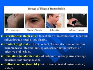
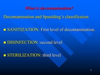
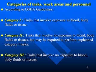
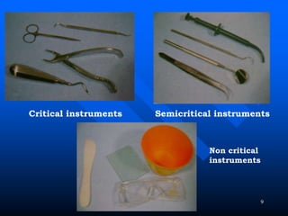
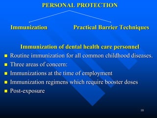
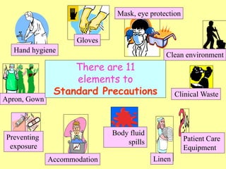
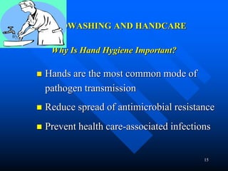
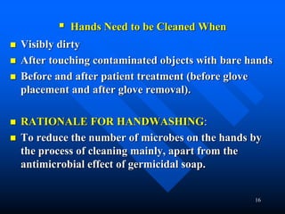
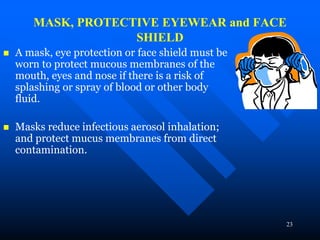
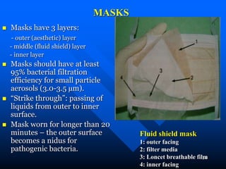
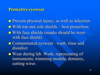
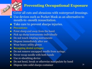
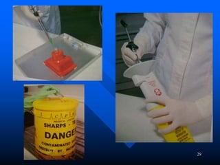
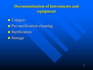
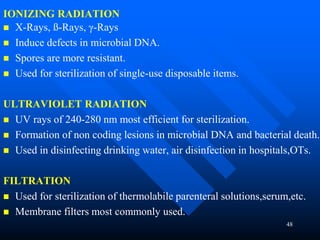
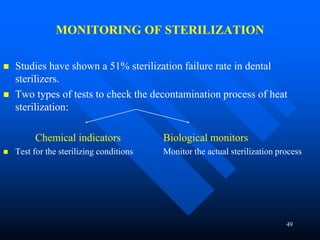
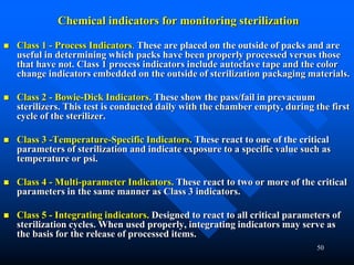
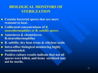
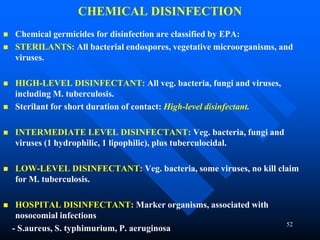
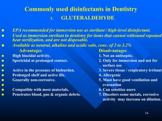
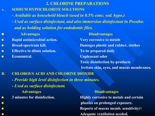
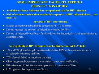
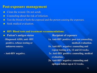
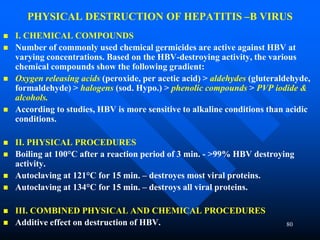
We can can minimize the risks of disease transmission to our self and to the patients in the dental office through carefully following the infection control and safety guidelines,
Dr. Hesham Dameer
Cross infection control in dentistry (Few basic points)Sumaiya Hasan
Recently, dentistry has been one of the few reasons of the spread of some major diseases such as hepatitis etc. If proper cross infection control is maintained by taking few precautions then this transfer of diseases can be stopped. This presentation contains only some basic precautions which should be taken to prevent cross infection.
The Indian Dental Academy is the Leader in continuing dental education , training dentists in all aspects of dentistry and
offering a wide range of dental certified courses in different formats.for more details please visit
www.indiandentalacademy.com
A concise and brief presentation on cleaning and shaping of root canals. Colorful and well pictured. Ideal for UG students and PG students to get a good understanding of BMP techniques.
STERILIZATION AND DISINFECTION IN A DENTAL CLINIC pptVineetha K
One of the basic things you need to know before starting a dental clinic. This presentation covers the basics of sterilization and disinfection in a dental setting.
Occupational Blood Borne Infections: Prevention is Better than CureApollo Hospitals
Viral infections like HIV, hepatitis Band C virus pose a big risk to the contacts of individuals with high risk behaviour as well as to the attending health care workers. Blood, semen, vaginal and other potentially infectious materials can transmit the infection to the susceptible contacts. Universal precautions should be strictly implemented during clinical examination, laboratory work and surgical procedures to prevent transmission to the health care providers. Health care workers should receive vaccination for hepatitis B infection. An inadvertent exposure should be managed with proper first aid and infectivity of the source and severity of exposure should be assessed. Severity of exposure is based on the nature and area of exposed surface, mode of injury and volume of infective material. Post-exposure prophylaxis (PEP) should be started as soon as possible after a proper counseling about the effectiveness of post-exposure prophylaxis, side effects and risk of carrying the infection to his familial contacts and its prevention.
this presentation involves the various sterilization and asepsis procedure that can be carried out in our dental clinics for proper maintenance of surgical as well as other procedures.
Safety precautions in the clinic and laboratory.pptxMustafa Al-Ali
Safety precautions in the clinic and laboratory.
Mustafa al-ali, 48
Safety precautions in the clinic and laboratory
Safety precautions in the dental clinic and laboratory are crucial to protect both patients and dental healthcare professionals. Here are some key safety measures to consider:
Personal Protective Equipment (PPE)
Hand Hygiene
Sterilization and Disinfection
Waste Management
Radiation Safety
Emergency Preparedness
Chemical Safety
Ergonomics
Personal Protective Equipment (PPE)
Personal protective equipment (PPE) should be selected based on risk assessment and tasks to be performed.
These items are designed to provide a protective barrier during dental procedures and through the sterilization process. PPE must also be considered for patients as they enter the facility and provided to administrative staff who may be screening them upon arrival.
Personal Protective Equipment (PPE)
Gown
Dental Hygiene Care Professionals (DHCP) should wear protective clothing (eg, gowns, jackets) to prevent contamination of scrubs and to protect the skin from exposure to blood and bodily fluids.
Sleeves should be long enough to protect the forearms.
Protective clothing should be changed after use or when it becomes visibly soiled by blood or other bodily fluids.
DHCP should remove protective clothing before leaving the work area.
Personal Protective Equipment (PPE)
Eyewear/Face Shields
Protective Eyewear
DHCP should wear protective eyewear with solid side shields or a face shield during procedures likely to generate splashes or sprays of blood or bodily fluids or the spatter of debris. Reusable protective eyewear should be cleaned with soap and water, and when visibly soiled, disinfected between patients.
Personal eyeglasses are not considered PPE.
Protective eyewear should be provided to patients.
Face Shields
Face shields provide full-face coverage.
Must be worn with a face mask.
Personal Protective Equipment (PPE)
Gloves
DHCP should wear gloves to prevent contamination of their hands when touching mucous membranes, blood, saliva, or other potentially infectious materials and to reduce the likelihood that microorganisms on their hands will be transmitted to patients during patient care.
Gloves should be used for one patient only and discarded appropriately after use.
Hand hygiene should be performed prior to donning gloves and immediately after glove removal.
Hand Hygiene
Hand hygiene is extremely important to prevent the spread of the SARS CoV-2 virus. It also interrupts the transmission of other viruses and bacteria, thus reducing the overall burden of disease, Dental healthcare facilities should ensure that hand hygiene supplies are readily available in every patient care location.
Pre-washing considerations
Remove jewelry, ring, watches, or bracelets
Remove artificial nails if present.
Cover skin cuts, abrasions, breaks or cracks with waterproof adhesive dressings.
Use running water; avoid dipping or washing hands in a basin of standing water
Rasamanikya is a excellent preparation in the field of Rasashastra, it is used in various Kushtha Roga, Shwasa, Vicharchika, Bhagandara, Vatarakta, and Phiranga Roga. In this article Preparation& Comparative analytical profile for both Formulationon i.e Rasamanikya prepared by Kushmanda swarasa & Churnodhaka Shodita Haratala. The study aims to provide insights into the comparative efficacy and analytical aspects of these formulations for enhanced therapeutic outcomes.
Title: Sense of Taste
Presenter: Dr. Faiza, Assistant Professor of Physiology
Qualifications:
MBBS (Best Graduate, AIMC Lahore)
FCPS Physiology
ICMT, CHPE, DHPE (STMU)
MPH (GC University, Faisalabad)
MBA (Virtual University of Pakistan)
Learning Objectives:
Describe the structure and function of taste buds.
Describe the relationship between the taste threshold and taste index of common substances.
Explain the chemical basis and signal transduction of taste perception for each type of primary taste sensation.
Recognize different abnormalities of taste perception and their causes.
Key Topics:
Significance of Taste Sensation:
Differentiation between pleasant and harmful food
Influence on behavior
Selection of food based on metabolic needs
Receptors of Taste:
Taste buds on the tongue
Influence of sense of smell, texture of food, and pain stimulation (e.g., by pepper)
Primary and Secondary Taste Sensations:
Primary taste sensations: Sweet, Sour, Salty, Bitter, Umami
Chemical basis and signal transduction mechanisms for each taste
Taste Threshold and Index:
Taste threshold values for Sweet (sucrose), Salty (NaCl), Sour (HCl), and Bitter (Quinine)
Taste index relationship: Inversely proportional to taste threshold
Taste Blindness:
Inability to taste certain substances, particularly thiourea compounds
Example: Phenylthiocarbamide
Structure and Function of Taste Buds:
Composition: Epithelial cells, Sustentacular/Supporting cells, Taste cells, Basal cells
Features: Taste pores, Taste hairs/microvilli, and Taste nerve fibers
Location of Taste Buds:
Found in papillae of the tongue (Fungiform, Circumvallate, Foliate)
Also present on the palate, tonsillar pillars, epiglottis, and proximal esophagus
Mechanism of Taste Stimulation:
Interaction of taste substances with receptors on microvilli
Signal transduction pathways for Umami, Sweet, Bitter, Sour, and Salty tastes
Taste Sensitivity and Adaptation:
Decrease in sensitivity with age
Rapid adaptation of taste sensation
Role of Saliva in Taste:
Dissolution of tastants to reach receptors
Washing away the stimulus
Taste Preferences and Aversions:
Mechanisms behind taste preference and aversion
Influence of receptors and neural pathways
Impact of Sensory Nerve Damage:
Degeneration of taste buds if the sensory nerve fiber is cut
Abnormalities of Taste Detection:
Conditions: Ageusia, Hypogeusia, Dysgeusia (parageusia)
Causes: Nerve damage, neurological disorders, infections, poor oral hygiene, adverse drug effects, deficiencies, aging, tobacco use, altered neurotransmitter levels
Neurotransmitters and Taste Threshold:
Effects of serotonin (5-HT) and norepinephrine (NE) on taste sensitivity
Supertasters:
25% of the population with heightened sensitivity to taste, especially bitterness
Increased number of fungiform papillae
Local Advanced Lung Cancer: Artificial Intelligence, Synergetics, Complex Sys...Oleg Kshivets
Overall life span (LS) was 1671.7±1721.6 days and cumulative 5YS reached 62.4%, 10 years – 50.4%, 20 years – 44.6%. 94 LCP lived more than 5 years without cancer (LS=2958.6±1723.6 days), 22 – more than 10 years (LS=5571±1841.8 days). 67 LCP died because of LC (LS=471.9±344 days). AT significantly improved 5YS (68% vs. 53.7%) (P=0.028 by log-rank test). Cox modeling displayed that 5YS of LCP significantly depended on: N0-N12, T3-4, blood cell circuit, cell ratio factors (ratio between cancer cells-CC and blood cells subpopulations), LC cell dynamics, recalcification time, heparin tolerance, prothrombin index, protein, AT, procedure type (P=0.000-0.031). Neural networks, genetic algorithm selection and bootstrap simulation revealed relationships between 5YS and N0-12 (rank=1), thrombocytes/CC (rank=2), segmented neutrophils/CC (3), eosinophils/CC (4), erythrocytes/CC (5), healthy cells/CC (6), lymphocytes/CC (7), stick neutrophils/CC (8), leucocytes/CC (9), monocytes/CC (10). Correct prediction of 5YS was 100% by neural networks computing (error=0.000; area under ROC curve=1.0).
Knee anatomy and clinical tests 2024.pdfvimalpl1234
This includes all relevant anatomy and clinical tests compiled from standard textbooks, Campbell,netter etc..It is comprehensive and best suited for orthopaedicians and orthopaedic residents.
NVBDCP.pptx Nation vector borne disease control programSapna Thakur
NVBDCP was launched in 2003-2004 . Vector-Borne Disease: Disease that results from an infection transmitted to humans and other animals by blood-feeding arthropods, such as mosquitoes, ticks, and fleas. Examples of vector-borne diseases include Dengue fever, West Nile Virus, Lyme disease, and malaria.
Basavarajeeyam is an important text for ayurvedic physician belonging to andhra pradehs. It is a popular compendium in various parts of our country as well as in andhra pradesh. The content of the text was presented in sanskrit and telugu language (Bilingual). One of the most famous book in ayurvedic pharmaceutics and therapeutics. This book contains 25 chapters called as prakaranas. Many rasaoushadis were explained, pioneer of dhatu druti, nadi pareeksha, mutra pareeksha etc. Belongs to the period of 15-16 century. New diseases like upadamsha, phiranga rogas are explained.
Adv. biopharm. APPLICATION OF PHARMACOKINETICS : TARGETED DRUG DELIVERY SYSTEMSAkankshaAshtankar
MIP 201T & MPH 202T
ADVANCED BIOPHARMACEUTICS & PHARMACOKINETICS : UNIT 5
APPLICATION OF PHARMACOKINETICS : TARGETED DRUG DELIVERY SYSTEMS By - AKANKSHA ASHTANKAR
2. 2
WHY IS INFECTION CONTROL IMPORTANT IN
DENTISTRY?
Infections present a significant hazard in the dental
environment.
Both patients and dental health care personnel
(DHCP) can be exposed to pathogens.
Contact with blood, oral and respiratory secretions,
and contaminated equipment occurs.
Proper procedures can prevent transmission of
infections among patients and DHCP.
3. 3
RATIONALE
Rationale for infection control is to control iatrogenic,
nosocomial infections among patients, and potential exposure
of care providers to disease causing microbes during provision
of care.
“Disease control or infection control”: means reducing the risk
of disease transmission.
“Occupational exposure”: Reasonably anticipated skin, eye,
mucous membrane, or parenteral contact with blood or OPIM
that can result from the performance of an employee's duties.
“Cross-infection” : disease transfer from one patient to the
other in the dental office.
4. 4
ROUTES OF DISEASE TRANSMISSION
Percutaneous (high-risk): Inoculation of microbes from blood and
saliva through needles and sharps.
Contact (high risk): Direct contact of non-intact skin or mucous
membranes to infected fluid, splash/spatter; tissue surfaces or
infective oral lesions.
Inhalation (moderate risk): of airborne microorganisms through
bioaerosols or droplet nuclei.
Indirect contact (low risk): with a contaminated instrument or
surface.
6. 6
What is decontamination?
Decontamination and Spaulding’s classification
SANITIZATION: First level of decontamination.
DISINFECTION :second level
STERILIZATION: third level
7. 7
Categories of tasks, work areas and personnel
According to OSHA Guidelines:
Category I : Tasks that involve exposure to blood, body
fluids or tissue.
Category II : Tasks that involve no exposure to blood, body
fluids or tissues, but may be required to perform unplanned
category I tasks.
Category III : Tasks that involve no exposure to blood,
body fluids or tissues.
8. 8
Adaptation of Spaulding’s Classification
Level Risks Control methods Materials/devices
Critical High Sterilization by:
Autoclave, Chemiclave
Dry heat, Full strength
gluteraldehyde
Items that pierce soft tissue, touch
bone. Scalpel blades, burs, extraction
forceps, elevators, needles, files, bone-
rongers, periodontal instruments, dental
explorers, biopsy punch, endodontic files
and reamers, and implants.
Semi-
critical
High Autoclave, Chemiclave
Dry heat, Full strength
gluteraldehyde
Items that enter the oral cavity, but do not
necessarily penetrate soft and hard tissue.
Mouth mirrors, handpiece, anesthetic
syringes, amalgam condensers, impression
trays, air/water syringe tips, high-volume
evacuator tips.
Non-
Critical
Moderate to
low
Surface disinfection by
intermediate level hospital
disinfectants –, phenols,
iodophors, quaternary
ammonium compounds
Items that do not enter the mouth or
penetrate soft tissues. Chair light handles,
instrument trays, high touch work
surfaces, bracket tables, chair controls,
Air/water syringes, hoses and dental
chairs.
Environm
ental
Low Disinfection with
intermediate to low level
disinfectant
Floors, walls and handles, not high touch
surfaces.
10. 10
PERSONAL PROTECTION
Immunization Practical Barrier Techniques
Immunization of dental health care personnel
Routine immunization for all common childhood diseases.
Three areas of concern:
Immunizations at the time of employment
Immunization regimens which require booster doses
Post-exposure
11. 11
Proposed protocol for HBV vaccine for Dental Health Care Workers in India
At the time of employment:
three 1 ml doses at 0, end of 1 month and 6 months i.m.
If a person is not immunized and is exposed to an infected patient’s body
fluids: A combination of Hepatitis B vaccine and an immunoglobulin (HBIg)
within 24 hours of exposure.
If exposed person does not want to take the immunization:
two doses of HBIg – dose 1 within 24 hours
- dose 2 25 to 30 days after exposure.
Proposed protocol for HBV vaccine for dental undergraduate and postgraduate
students and dental teaching faculty in India
Mandatory to be vaccinated against Hepatitis B with a three- dose regimen
and a booster.
Dental assistants, hygienists, mechanics and all those who come in direct
contact with patient care, cleaning and sterilization of instruments in the
institution.
Mandatory for institutions to provide free vaccinations.
13. 13
STANDARD / UNIVERSAL PRECAUTIONS
CDC recommends that all patients be treated as potentially
infectious.
Appropriate level of infection control measures apply to all
patients.
Level of infection control should be based upon the clinical
procedure to be carried out and reasonably anticipated risk.
Standard Precautions apply to blood and also
- Body fluids, secretions, and excretions except sweat,
whether or not they contain blood
– Non-intact (broken) skin
– Mucous membranes
– Saliva has always been considered a potentially infectious
material in dental infection control
14. Hand hygiene
Gloves
Mask, eye protection
Clean environment
Clinical Waste
Patient Care
Equipment
Linen
Body fluid
spills
Accommodation
Preventing
exposure
Apron, Gown
There are 11
elements to
Standard Precautions
15. 15
HANDWASHING AND HANDCARE
Why Is Hand Hygiene Important?
Hands are the most common mode of
pathogen transmission
Reduce spread of antimicrobial resistance
Prevent health care-associated infections
16. 16
Hands Need to be Cleaned When
Visibly dirty
After touching contaminated objects with bare hands
Before and after patient treatment (before glove
placement and after glove removal).
RATIONALE FOR HANDWASHING:
To reduce the number of microbes on the hands by
the process of cleaning mainly, apart from the
antimicrobial effect of germicidal soap.
17. 17
What should one use?
Washing hands with plain soap and water
Washing hands with water and antimicrobial soap
Chlorhexidine gluconate – 0.75% to 4% concentration (CHG) –
dispensed as liquid soap or foam
Parachlorometaxylenol (PCMX) liquid
Iodine liquid or Triclosan liquid, gel or foam
CHG 4% – as surgical scrub.
Hand –sanitizers : alcohol-based
: without alcohol
Recommended only when handwashing is impractical or cannot be
done.
Limitation: do not clean hands adequately.
18. 18
Surgical soaps should have following properties:
Substantivity or residual action
Broad-spectrum antimicrobial activity
Good kill-rate (i.e., fast or rapid action and reduce the
bacterial load efficiently)
Good detergent
Non-irritating to skin
Fast-acting
20. 20
GLOVES
Pathogenic microorganisms in blood, saliva and plaque can
contaminate the hands of DHCP;
These microorganisms can infect the host by passing through
dermal defects, and they can contaminate instruments and
surfaces.
Gloves prevent cross-contamination to patients and also protect
the hands of oral health care providers.
Gloving is not a substitute for handwashing!
22. 22
Recommendations for gloving
Hands must be washed before gloving and after removing gloves.
Check gloves for cuts or defects.
Double gloving reduces the chances of contamination through
inherent pin-holes.
Reuse of gloves increases risks. Microorganisms may enter
inherent pin-holes or tears.
Washing gloves weakens them, makes them tacky.
Gloves must be worn (clean, non-sterile gloves are adequate for non
invasive procedures) when in contact with blood, body fluids, secretions,
excretions and contaminated items / equipment; before touching mucus
membranes and non intact skin.
Gloves may need to be changed between tasks and procedures on the same
patient.
23. 23
MASK, PROTECTIVE EYEWEAR and FACE
SHIELD
A mask, eye protection or face shield must be
worn to protect mucous membranes of the
mouth, eyes and nose if there is a risk of
splashing or spray of blood or other body
fluid.
Masks reduce infectious aerosol inhalation;
and protect mucus membranes from direct
contamination.
24. 24
MASKS
Masks have 3 layers:
- outer (aesthetic) layer
- middle (fluid shield) layer
- inner layer
Masks should have at least
95% bacterial filtration
efficiency for small particle
aerosols (3.0-3.5 µm).
“Strike through”: passing of
liquids from outer to inner
surface.
Mask worn for longer than 20
minutes – the outer surface
becomes a nidus for
pathogenic bacteria.
Fluid shield mask
1: outer facing
2: filter media
3: Loncet breathable film
4: inner facing
25. 25
Protective eyewear
Prevent physical injury, as well as infection.
With top and side shields – best protection;
With face shields (masks should be worn
with face shield)
Contaminated eyewear : wash, rinse and
disinfect
Wear during lab. Work, reprocessing of
instruments, trimming models, dentures,
cutting wires
27. 27
Protective clothing: Apron / Gown / Footwear
Fluid resistant full-sleeved gown to protect the uniform from:
– soiling during procedures and patient care activities that are
likely to generate splashes or sprays of blood or body fluids.
– contamination with micro-organisms when bed-making, any
direct patient care or direct contact with the environment of
an isolated patient.
When removing visibly contaminated clothing, fold the soiled
area inside.
Send to commercial laundry or wash with hot water at 80º C for
10 minutes using strong detergent and bleach, if possible.
Always remove protective clothing before leaving the surgery.
Use protective footwear, to prevent contamination of the feet,
e.g. during operations. Remove contaminated footwear when
procedure is finished.
28. 28
Preventing Occupational Exposure
Cover all cuts and abrasions with waterproof dressings.
Use devices such as Pocket Mask as an alternative to
mouth-to -mouth resuscitation.
Take care to prevent sharps injuries.
Precautions:
Point sharp end away from the hand.
Pick up sharp instruments individually
Do not touch rotating instruments
Dispose immediately after use
Wear heavy utility gloves
Recapping dental syringes:
Do not remove uncapped needle from syringe.
Never recap needle with both hands
Use re-sheathing device
Do not bend, break or otherwise manipulate by hand
Dispose into solid sharps container
30. 30
Blood and Body Fluid Spillages
Disinfect all blood and body fluid spillages immediately
wearing protective clothing (gloves, apron and if risk of
splash, goggles).
Completely cover either by disposable towels, which are then
treated with 10,000 ppm sodium hypochlorite solution or by
sodium dichloroisocyanurate granules. At least 5 minutes must
elapse before the towels etc are cleared and disposed of as
clinical waste.
Wear appropriate protective clothing, which will include
household gloves, protective eyewear and a disposable apron
and, in the case of an extensive floor spillage, protective
footwear. Good ventilation is essential.
31. 31
Ensure that the clinical areas are clean.
Particular attention must be paid to cleaning of
horizontal surfaces, floors, beds, bed-side equipment
and other frequently touched surfaces.
If areas are not clean the domestic supervisor must be
informed.
Environment
32. Clinical Waste: Recommended labeling and
color coding
COLOR
CODING
TYPE OF
CONTAINER
WASTE CATEGORY TREATMENT
OPTIONS
YELLOW Plastic bag Human anatomical waste, animal
waste, microbiological & biotech.
waste. Solid waste (items
contaminated with blood/body
fluids, eg. Cotton, soiled dressing,
etc.)
Incineration / deep
burial
RED Disinfected
container /
plastic bag
Microbiological & biotech waste,
disposable items other than waste
sharps, such as tubings, catheters,
i.v sets, solid waste.
Autoclaving /
microwaving / chemical
treatment
BLUE / WHITE
TRANSULECT
Plastic bag /
puncture proof
container
Waste sharps, disposable items. Autoclave / microwave
/ chemical treatment &
destruction / shredding
BLACK Plastic bag Discarded medicines, cytotoxic
drugs, incineration ash, chemical
waste.
Disposal in secured
landfill.
33. 33
Additional Measures
Rubber dams
Surface covers
High volume aspiration
Pre operative patient rinse
Pre treatment tooth brushing
Use of rubber cups instead of
bristle brushes during polishing
Antiretraction valves
Flushing water through the
handpiece between patients
34. 34
Surface covers
Single-use disposable, water proof barriers
Light handles, hand operated chair controls,
suction hoses, chairs, bracket tables
clear plastic wrap, aluminium foil, paper
with impervious plastic backing,
polyethylene sheets and tubing.
Must be replaced after each patient and
disposed of as contaminated waste
39. 39
Pre-sterilization cleaning
Separation of waste and instruments.
Instruments securely taken to the reprocessing area –
handpieces removed, all other items need to be cleaned first.
Cleaning of instruments:
To reduce the bioburden, remove accumulated debris.
» Hand Scrubbing Ultrasonic Cleaning
Inspection and packaging of cleaned instruments.
40. 40
Inspection and packaging of cleaned instruments
After sonication – rinse, dry, inspect for residual debris.
Packing of instruments prior to sterilization will depend
upon the type of sterilization, and nature of items to be
sterilized.
Packaged instruments can be stored; non packaged
instruments have to be used immediately.
Clearview sterilization pouches – single-wall paper, sealed
nylon, and paper/plastic pouches.
If instruments are to be cold sterilized, they should not be
packaged.
41. 41
INSTRUMENT STERILIZATION
PHYSICAL AGENTS CHEMICAL AGENTS
• Heat – moist 1. Agents acting on the cell membrane
- dry - Surface acting agents (quaternary
ammonium compounds)
• Ionizing radiation - Phenols
X-rays - Organic solvents (alcohol, chloroform)
ß-rays 2. Agents that denature proteins
Gamma rays - Acids and alkalies
• Ultraviolet rays 3. Agents acting on functional groups of
• Filtration proteins
- Heavy metals (copper, silver, mercury)
- Oxidizing agents (iodine, chlorine, hydrogen
peroxide)
- Alkylating agents (formaldehyde, ethylene
oxide)
42. 42
MOIST HEAT
I. Temp. below 100°C : a. HOLDER METHOD
b. FLASH METHOD
II. Temp. around 100°C : a. TYNDALLIZATION
b. HOT WATER BOILERS
III. Temp. above 100°C : AUTOCLAVES
Mechanism of microbial inactivation by moist heat:
Structural damage to cell membrane.
Coagulation of proteins, and denaturation of spore enzymes.
Damage to bacterial chromosomes.
43. 43
STEAM AUTOCLAVE
Charles Chamberland in 1879.
Sterilization using steam under pressure
Temp.-time combinations:
Temp. °C (Pressure) Minimum hold time
134-138 ( 30 psi) 3-5 min.
121-124 ( 15 psi) 15-20 min.
Characteristics :
Destroys all forms of microbial life, including bacterial endospores in the
recommended time;
Additional ‘safety factor’ interval must be allowed…. Reach and maintain
121°C for 45 min.
Sterilization intervals vary with load size, nature of materials, instruments;
and packaging.
ADVANTAGES DISADVANTAGES
-short cycle time - Corrosion of unprotected carbon steel
-Good penetration - Dulling of cutting edges
-Wide range - May destroy heat sensitive materials.
44. 44
Areas of problems:
- Faulty preparation of materials (packaging)
- Improper functioning of sterilizer (temp. / pressure)
- Presence of air in chamber ( delay upto 10 times longer)
- Excess water in steam ( passageway for microorganisms)
- Corrosion of carbon steel instruments (1% sod. Nitrite)
Acceptable materials:
- cloth goods, high quality S/S instr., glass slabs, stones, dishes, heat resistant
plastics, handpieces that can be autoclaved.
Limitations of use:
- rusting/corrosion of carbon steel instr.
- needles, oil, wax, dry powder should not be autoclaved
Other types:
Statim cassette autoclave Autoclave with pre-vacuum &
post-vacuum features
46. 46
Chemical Vapour Sterilizer Ethylene oxide sterilizer
Parameters: 132°C at 20-40 psi, 20 min. Room temp. (25°C), 10-16 hrs.
Chemical: Deodorized alcohol, formald., Ethylene oxide gas
ethyl methyl ketone soln.
Advantages: Short cycle High penetrability.
No rusting Suitable for heat labile materials.
Instr. Dry at the end of cycle No residue on evaporation.
Does not dull cutting edges Suitable for materials that cannot
be exposed to moisture.
Suitable for Ortho. s/s wires
Disadv.: Instr. Must be completely dried Long cycle time.
Destroy heat sensitive plastics Tissue irritation
Chemical odor Explosive (“spark shield”)
Acceptable Metal instruments Suction tubing, handpieces, radio
materials graphic film holdrs, prosth. Appl.
47. 47
DRY HEAT
Less efficient than moist heat; bacterial spores are more resistant – may require
temp. of 140°C for 3 hours to get killed.
Two methods:
Flaming Hot air oven
Mechanism of action: oxidation, protein denaturation
Temperature Holding time
› 160°C › 120 min.
› 170°C › 60 min.
› 180°C › 30 min.
Suitable materials: glassware, glass syringes, oils, oily injections, metal instr, mirror
Advantages: - Does not dull cutting edges
- Does not rust or corrode.
Disadv. : - Long cycle
- Poor penetration
- May discolor and char fabric
- Destroys heat labile items.
48. 48
IONIZING RADIATION
X-Rays, ß-Rays, γ-Rays
Induce defects in microbial DNA.
Spores are more resistant.
Used for sterilization of single-use disposable items.
ULTRAVIOLET RADIATION
UV rays of 240-280 nm most efficient for sterilization.
Formation of non coding lesions in microbial DNA and bacterial death.
Used in disinfecting drinking water, air disinfection in hospitals,OTs.
FILTRATION
Used for sterilization of thermolabile parenteral solutions,serum,etc.
Membrane filters most commonly used.
49. 49
MONITORING OF STERILIZATION
Studies have shown a 51% sterilization failure rate in dental
sterilizers.
Two types of tests to check the decontamination process of heat
sterilization:
Chemical indicators Biological monitors
Test for the sterilizing conditions Monitor the actual sterilization process
50. 50
Chemical indicators for monitoring sterilization
Class 1 - Process Indicators. These are placed on the outside of packs and are
useful in determining which packs have been properly processed versus those
that have not. Class 1 process indicators include autoclave tape and the color
change indicators embedded on the outside of sterilization packaging materials.
Class 2 - Bowie-Dick Indicators. These show the pass/fail in prevacuum
sterilizers. This test is conducted daily with the chamber empty, during the first
cycle of the sterilizer.
Class 3 -Temperature-Specific Indicators. These react to one of the critical
parameters of sterilization and indicate exposure to a specific value such as
temperature or psi.
Class 4 - Multi-parameter Indicators. These react to two or more of the critical
parameters in the same manner as Class 3 indicators.
Class 5 - Integrating indicators. Designed to react to all critical parameters of
sterilization cycles. When used properly, integrating indicators may serve as
the basis for the release of processed items.
51. 51
BIOLOGICAL MONITORS OF
STERILZATION
Contain bacterial spores that are more
resistant to heat.
Calibrated concentrations of B.
stearothermophilus or B. subtilis spores.
Autoclaves & chemiclaves:
B.stearothermophilus
B. subtilis: dry heat ovens & ethylene oxide
Intra-office biological monitoring highly
recommended.
Positive culture results indicate that not all
spores were killed, and items sterilized may
not be sterile.
52. 52
CHEMICAL DISINFECTION
Chemical germicides for disinfection are classified by EPA:
STERILANTS: All bacterial endospores, vegetative microorganisms, and
viruses.
HIGH-LEVEL DISINFECTANT: All veg. bacteria, fungi and viruses,
including M. tuberculosis.
Sterilant for short duration of contact: High-level disinfectant.
INTERMEDIATE LEVEL DISINFECTANT: Veg. bacteria, fungi and
viruses (1 hydrophilic, 1 lipophilic), plus tuberculocidal.
LOW-LEVEL DISINFECTANT: Veg. bacteria, some viruses, no kill claim
for M. tuberculosis.
HOSPITAL DISINFECTANT: Marker organisms, associated with
nosocomial infections
- S.aureus, S. typhimurium, P. aeruginosa
53. 53
Properties of an ideal disinfectant:
- Broad spectrum antimicrobial
- Fast acting
- Not affected by physical factors
- Non toxic, non irritating
- Surface compatibility
- Residual effect
- Easy to use
- Odorless
- Economical.
Types of Disinfectants:
Immersion disinfectants Surface disinfectants
54. 54
Commonly used disinfectants in Dentistry
1. GLUTERALDEHYDE
EPA recommended for immersion use as sterilant / high-level disinfectant.
Used as immersion sterilant in dentistry for items that cannot withstand repeated
heat sterilization, and are not disposable.
Available as neutral, alkaline and acidic soln, conc. of 2 to 3.2%
Advantages Disadvantages
High biocidal activity. 1. Not an antiseptic.
Sporicidal at prolonged contact. 2. Only for immersion and not for
surface use
Active in the presence of bioburden. 3. Severe tissue / respiratory irritant
Prolonged shelf and active life. 4. Allergenic
Generally non-corrosive. 5. Must have good ventilation and
evacuation
Compatible with most materials. 6. Can sensitize users
Penetrates blood, pus & organic debris. 7. Discolors some metals, corrosive
activity may increase on dilution.
55. 55
2. CHLORINE PREPARATIONS
A. SODIUM HYPOCHLORITE SOLUTIONS
- Available as household bleach (used in 0.5% conc. sod. hypo.)
- Used as surface disinfectant, and also immersion disinfectant in Prostho.
and as holding solution for endodontic files.
Advantages Disadvantages
Rapid antimicrobial action. Very corrosive to metals
Broad-spectrum kill. Damages plastic and rubber, clothes
Effective in dilute solution. To be prepared daily
Economical. Unpleasant odor
Toxic disinfection by-products
Irritate skin, eyes, and mucus membranes.
B. CHLOROUS ACID AND CHLORINE DIOXIDE
- Provide high level disinfection in three minutes.
- Used as surface disinfectant.
Advantages Disadvantages
3 minutes for disinfection. Highly corrosive to metals and certain
plastics on prolonged exposure.
Reports of mucus memb. sensitivity.
Adequate ventilation needed.
56. 56
3. PHENOLS AND DERIVATIVES
- Carbolic acid – classical antiseptic for surgical procedures.
- Synthetic phenols – currently approved by EPA: Biphenols and triphenols.
- Used as surface and immersion disinfectants.
Advantages Disadvantages
Triphenols are better than Dual Phenols May affect some polymers.
Broad Spectrum Kill. Some have film accumulation.
Compatible with most materials. May not be used in neonatal and
pediatric practices due to possible
adverse reaction.
Residual biocidal action. Should be prepared freshly.
Fast acting Epithelial toxicity in exposed tissues.
4. IODINE AND IODOPHORS
- Iodine: Oldest skin antiseptic.
- skin irritation, hypersensitivity, corrosion of metals, staining of skin and
clothing.
- Iodophors; complex of elemental iodine or triiodide, with a carrier. Used for
skin preparation for surgery, effective handwashing antiseptics.
Advantages Disadvantages
Broad spectrum Unstable at high temperatures
Short biocidal activity Dilution and contact time critical
Few reactions · Solution to be prepared daily
Residual biocidal action May discolor some surfaces
57. 57
5. Hydrogen Peroxide (0.05%)
- Recently introduced disinfectant.
-Release nascent oxygen, may be of use for surface disinfection.
- Not currently recommended by EPA.
Advantages Disadvantages
Rapid antimicrobial action Not many reported disadvantages as it
Broad-spectrum kill is still new in the market
Effective in dilute solution Can be corrosive on metals, and dangerous
to skin (burns) if used in high conc.
Prolonged shelf and active life.
Compatible with metals, plastics
and impression materials.
Good for use in dental laboratories.
58. 58
6. ALCOHOLS
- Not recommended by ADA or CDC as disinfectant for dental practice.
- Synergistic action with phenolics. May be used for surface disinfection.
Advantages Disadvantages
70% Isopropyl alcohol, ethyl alcohol- Not recommended for use as surface disinfectants.
Bactericidal, virucidal, tuberculocidal. Activity rapidly diminishes in presence of blood
and saliva.
Corrosive on metals, destroy rubbers & plastics.
No sporicidal activity.
7. QUARTERNARY AMMONIA COMPOUNDS
- Cationic surface-active disinfectants.
- Not approved by ADA for instrument or surface disinfection.
Advantages Disadvantages
Bactericidal at low conc. Not tuberculocidal, sporicidal or virucidal against
Particularly active against G +ve bacteria. hydrophilic viruses.
Good cleaners Inactivated by hard water, inactivated by organic
matter.
Some have M.tuberculosis kill claim Alcohol based quats may affect low viscosity
impression dimensional stability.
60. 60
Dental Handpieces
Contamination:
- Surface contamination by direct contact
- Internal – through cartridge chamber
- Water retraction
Current Guidelines:
- Sterilization by autoclave, chemiclave or newer generation, shorter
cycle dry heat ovens
- Disinfection – as per manufacturer’s recommendations
Precautions:
- Proper lubrication prior to sterilization.
- Do not sterilize with other instruments.
- Do not operate handpiece without bur; do not sterilize with the bur
installed.
- Run the handpiece for 20 sec. after use.
- Do not immerse in disinfectant solution, gluteraldehyde should not
be used
61. 61
Newer handpieces
- Can withstand repeated heat sterilization
- Solid fibre-optic rod
- New heat resistant cartridges
- Ceramic bearings
- Lubrication-free handpieces
Two new systems:
The turbonet system Decident disposable
disinfectant sleeve
62. 62
Rotary instruments:
- Diamond and carbide burs – autoclave
- Carbon-steel burs – chemical vapour sterilization
- glass bead sterilizer
- Retentive pin-twist drills – steam autoclave, chemiclave
Visible light-curing units
Triple syringe / air-water syringe
Ultrasonic scaler
Compressor:
- Clean, oil-free air
- Good quality filter
- Drain the air receiver daily
- Service regularly
63. 63
Dental Unit Water Supply
Contamination:
- Retraction valves
- Internal contamination
- Water supply
Acceptable microbial counts < 500 CFU/ml
Precautions:
- Check/ Anti-retraction valves
- Sterilization
- Flushing the air/water lines
Disinfection of the Dental unit:
Within the dental unit:
- Hydrogen peroxide, hypo, gluteraldehyde.
Disinfection of the unit water line:
- Povidone iodine, sterile water.
Disinfection of the mains water supply:
- 15% Sod. Hypo. for 10 min. each day.
64. 64
INFECTION CONTROL IN PROSTHETICS
Semi-critical instruments Non-critical:
and items:
- Impressions - Articulators and face bows
- Prosthesis which have been worn - Mixing bowls and spatulas
- Face-bow fork - Shade and mold guides
- Wax knife - Prosthetic rulers
- Prosthesis at try-in stage - Wax rims (discard)
- Metal dispensing syringes
for impressions
- Bite blocks
- Polishing stones and rag wheels
- Impression trays returned from the lab
( Al. or Cr. Plated-heat sterilization,
plastic trays-discard)
65. 65
Impressions
Rinse under running water
Alginate impressions: hypo 1:5 or 1:10; 2% gluteraldehyde
Results: insignificant distortion for study casts, not for master casts;
surface quality not adversely affected
Reversible hydrocolloid impressions: iodophor (1:213), bleach
(1:10), 2% gluteraldehyde
ZOE and compound impressions: limited data available
ZOE – 2% gluteraldehyde or iodophor; compound – hypo 1:10
Elastomeric impressions: gluteraldehyde 1 or 2 % ,iodophor, 5.2%
sodium hypochlorite, chlorine dioxide (diluted)
Prosthesis:
Metal dentures Acrylic dentures
- Iodophors – 1st choice - Sodium hypochlorite
- Hypo 1:5 for 5 min. - Gluteraldehyde with phenolic
buffer (sporicidin) should not be used
66. 66
Orthodontics
Orthodontists have second highest incidence of Hepatitis B among
dental personnel.
Orthodontic pliers – damage after repeated autoclaving
- lubricate the hinges
- dip in 1% sodium nitrite
Chemical vapour sterilization – minimal damage
Convection heat, rapid heat sterilization
Bands, wires, brackets – band cassettes – rapid dry heat, steam or
chemical vapour sterilization
2% gluteraldehyde overnight
67. 67
Endodontics
Precleaning disinfection: holding
solution; synthetic phenols, hypo
5.2%
Ultrasonic cleaning
Sterilization: instrument trays –
chemiclave or autoclave
Gutta percha points: hypo 1:5 for 5
minutes
Glass-bead/hot salt sterilizers
For endodontic files and rotary instruments.
Temp. 218-246°C for a minimum 15 sec.
immersion
Hot salt is preferable to glass beads
68. 68
Dental Radiology
X-ray equipment – surfaces should be
covered or disinfected after use
- X-ray cone, tube head, exposure control and
panels
- Non-disposable film holders , panoramic bite
blocks
– autoclave, chemiclave
Wear gloves while taking radiographs
Contamination of radiographs: saliva, blood
- Plastic envelopes or cling films
Dark room and radiographic processing
equipment:
- Disinfect – counter tops, shelves, process tank
covers
- Wear gloves while processing films
- Loading compartment should be disinfected
69. 69
Oral Surgery / Implantology
Additional measures
Precautions-
- Thorough hand-scrubbing
- Sterile disposable gowns,masks, head covers, eye protection
- Contamination of surfaces should be minimized
- Pre-operative rinsing, swab the incision area
- Sterile irrigant or coolant
- Proper reprocessing of instruments
Extracted teeth, biopsy specimens and tissues
- Potentially infectious, medical waste
- Decontamination by heat sterilization, immersion in 5000ppm bleach,
7% H2O2 or gluteraldehyde; storage – 10% formalin.
- Research purpose – 0.05% thymol solution
70. 70
Electrosurgery and LASER
Smoke and by-products
Toxic gases – HCN, Benzene, Formaldehyde
Tissue debris, microbes and viruses
Full PPE, high speed suction, improve air circulation
New emerging diseases – Creutfeldt Jakob Disease (CJD)
and Prion related diseases
A type of a fatal degenerative disease of central nervous system
Caused by abnormal “prion” protein
also been identified in the tonsils, eye tissue, and pituitary glandular
tissue.
One case per million population worldwide
Prions are resistant to conventional physical and chemical methods of
decontamination.
71. 71
If a patient with known status, only treat urgent
condition with following additional precautions:
Use single-use disposable items and equipment
Consider items difficult to clean (e.g., endodontic
files, broaches) as single-use disposable
Keep instruments moist until cleaned
Clean and autoclave for longest cycle; upto 1
hour sterilization time.
Do not use flash sterilization
72. Toothbrush disinfection
What is the need for toothbrush disinfection?
We, as clinicians talk so much about daily disinfection procedures
for our instruments and working environment…
However, we neglect to disinfect the one thing that we use to clean
our mouth daily…..our toothbrush.
Remember….the most fertile breeding ground for the microorganisms
in our bathroom is our “toothbrush”.
Microorganisms including Streptococcus mutans, Bacteroids,
Clostridium, Staphylococcus, alpha hemolytic streptococci,
Candida albicans, and others have been isolated from
toothbrushes.
Studies have shown intra-oral translocation of these bacteria. Oral
hygiene aids can harbor a wide range of microorganisms, also
contribute to bacteremia, especially in patients with severe
periodontitis.
73. Options for toothbrush disinfection
Chemical Disinfectants: 1% sod. hypochlorite and 0.12% CHX for
20 hours
Listerine for 20 minutes after brushing.
Brush sprays: Solution consisting of activated ethanol 40% v/v with
a biocide (parabens).
UV light sanitizers: Constant stream of UV light for 3 min.
Modified brushes:
Ozone toothbrush: Perforated brush head for improved hygiene.
- Venturi and coanda effects.
Coated toothbrush filaments:
- Zeolithic crystals: crystals with Ag and
Zn ions; long-term contact antibacterial
activity.
- CHX Coatings
74. 74
NEWER TECHNOLOGIES ON THE HORIZON
E-BEAM STERILIZATION
E-beam, a concentrated, highly charged stream of electrons, generated by the
acceleration and conversion of electricity. High-energy electron beams are
typically required to achieve penetration of the product and packaging.
Mechanism of action: High energy electrons alter various chemical and
molecular bonds, including the reproductive cells of microorganisms.
Adv. – short exposure time; compatibility with most materials, including
plastics and resins; no residues.
Method of choice for processing for products of high volume/low value such as
syringes, or low volume/high value such as implants.
One of the cheapest methods for terminal sterilization of products and
packages.
75. 75
HYDROGEN PEROXIDE GAS PLASMA
The system injects and vaporizes a solution of 59% hydrogen peroxide into
the chamber, killing any bacteria on any package and product surfaces the
vapor can reach.
Next, an electromagnetic field is created in the chamber, creating a
plasma cloud that generates free radicals that kill any remaining
bacteria. At the end of the process, the free radicals lose their high
energy, and the hydrogen peroxide converts to water and oxygen
molecules.
Low-volume, high-value devices, particularly biological tissues, and implants.
Problems: Small sterilizer volume chamber; expensive; low
penetrating ability; not effective with paper, cellulose, linen.
76. 76
BRIGHT LIGHT
Another emerging technology not currently in wide use but still promising is
the use of bright light.
Short pulses of high-intensity, broad-spectrum white light to kill
microorganisms without heat, chemicals, or ionizing radiation. The
light lasts for a few hundred millionths of a second and is 20,000 times
brighter than sunlight.
The light can go through any material that can transmit the appropriate
wavelengths, such as polypropylene and polyethylene.
The pulsed light offers the potential to perform terminal sterilization
on top of aseptic processing for injectable and parenteral fluids.
77. 77
OZONE
For instruments sensitive to repeated heat and moisture cycles, molecular
ozone is a low-temperature sterilization process.
oxygen gaseous ozone.
electric current
70 to 90% humidification phase.
The resultant gas is then vented into a sterilization chamber where the
microbes are eliminated through oxidation.
This system is both non-toxic and environmentally sound.
Disadvantages: limited penetrability, possible degradation of some plastics
and possible corrosion of metals.
This alternative is still in research and development and is not available at
this time.
78. 78
SOME IMPORTANT FACTS RELATED TO
DISINFECTION OF HIV
Available evidence indicates low occupational risk for HIV infection.
Risk of seroconversion after needlestick exposure to HIV infected blood….less
than 1%.
Survival of HIV after drying
Studies carried out using highly concentrated HIV samples.
Drying reduces the amount of infectious virus by 90-99%.
Drying of infected blood/body fluids reduces the theoretical risk of transmission to
essentially zero.
Susceptibility of HIV to disinfection by disinfectants & U.V. light
1% and 2% gluteraldehyde inactivated cell-free HIV within one minute; cell-
associated virus more resilient.
70% alcohol failed to inactivate the virus.
Chlorine, phenols, quaternary ammonium compounds –effective.
Effectiveness of disinfectants compromised in presence of blood.
U.V light and boiling water – effective.
79. 79
Post-exposure management
Clean the wound. Do not scrub.
Counseling about the risk of infection.
Test the blood of both the exposed and the person causing the exposure.
Seek medical evaluation.
HIV Blood tests and treatment recommendations:
Patient’s antigen status Recipient of exposure
- Diagnosed AIDS, anti- HIV 1a. Anti-HIV positive: post test counseling,
positive, refuses testing or medical evaluation.
unknown source. 1b. Anti-HIV negative: counseling and
repeat testing at 6, 12 and 24 weeks.
- Anti-HIV negative. 2a. Anti-HIV positive: counseling, medical
evaluation.
2b. Anti-HIV negative: counseling and
optional follow-up at 12 weeks.
80. 80
PHYSICAL DESTRUCTION OF HEPATITIS –B VIRUS
I. CHEMICAL COMPOUNDS
Number of commonly used chemical germicides are active against HBV at
varying concentrations. Based on the HBV-destroying activity, the various
chemical compounds show the following gradient:
Oxygen releasing acids (peroxide, per acetic acid) > aldehydes (gluteraldehyde,
formaldehyde) > halogens (sod. Hypo.) > phenolic compounds > PVP iodide &
alcohols.
According to studies, HBV is more sensitive to alkaline conditions than acidic
conditions.
II. PHYSICAL PROCEDURES
Boiling at 100°C after a reaction period of 3 min. - >99% HBV destroying
activity.
Autoclaving at 121°C for 15 min. – destroyes most viral proteins.
Autoclaving at 134°C for 15 min. – destroys all viral proteins.
III. COMBINED PHYSICAL AND CHEMICAL PROCEDURES
Additive effect on destruction of HBV.
81. 81
Status of Dental Infection Control and Safety in India
Level of infection control in India still far behind.
Requires more efforts and development of formal programs.
Policy through grass-roots education should address the following:
- Training for dental students & practitioners.
- Introduction and provision of instruments and equipment needed.
- Craft the recommendations.
- Surveillance of safe practices.
- Dissemination of information for patients.
- Setting up HIV and blood borne disease dental care centers.
- Expanding the duties of Public Health Dentistry/ Community
Dentistry Departments to provide out-reach dental care to rural HIV
and other BBP infected patients.
82. 82
CONCLUSION
Level of infection control in India still far behind, and a number of questions
regarding infection-control practices and their effectiveness still remain
unanswered.
Requires more efforts and development of formal programs.
Policy through grass-roots education should address the following:
- Training for dental students & practitioners.
- Surveillance of safe practices.
- Establish routine evaluation of the infection-control program, including
evaluation of performance indicators.
- Setting up HIV and blood borne disease dental care centers.
- Expanding the duties of Public Health Dentistry/ Community Dentistry
Departments to provide out-reach dental care to rural HIV and other BBP
infected patients.
83. 83
REFERENCES
1. Wood PR. Cross Infection Control in Dentistry – A practical illustrated guide.
Mosby publishers.
2. Cottone JA, Terezhalmy GT, Molinari JA, editors. Practical Infection Control in
Dentistry. 2nd Ed. Williams & Wilkins, Philadelphia, PA. 1995.
3. Kohli A, Puttaiah R. Infection Control and Occupational Safety
Recommendations for Oral Health Professionals. Dental Council of India, New
Delhi.
4. Vishwanathan R, Ranganathan K. HIV disease in India – Handbook for Dental
Professionals. Produced by Ragas Dental College Chennai in collaboration with
YRG Care – Chennai. Dental Council of India, New Delhi.
5. Prabhu SR, Rao B, Kohli A. HIV and AIDS in Dental Practice – Handbook for
Dental Practitioners. Dental Council of India, New Delhi.
84. 84
6. World Health Organization. SEARO Regional Health Papers no. 18. A Manual on
Infection Control in Health Facilities. World Health Organization. Regional Office
for South-East Asia. New Delhi, 1988.
7. Kohn WJ et al. (2003). Guidelines for Infection Control in Dental Health-Care
Settings – 2003. [Online] MMWR Recommendations and Reports 2003 Dec 19; 52
(No. RR—17). Available: http://www.cdc.gov/mmwR/PDF/rr/rr5217.pdf [20th May
2008]
8. Sterigenics. Sterilization Alternatives. Electron Beam Radiation. [Online]. Available:
http://www.sterigenics.com/services/medical_sterilization/contract_sterilization/elec
tron_beam_radiation/sterilization_alternatives__electron_beam_radiation.pdf [29th
May 2008]
9. Allen D (1998). The Changing Face of Sterilization. [Online] Pharmaceutical and
Medical News Packaging Magazine 1998 Nov; 35-46. Available:
http://www.devicelink.com/pmpn/archive/98/11/004.html [29th May 2008]
10. Molinari JA. Infection control: Its evolution to the current standard precautions. J
Am Dent Assoc 2003; 134: 569-574.
85. 85
11. Park K. Park’s Textbook of Preventive and Social Medicine. 19th ed. Bhanot
Publishers, Jabalpur.
12. Thraenhart O, Jursch C. Measures for Disinfection and Control of Viral Hepatitis.
In: Block SS. Disinfection, Sterilization and Preservation. 5th Ed. Lippincott
Williams & Wilkins, 2001.
13. Curran E 2003. A Self-directed Learning Unit ON Standard Precautions. [Online]
developed from NHS Greater Glasgow Control of Infection Policy 2003. Available:
shscintranet.gcal.ac.uk/student/Documents/PodDocs/sdlu_sp1.ppt [29th December
2008]