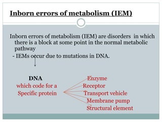
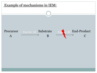
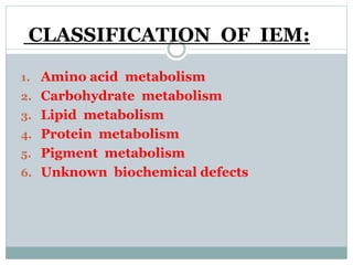
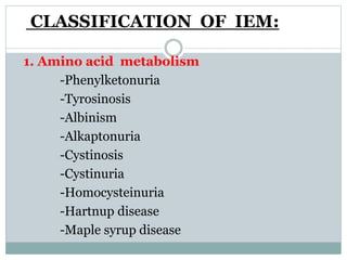
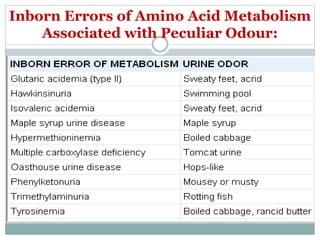
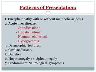
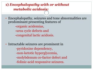
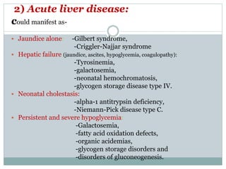
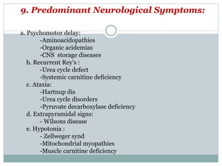
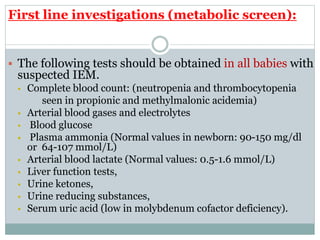
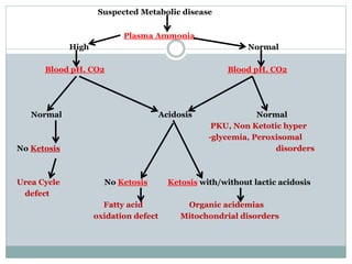
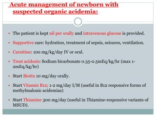
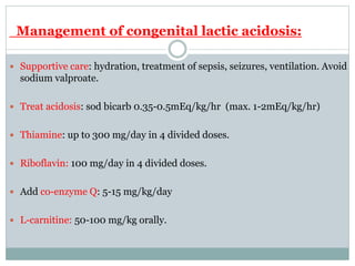
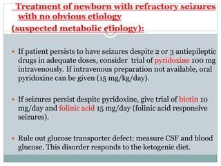
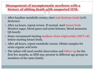
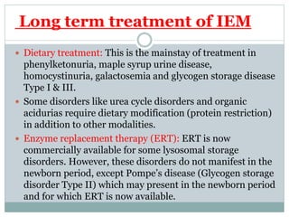
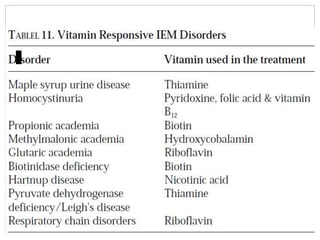
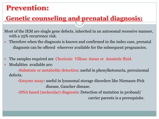
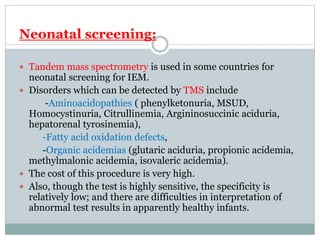
INBORN ERROR OF METABOLISM
Dr. BK Kalakheti, MD
METABOLISM
 Metabolism Catabolism (Breaking down)
Anabolism (Building up)
 Enzymes play an important role in facilitating
the process by serving as catalysts in the
conversion of one chemical (metabolite) to
another.
Catabolism :
 Catabolism is the set of metabolic pathways that
break down molecules into smaller units and release
energy.
Anabolism :
 Anabolism is the set of metabolic pathways that
construct molecules from smaller units. These
reactions require energy.
Inborn errors of metabolism (IEM)
Inborn errors of metabolism (IEM) are disorders in which
there is a block at some point in the normal metabolic
pathway
- IEMs occur due to mutations in DNA.
DNA Enzyme
which code for a Receptor
Specific protein Transport vehicle
Membrane pump
Structural element
Example of mechanisms in IEM:
Precursor Substrate End-Product
A B C
 Example of mechanisms in IEM:
Precursor Substrate End-Product
A B C
Abnormal metabolic
pathway
Toxic Metabolite D
CLASSIFICATION OF IEM:
1. Amino acid metabolism
2. Carbohydrate metabolism
3. Lipid metabolism
4. Protein metabolism
5. Pigment metabolism
6. Unknown biochemical defects
CLASSIFICATION OF IEM:
1. Amino acid metabolism
-Phenylketonuria
-Tyrosinosis
-Albinism
-Alkaptonuria
-Cystinosis
-Cystinuria
-Homocysteinuria
-Hartnup disease
-Maple syrup disease
CLASSIFICATION OF IEM:
2. Carbohydrate synthesis:
-Congenital lactose intolerance
-Galactosemia
-Glycogen storage disease
-Diabetes mellitus
-Scurvy
CLASSIFICATION OF IEM:
3. Lipid metabolism:
-Abetalipoproteinemia
-Progressive lipodystrophy
-Lipid storage disorders
* Gaucher
* Neimann-Pick
* Tay-sachs
* Hyperlipoproteinemia
CLASSIFICATION OF IEM:
4. Protein Metabolism:
-Immunoglobulin deficiencies
-Absent clotting factors
(Hemophilia, Christmas dis,
Hypoprothrombinemia)
-Metal binding protein deficiency
(Wilson hepatolenticular degeneration)
-Alpha-1 anti trypsin deficiency
CLASSIFICATION OF IEM:
5. Pigment metabolism:
-Porphyrias
-Methemoglobinemias
-Albinism
-Crigler-Najjar dis
-Dubin-Johnson dis
-Gilbert dis
-Rotor synd
-Primary hemochromatosis
CLASSIFICATION OF IEM:
6. Unknown biochemical defect:
-Osteogenesis imperfecta
-Marfan syndrome
-Achondroplasia
-Ehler danlos syndrome
Suspecting Inborn Errors:
HISTORY:
(1) Symptoms accompany starting/changes in diet,
(2) Children with seizures,
(3) Developmental delay,
(4) Recurrent vomiting,
(5) Unusual odour of urine,
(6) Parental consanguinity,
(7) Problems suggestive of as retardation of previously
acquired dev milestones
(8) Unexplained deaths in first and second degree relatives.
Suspecting Inborn Errors:
CLINICALLY:
IEM must also be considered in the differential diagnosis of
 Critically ill newborns,
 Neurodegeneration,
 Mental retardation ,
 Coarse facies / dysmorphic features,
 Parenchymal liver disease,
 Cardiomyopathy,
 Organomegaly
 Unexplained acidosis,
 Corneal opacity, cataract or dislocation of lens,
 Hyperammonemia and
 Hypoglycemia
Clinical pointers towards specific IEMs:
Inborn Errors of Amino Acid Metabolism
Associated with Peculiar Odour:
Patterns of Presentation:
1. Encephalopathy with or without metabolic acidosis
2. Acute liver disease:
- Jaundice alone
- Hepatic failure
- Neonatal cholestasis
- Hypoglycemia
3. Dysmorphic features.
4. Cardiac disease.
5. Diarrhea
6. Hepatomegaly +/- Splenomegaly
7. Predominant Neurological symptoms
1) Encephalopathy with or without
metabolic acidosis:
 Encephalopathy, seizures and tone abnormalities are
predominant presenting features of
-organic acidemias,
-urea cycle defects and
-congenital lactic acidosis.
 Intractable seizures are prominent in
-pyridoxine dependency,
-non-ketotic hyperglycemia,
-molybdenum co-factor defect and
-folinic-acid responsive seizures.
2) Acute liver disease:
could manifest as-
 Jaundice alone: -Gilbert syndrome,
-Criggler-Najjar syndrome
 Hepatic failure (jaundice, ascites, hypoglycemia, coagulopathy):
-Tyrosinemia,
-galactosemia,
-neonatal hemochromatosis,
-glycogen storage disease type IV.
 Neonatal cholestasis:
-alpha-1 antitrypsin deficiency,
-Niemann-Pick disease type C.
 Persistent and severe hypoglycemia:
-Galactosemia,
-fatty acid oxidation defects,
-organic acidemias,
-glycogen storage disorders and
-disorders of gluconeogenesis.
3) Dysmorphic features:
 seen in
-Peroxismal disorders,
-Pyruvate dehydrogenase deficiency,
-Congenital disorders of glycosylation and
-Lysosomal storage diseases.
Some IEMs may present with
non-immune hydrops fetalis
-lysosomal storage disorders and
-Congenital disorders of glycosylation.
4) Cardiac disease:
Cardiomyopathy is a prominent feature in some IEM
like in:
-Fatty acid oxidation defects,
-Glycogen storage disease type II and
-Mitochondrial electron transport chain defects.
6. Diarrhea :
a. Severe watery diarrhea:
- Congenital chloride diarrhea
- Galactosemia
- Primary lactase, sucrase, isomaltase deficiency.
b. Chronic diarrhea:
-Bile acid disorders
-Infantile Refsum disease
-Respiratory chain disorders asso with
steatorrhea
-Vitamin D deficient osteopenia
-Hypocholesterolemia
c. Diarrhea, Failure to thrive, hypotonia, hepatomegaly:
-Glycogen storage dis 1
- Wolmans disease
7. Hepatomegaly :
-Tyrosinemia
-Galactosemia
-Fructosemia
-Alpha 1 antitrypsin deficiency.
 8. Hepatomegaly + Splenomegaly:
-Mucolipidosis
-Gaucher’s dis
-Niemann-Pick type A
9. Predominant Neurological Symptoms:
a. Psychomotor delay:
-Aminoacidopathies
-Organic acidemias
-CNS storage diseases
b. Recurrent Rey’s :
-Urea cycle defect
-Systemic carnitine deficiency
c. Ataxia:
-Hartnup dis
-Urea cycle disorders
-Pyruvate decarboxylase deficiency
d. Extrapyramidal signs:
- Wilsons disease
e. Hypotonia :
- Zellweger synd
-Mitochondrial myopathies
-Muscle carnitine deficiency
INVESTIGATIONS:
 Metabolic investigations should be initiated as
soon as the possibility is considered.
 The outcome of treatment of many IEM especially
those associated with hyperammonemia is directly
related to the rapidity with which problems are
detected and treated.
First line investigations (metabolic screen):
 The following tests should be obtained in all babies with
suspected IEM.
 Complete blood count: (neutropenia and thrombocytopenia
seen in propionic and methylmalonic acidemia)
 Arterial blood gases and electrolytes
 Blood glucose
 Plasma ammonia (Normal values in newborn: 90-150 mg/dl
or 64-107 mmol/L)
 Arterial blood lactate (Normal values: 0.5-1.6 mmol/L)
 Liver function tests,
 Urine ketones,
 Urine reducing substances,
 Serum uric acid (low in molybdenum cofactor deficiency).
Suspected Metabolic disease
Plasma Ammonia
High Normal
Blood pH, CO2 Blood pH, CO2
Normal Acidosis Normal
PKU, Non Ketotic hyper
-glycemia, Peroxisomal
No Ketosis disorders
Urea Cycle No Ketosis Ketosis with/without lactic acidosis
defect
Fatty acid Organic acidemias
oxidation defect Mitochondrial disorders
Categorization of neonatal IEM using metabolic
screening tests:
Second line investigations
(ancillary and confirmatory tests)
 These tests need to be performed in a targeted manner, based on
presumptive diagnosis after first line investigations:
1) Gas chromatography mass spectrometry (GCMS) of urine- for
diagnosis of organic acidemias.
2) Plasma amino acids and acyl carnitine profile: by Tandem mass
spectrometry (TMS)- for diagnosis of organic acidemias, urea cycle
defects, aminoacidopathies and fatty acid oxidation defects.
3) High performance liquid chromatography (HPLC): for
quantitative analysis of amino acids in blood and urine; required
for diagnosis of organic acidemias and aminoacidopathies.
4) Lactate/pyruvate ratio- in cases with elevated lactate.
5) Urinary orotic acid- in cases with hyperammonemia for
classification of urea cycle defect.
6) Enzyme assay: This is required for definitive diagnosis, but
not available for most IEM’s.
Available enzyme assays include:
-Biotinidase assay- in cases with suspected biotinidase
deficiency (intractable seizures, seborrheic rash, alopecia);
-GALT (galactose 1-phosphate uridyl transferase assay)-
in cases with suspected galactosemia (hypoglycemia,
cataracts, reducing sugars in urine).
7) Neuroimaging: MRI
 Some IEM may be associated with structural malformations .
Examples:
-Zellweger syndrome has diffuse cortical migration and
sulcation abnormalities.
-Agenesis of corpus callosum has been reported in Menke’s
disease, pyruvate decarboxylase deficiency and nonketotic
hyperglycinemia.
-Maple syrup urine disease (MSUD): brainstem and
cerebellar edema.
-Propionic & methylmalonic acidemia: basal ganglia signal
change.
-Glutaric aciduria: frontotemporal atrophy, subdural
hematomas.
8) Magnetic resonance spectroscopy (MRS): may be helpful in
selected disorders
 E.g. lactate peak elevated in mitochondrial disorders,
leucine peak elevated in MSUD.
9) Electroencephalography (EEG):
 Comb-like rhythm in MSUD,
 Burst suppression in Non Ketotic Hyperglycemia and
holocarboxylase synthetase deficiency.
10) Plasma very long chain fatty acid (VLCFA) levels:
elevated in peroxisomal disorders.
11)Mutation analysis when available.
12) CSF aminoacid analysis: CSF Glycine levels elevated in
NKH.
TREATMENT:
 In most cases, treatment needs to be instituted empirically
without a specific diagnosis.
 The metabolic screen helps to broadly categorize the
patient’s IEM (e.g. urea cycle defect, organic academia,
congenital lactic acidosis etc), on the basis of which,
empirical treatment can be instituted
Aims of treatment
1. To reduce the formation of toxic metabolites by decreasing
substrate availability (by stopping feeds and preventing
endogenous catabolism)
2. To provide adequate calories.
3. To enhance the excretion of toxic metabolites.
4. To institute co-factor therapy for specific disease and also
empirically if diagnosis not established.
5. Supportive care-
-Treatment of seizures (avoid sodium valproate –
may increase ammonia levels),
-Maintain euglycemia and normothermia,
-Fluid, electrolyte & acid-base balance,
-Treatment of infection,
-Mechanical ventilation if required.
Management of hyperammonemia:
1) Discontinue all feeds. Provide adequate calories by
intravenous glucose and lipids. Maintain glucose
infusion rate 8-10mg/kg/min. Start intravenous lipid
0.5 g/kg/day (up to 3 g/kg/day). After stabilization
gradually add protein 0.25 g/kg till 1.5 g/kg/day.
2) Dialysis is the only means for rapid removal of
ammonia and hemodialysis is more effective and faster
than peritoneal dialysis. Exchange transfusion is not
useful.
3) Alternative pathways for nitrogen excretion-:
-Sodium benzoate (IV/oral)- loading dose 250
mg/kg then 250-400 mg/kg/day in 4 divided doses. (I/V
-Sodium phenylbutyrate; loading dose 250 mg/kg
followed by 250-500 mg/kg/day.
-L-arginine (oral or IV)- 300 mg/kg/day (IV)
-L-carnitine (oral or IV)- 200 mg/kg/day
4) Supportive care: Treatment of sepsis,seizures, ventilation.
Avoid sodium valproate.
Acute management of newborn with
suspected organic acidemia:
 The patient is kept nil per orally and intravenous glucose is provided.
 Supportive care: hydration, treatment of sepsis, seizures, ventilation.
 Carnitine: 100 mg/kg/day IV or oral.
 Treat acidosis: Sodium bicarbonate 0.35-0.5mEq/kg/hr (max 1-
2mEq/kg/hr)
 Start Biotin 10 mg/day orally.
 Start Vitamin B12; 1-2 mg/day I/M (useful in B12 responsive forms of
methylmalonic acidemias)
 Start Thiamine 300 mg/day (useful in Thiamine-responsive variants of
MSUD).
Management of congenital lactic acidosis:
 Supportive care: hydration, treatment of sepsis, seizures, ventilation. Avoid
sodium valproate.
 Treat acidosis: sod bicarb 0.35-0.5mEq/kg/hr (max. 1-2mEq/kg/hr)
 Thiamine: up to 300 mg/day in 4 divided doses.
 Riboflavin: 100 mg/day in 4 divided doses.
 Add co-enzyme Q: 5-15 mg/kg/day
 L-carnitine: 50-100 mg/kg orally.
Treatment of newborn with refractory seizures
with no obvious etiology
(suspected metabolic etiology):
 If patient persists to have seizures despite 2 or 3 antiepileptic
drugs in adequate doses, consider trial of pyridoxine 100 mg
intravenously. If intravenous preparation not available, oral
pyridoxine can be given (15 mg/kg/day).
 If seizures persist despite pyridoxine, give trial of biotin 10
mg/day and folinic acid 15 mg/day (folinic acid responsive
seizures).
 Rule out glucose transporter defect: measure CSF and blood
glucose. This disorder responds to the ketogenic diet.
Management of asymptomatic newborn with a
history of sibling death with suspected IEM:
 After baseline metabolic screen, start oral dextrose feeds (10%
dextrose).
 After 24 hours, repeat screen. If normal, start breast feeds.
Monitor sugar, blood gases and urine ketones, blood ammonia
Q6 hourly.
 Some recommend starting medium chain triglycerides (MCT oil)
before starting breast feeds,
 After 48 hours, repeat metabolic screen. Obtain samples for
urine organic acid tests.
 The infant will need careful observation and follow-up for the
first few months, as IEM may present in different age groups in
members of the same family.
Long term treatment of IEM
 Dietary treatment: This is the mainstay of treatment in
phenylketonuria, maple syrup urine disease,
homocystinuria, galactosemia and glycogen storage disease
Type I & III.
 Some disorders like urea cycle disorders and organic
acidurias require dietary modification (protein restriction)
in addition to other modalities.
 Enzyme replacement therapy (ERT): ERT is now
commercially available for some lysosomal storage
disorders. However, these disorders do not manifest in the
newborn period, except Pompe’s disease (Glycogen storage
disorder Type II) which may present in the newborn period
and for which ERT is now available.
Prevention:
Genetic counseling and prenatal diagnosis:
Most of the IEM are single gene defects, inherited in an autosomal recessive manner,
with a 25% recurrence risk.
 Therefore when the diagnosis is known and confirmed in the index case, prenatal
diagnosis can be offered wherever available for the subsequent pregnancies.
 The samples required are Chorionic Villous tissue or Amniotic fluid.
 Modalities available are:
-Substrate or metabolite detection: useful in phenylketonuria, peroxisomal
defects.
-Enzyme assay: useful in lysosomal storage disorders like Niemann-Pick
disease, Gaucher disease.
-DNA based (molecular) diagnosis: Detection of mutation in proband/
carrier parents is a prerequisite.
Neonatal screening:
 Tandem mass spectrometry is used in some countries for
neonatal screening for IEM.
 Disorders which can be detected by TMS include
-Aminoacidopathies ( phenylketonuria, MSUD,
Homocystinuria, Citrullinemia, Argininosuccinic aciduria,
hepatorenal tyrosinemia),
-Fatty acid oxidation defects,
-Organic acidemias (glutaric aciduria, propionic acidemia,
methylmalonic acidemia, isovaleric acidemia).
 The cost of this procedure is very high.
 Also, though the test is highly sensitive, the specificity is
relatively low; and there are difficulties in interpretation of
abnormal test results in apparently healthy infants.
Inborn error approach.pptx

Inborn error approach.pptx

  • 1.
    INBORN ERROR OFMETABOLISM Dr. BK Kalakheti, MD
  • 2.
    METABOLISM  Metabolism Catabolism(Breaking down) Anabolism (Building up)  Enzymes play an important role in facilitating the process by serving as catalysts in the conversion of one chemical (metabolite) to another.
  • 3.
    Catabolism :  Catabolismis the set of metabolic pathways that break down molecules into smaller units and release energy.
  • 4.
    Anabolism :  Anabolismis the set of metabolic pathways that construct molecules from smaller units. These reactions require energy.
  • 5.
    Inborn errors ofmetabolism (IEM) Inborn errors of metabolism (IEM) are disorders in which there is a block at some point in the normal metabolic pathway - IEMs occur due to mutations in DNA. DNA Enzyme which code for a Receptor Specific protein Transport vehicle Membrane pump Structural element
  • 6.
    Example of mechanismsin IEM: Precursor Substrate End-Product A B C
  • 7.
     Example ofmechanisms in IEM: Precursor Substrate End-Product A B C Abnormal metabolic pathway Toxic Metabolite D
  • 8.
    CLASSIFICATION OF IEM: 1.Amino acid metabolism 2. Carbohydrate metabolism 3. Lipid metabolism 4. Protein metabolism 5. Pigment metabolism 6. Unknown biochemical defects
  • 9.
    CLASSIFICATION OF IEM: 1.Amino acid metabolism -Phenylketonuria -Tyrosinosis -Albinism -Alkaptonuria -Cystinosis -Cystinuria -Homocysteinuria -Hartnup disease -Maple syrup disease
  • 10.
    CLASSIFICATION OF IEM: 2.Carbohydrate synthesis: -Congenital lactose intolerance -Galactosemia -Glycogen storage disease -Diabetes mellitus -Scurvy
  • 11.
    CLASSIFICATION OF IEM: 3.Lipid metabolism: -Abetalipoproteinemia -Progressive lipodystrophy -Lipid storage disorders * Gaucher * Neimann-Pick * Tay-sachs * Hyperlipoproteinemia
  • 12.
    CLASSIFICATION OF IEM: 4.Protein Metabolism: -Immunoglobulin deficiencies -Absent clotting factors (Hemophilia, Christmas dis, Hypoprothrombinemia) -Metal binding protein deficiency (Wilson hepatolenticular degeneration) -Alpha-1 anti trypsin deficiency
  • 13.
    CLASSIFICATION OF IEM: 5.Pigment metabolism: -Porphyrias -Methemoglobinemias -Albinism -Crigler-Najjar dis -Dubin-Johnson dis -Gilbert dis -Rotor synd -Primary hemochromatosis
  • 14.
    CLASSIFICATION OF IEM: 6.Unknown biochemical defect: -Osteogenesis imperfecta -Marfan syndrome -Achondroplasia -Ehler danlos syndrome
  • 15.
    Suspecting Inborn Errors: HISTORY: (1)Symptoms accompany starting/changes in diet, (2) Children with seizures, (3) Developmental delay, (4) Recurrent vomiting, (5) Unusual odour of urine, (6) Parental consanguinity, (7) Problems suggestive of as retardation of previously acquired dev milestones (8) Unexplained deaths in first and second degree relatives.
  • 16.
    Suspecting Inborn Errors: CLINICALLY: IEMmust also be considered in the differential diagnosis of  Critically ill newborns,  Neurodegeneration,  Mental retardation ,  Coarse facies / dysmorphic features,  Parenchymal liver disease,  Cardiomyopathy,  Organomegaly  Unexplained acidosis,  Corneal opacity, cataract or dislocation of lens,  Hyperammonemia and  Hypoglycemia
  • 17.
  • 18.
    Inborn Errors ofAmino Acid Metabolism Associated with Peculiar Odour:
  • 19.
    Patterns of Presentation: 1.Encephalopathy with or without metabolic acidosis 2. Acute liver disease: - Jaundice alone - Hepatic failure - Neonatal cholestasis - Hypoglycemia 3. Dysmorphic features. 4. Cardiac disease. 5. Diarrhea 6. Hepatomegaly +/- Splenomegaly 7. Predominant Neurological symptoms
  • 20.
    1) Encephalopathy withor without metabolic acidosis:  Encephalopathy, seizures and tone abnormalities are predominant presenting features of -organic acidemias, -urea cycle defects and -congenital lactic acidosis.  Intractable seizures are prominent in -pyridoxine dependency, -non-ketotic hyperglycemia, -molybdenum co-factor defect and -folinic-acid responsive seizures.
  • 21.
    2) Acute liverdisease: could manifest as-  Jaundice alone: -Gilbert syndrome, -Criggler-Najjar syndrome  Hepatic failure (jaundice, ascites, hypoglycemia, coagulopathy): -Tyrosinemia, -galactosemia, -neonatal hemochromatosis, -glycogen storage disease type IV.  Neonatal cholestasis: -alpha-1 antitrypsin deficiency, -Niemann-Pick disease type C.  Persistent and severe hypoglycemia: -Galactosemia, -fatty acid oxidation defects, -organic acidemias, -glycogen storage disorders and -disorders of gluconeogenesis.
  • 22.
    3) Dysmorphic features: seen in -Peroxismal disorders, -Pyruvate dehydrogenase deficiency, -Congenital disorders of glycosylation and -Lysosomal storage diseases. Some IEMs may present with non-immune hydrops fetalis -lysosomal storage disorders and -Congenital disorders of glycosylation.
  • 23.
    4) Cardiac disease: Cardiomyopathyis a prominent feature in some IEM like in: -Fatty acid oxidation defects, -Glycogen storage disease type II and -Mitochondrial electron transport chain defects.
  • 24.
    6. Diarrhea : a.Severe watery diarrhea: - Congenital chloride diarrhea - Galactosemia - Primary lactase, sucrase, isomaltase deficiency. b. Chronic diarrhea: -Bile acid disorders -Infantile Refsum disease -Respiratory chain disorders asso with steatorrhea -Vitamin D deficient osteopenia -Hypocholesterolemia c. Diarrhea, Failure to thrive, hypotonia, hepatomegaly: -Glycogen storage dis 1 - Wolmans disease
  • 25.
    7. Hepatomegaly : -Tyrosinemia -Galactosemia -Fructosemia -Alpha1 antitrypsin deficiency.  8. Hepatomegaly + Splenomegaly: -Mucolipidosis -Gaucher’s dis -Niemann-Pick type A
  • 26.
    9. Predominant NeurologicalSymptoms: a. Psychomotor delay: -Aminoacidopathies -Organic acidemias -CNS storage diseases b. Recurrent Rey’s : -Urea cycle defect -Systemic carnitine deficiency c. Ataxia: -Hartnup dis -Urea cycle disorders -Pyruvate decarboxylase deficiency d. Extrapyramidal signs: - Wilsons disease e. Hypotonia : - Zellweger synd -Mitochondrial myopathies -Muscle carnitine deficiency
  • 27.
    INVESTIGATIONS:  Metabolic investigationsshould be initiated as soon as the possibility is considered.  The outcome of treatment of many IEM especially those associated with hyperammonemia is directly related to the rapidity with which problems are detected and treated.
  • 28.
    First line investigations(metabolic screen):  The following tests should be obtained in all babies with suspected IEM.  Complete blood count: (neutropenia and thrombocytopenia seen in propionic and methylmalonic acidemia)  Arterial blood gases and electrolytes  Blood glucose  Plasma ammonia (Normal values in newborn: 90-150 mg/dl or 64-107 mmol/L)  Arterial blood lactate (Normal values: 0.5-1.6 mmol/L)  Liver function tests,  Urine ketones,  Urine reducing substances,  Serum uric acid (low in molybdenum cofactor deficiency).
  • 29.
    Suspected Metabolic disease PlasmaAmmonia High Normal Blood pH, CO2 Blood pH, CO2 Normal Acidosis Normal PKU, Non Ketotic hyper -glycemia, Peroxisomal No Ketosis disorders Urea Cycle No Ketosis Ketosis with/without lactic acidosis defect Fatty acid Organic acidemias oxidation defect Mitochondrial disorders
  • 33.
    Categorization of neonatalIEM using metabolic screening tests:
  • 34.
    Second line investigations (ancillaryand confirmatory tests)  These tests need to be performed in a targeted manner, based on presumptive diagnosis after first line investigations: 1) Gas chromatography mass spectrometry (GCMS) of urine- for diagnosis of organic acidemias. 2) Plasma amino acids and acyl carnitine profile: by Tandem mass spectrometry (TMS)- for diagnosis of organic acidemias, urea cycle defects, aminoacidopathies and fatty acid oxidation defects. 3) High performance liquid chromatography (HPLC): for quantitative analysis of amino acids in blood and urine; required for diagnosis of organic acidemias and aminoacidopathies.
  • 35.
    4) Lactate/pyruvate ratio-in cases with elevated lactate. 5) Urinary orotic acid- in cases with hyperammonemia for classification of urea cycle defect. 6) Enzyme assay: This is required for definitive diagnosis, but not available for most IEM’s. Available enzyme assays include: -Biotinidase assay- in cases with suspected biotinidase deficiency (intractable seizures, seborrheic rash, alopecia); -GALT (galactose 1-phosphate uridyl transferase assay)- in cases with suspected galactosemia (hypoglycemia, cataracts, reducing sugars in urine).
  • 36.
    7) Neuroimaging: MRI Some IEM may be associated with structural malformations . Examples: -Zellweger syndrome has diffuse cortical migration and sulcation abnormalities. -Agenesis of corpus callosum has been reported in Menke’s disease, pyruvate decarboxylase deficiency and nonketotic hyperglycinemia. -Maple syrup urine disease (MSUD): brainstem and cerebellar edema. -Propionic & methylmalonic acidemia: basal ganglia signal change. -Glutaric aciduria: frontotemporal atrophy, subdural hematomas.
  • 37.
    8) Magnetic resonancespectroscopy (MRS): may be helpful in selected disorders  E.g. lactate peak elevated in mitochondrial disorders, leucine peak elevated in MSUD. 9) Electroencephalography (EEG):  Comb-like rhythm in MSUD,  Burst suppression in Non Ketotic Hyperglycemia and holocarboxylase synthetase deficiency. 10) Plasma very long chain fatty acid (VLCFA) levels: elevated in peroxisomal disorders. 11)Mutation analysis when available. 12) CSF aminoacid analysis: CSF Glycine levels elevated in NKH.
  • 38.
    TREATMENT:  In mostcases, treatment needs to be instituted empirically without a specific diagnosis.  The metabolic screen helps to broadly categorize the patient’s IEM (e.g. urea cycle defect, organic academia, congenital lactic acidosis etc), on the basis of which, empirical treatment can be instituted
  • 39.
    Aims of treatment 1.To reduce the formation of toxic metabolites by decreasing substrate availability (by stopping feeds and preventing endogenous catabolism) 2. To provide adequate calories. 3. To enhance the excretion of toxic metabolites. 4. To institute co-factor therapy for specific disease and also empirically if diagnosis not established. 5. Supportive care- -Treatment of seizures (avoid sodium valproate – may increase ammonia levels), -Maintain euglycemia and normothermia, -Fluid, electrolyte & acid-base balance, -Treatment of infection, -Mechanical ventilation if required.
  • 40.
    Management of hyperammonemia: 1)Discontinue all feeds. Provide adequate calories by intravenous glucose and lipids. Maintain glucose infusion rate 8-10mg/kg/min. Start intravenous lipid 0.5 g/kg/day (up to 3 g/kg/day). After stabilization gradually add protein 0.25 g/kg till 1.5 g/kg/day. 2) Dialysis is the only means for rapid removal of ammonia and hemodialysis is more effective and faster than peritoneal dialysis. Exchange transfusion is not useful.
  • 41.
    3) Alternative pathwaysfor nitrogen excretion-: -Sodium benzoate (IV/oral)- loading dose 250 mg/kg then 250-400 mg/kg/day in 4 divided doses. (I/V -Sodium phenylbutyrate; loading dose 250 mg/kg followed by 250-500 mg/kg/day. -L-arginine (oral or IV)- 300 mg/kg/day (IV) -L-carnitine (oral or IV)- 200 mg/kg/day 4) Supportive care: Treatment of sepsis,seizures, ventilation. Avoid sodium valproate.
  • 42.
    Acute management ofnewborn with suspected organic acidemia:  The patient is kept nil per orally and intravenous glucose is provided.  Supportive care: hydration, treatment of sepsis, seizures, ventilation.  Carnitine: 100 mg/kg/day IV or oral.  Treat acidosis: Sodium bicarbonate 0.35-0.5mEq/kg/hr (max 1- 2mEq/kg/hr)  Start Biotin 10 mg/day orally.  Start Vitamin B12; 1-2 mg/day I/M (useful in B12 responsive forms of methylmalonic acidemias)  Start Thiamine 300 mg/day (useful in Thiamine-responsive variants of MSUD).
  • 43.
    Management of congenitallactic acidosis:  Supportive care: hydration, treatment of sepsis, seizures, ventilation. Avoid sodium valproate.  Treat acidosis: sod bicarb 0.35-0.5mEq/kg/hr (max. 1-2mEq/kg/hr)  Thiamine: up to 300 mg/day in 4 divided doses.  Riboflavin: 100 mg/day in 4 divided doses.  Add co-enzyme Q: 5-15 mg/kg/day  L-carnitine: 50-100 mg/kg orally.
  • 44.
    Treatment of newbornwith refractory seizures with no obvious etiology (suspected metabolic etiology):  If patient persists to have seizures despite 2 or 3 antiepileptic drugs in adequate doses, consider trial of pyridoxine 100 mg intravenously. If intravenous preparation not available, oral pyridoxine can be given (15 mg/kg/day).  If seizures persist despite pyridoxine, give trial of biotin 10 mg/day and folinic acid 15 mg/day (folinic acid responsive seizures).  Rule out glucose transporter defect: measure CSF and blood glucose. This disorder responds to the ketogenic diet.
  • 45.
    Management of asymptomaticnewborn with a history of sibling death with suspected IEM:  After baseline metabolic screen, start oral dextrose feeds (10% dextrose).  After 24 hours, repeat screen. If normal, start breast feeds. Monitor sugar, blood gases and urine ketones, blood ammonia Q6 hourly.  Some recommend starting medium chain triglycerides (MCT oil) before starting breast feeds,  After 48 hours, repeat metabolic screen. Obtain samples for urine organic acid tests.  The infant will need careful observation and follow-up for the first few months, as IEM may present in different age groups in members of the same family.
  • 46.
    Long term treatmentof IEM  Dietary treatment: This is the mainstay of treatment in phenylketonuria, maple syrup urine disease, homocystinuria, galactosemia and glycogen storage disease Type I & III.  Some disorders like urea cycle disorders and organic acidurias require dietary modification (protein restriction) in addition to other modalities.  Enzyme replacement therapy (ERT): ERT is now commercially available for some lysosomal storage disorders. However, these disorders do not manifest in the newborn period, except Pompe’s disease (Glycogen storage disorder Type II) which may present in the newborn period and for which ERT is now available.
  • 48.
    Prevention: Genetic counseling andprenatal diagnosis: Most of the IEM are single gene defects, inherited in an autosomal recessive manner, with a 25% recurrence risk.  Therefore when the diagnosis is known and confirmed in the index case, prenatal diagnosis can be offered wherever available for the subsequent pregnancies.  The samples required are Chorionic Villous tissue or Amniotic fluid.  Modalities available are: -Substrate or metabolite detection: useful in phenylketonuria, peroxisomal defects. -Enzyme assay: useful in lysosomal storage disorders like Niemann-Pick disease, Gaucher disease. -DNA based (molecular) diagnosis: Detection of mutation in proband/ carrier parents is a prerequisite.
  • 49.
    Neonatal screening:  Tandemmass spectrometry is used in some countries for neonatal screening for IEM.  Disorders which can be detected by TMS include -Aminoacidopathies ( phenylketonuria, MSUD, Homocystinuria, Citrullinemia, Argininosuccinic aciduria, hepatorenal tyrosinemia), -Fatty acid oxidation defects, -Organic acidemias (glutaric aciduria, propionic acidemia, methylmalonic acidemia, isovaleric acidemia).  The cost of this procedure is very high.  Also, though the test is highly sensitive, the specificity is relatively low; and there are difficulties in interpretation of abnormal test results in apparently healthy infants.

Editor's Notes

  • #18 Zellweger- peroxisomal import disorder. Menkes disease (kinky hair disease) is a progressive neurodegenerative condition inherited as a sex-linked recessive trait. The Menkes gene codes for a copper transporting P-type ATPase, and mutations in the protein are associated with low serum copper and ceruloplasmin levels as well as a defect in copper absorption and transport across the intestines.
  • #25 Wolman- lipd storage disorder
  • #32 THAN Transient hyperammonemia of newborn