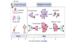
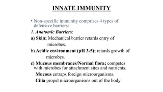
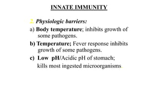
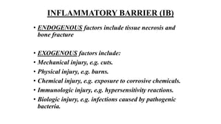
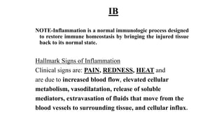
IMMUNOLOGY 2
• INNATE –always present in HEALTHY
INDIVIDUALS, prepared to BLOCK the entry of
microbes and to rapidly ELIMINATE microbes that do
succeed in entering host tissues.
• ADAPTIVE-is STIMULATED by microbes that invade
tissues, or ADAPTS to the presence of microbial
invaders.
IMMUNITY
• TEACHING OBJECTIVE
• Recognize the significance of the immune system in
combating infection and disease.
• Distinguish between non-specific (innate) and
specific (adaptive) immune systems.
• Understand the mechanisms combating
infection/disease (killing pathogens).
IMMUNITY
•Know the humoral and cellular
components of the non-specific
immunity.
•Comprehend the mechanism of
action of the humoral and cellular
components of non-specific
immunity.
INNATE IMMUNITY
• Innate (Non-Specific) defense mechanisms provide
the 1st line of host defense against invading
pathogens until an acquired immune response
develops.
• Non-specific defense mechanisms are present in
all individuals.
• They are effective at birth and function without
requiring prior exposure to a microorganism or its
Ags.
INNATE IMMUNITY
• Non-specific immunity comprises 4 types of
defensive barriers:
1. Anatomic Barriers:
a) Skin; Mechanical barrier retards entry of
microbes.
b) Acidic environment (pH 3-5); retards growth of
microbes.
c) Mucous membranes/Normal flora; competes
with microbes for attachment sites and nutrients.
Mucous entraps foreign microorganisms.
Cilia propel microorganisms out of the body.
INNATE IMMUNITY
2. Physiologic barriers:
a) Body temperature; inhibits growth of
some pathogens.
b) Temperature; Fever response inhibits
growth of some pathogens.
c) Low pH/Acidic pH of stomach;
kills most ingested microorganisms.
INNATE IMMUNITY
2. Chemical Mediators:
d) Lysozyme; in serum and tears, breaks
down the bacterial cell wall
(peptidoglycan).
e) Interferon; induces antiviral state in
uninfected cells and activates other
cells which kill pathogens.
INNATE IMMUNITY
f) Complement components and their products cause
destruction of microorganism directly or with help
of phagocytic cells.
Acute phase proteins (such as CRP) interact with the
complement system proteins to combat infections.
INNATE IMMUNITY
3. Phagocytic/endocytic barriers;
a) Various cells internalize (endocytose) and
break down foreign macromolecules.
b) Specialized cells (blood monocytes,
neutrophils, tissue macrophages) internalize
(phagocytose), kill and digest whole
microorganisms.
INTRACELLULAR AND EXTRACELLULAR
KILLING OF MICROORGANISMS
Once the invading organism has penetrated the physical
and chemical barriers that constitute the 1st line of defense,
it encounters the next line of defense, the specialized cells.
These include the polymorphonuclear leucocytes
(PMN), monocytes, macrophages.
ENDOCYTOSIS
Endocytosis is the ingestion by cells of macromolecules
present in extracellular fluid. This can occur by
pinocytosis, which involves non-specific invagination,
or by receptor-mediated endocytosis, a process
involving the selective binding of macromolecules to
specific membrane receptors.
In pinocytosis, non-specific membrane invagination
internalizes macromolecules in proportion to their
extracellular concentration.
ENDOCYTOSIS
In receptor-mediated endocytosis, macromolecules are
selectively internalized after binding to specific
membrane receptors.
The endocytic vesicles formed by either process fuse
with each other and are delivered to endosomes.
The acidic interior of endosomes facilitates dissociation
of macromolecules ligands from their receptors; the
latter are then recycled back to the cell surface.
ENDOCYTOSIS
Endosomes them fuse with primary lysosomes to form
secondary lysosomes to reduce the ingested
macromolecules to small breakdown products including
nucleotides, sugars, and peptides.
Primary lysosomes are derived from the Golgi complex
and contains degrading enzymes (proteases, lipases,
nucleases and other hydrolytic enzymes).
PHAGOCYTOSIS
Phagocytosis, is the ingestion of invading particles, e.g.
bacteria by phagocytic cells.
It is a critical protective mechanism of the immune
system.
Many microorganisms release substances that attract
phagocytic cells.
Phagocytosis can be enhanced by a variety of factors
(OPSONINS-Abs, complements) that make the
foreign particles an easier target.
PHAGOCYTOSIS
After ingestion, the foreign particle is entrapped in a
phagocytic vacuole (phagosome), which fuses with
lysosomes to form phagolysosome.
The phagolysome releases its degradative enzymes, which
digest the particle.
Phagocytes can also damage invading pathogens through
the generation of toxic products in a process known as
the RESPIRATORY BURST.
PHAGOCYTOSIS
The toxic products produced by the respiratory burst
include nitric oxide, hydrogen peroxide, superoxide
anion, and hypochlorous acid.
The toxic products produced by the RB can also
damage host cells.
Series of protective enzymes produced by phagocytes
limits their microbicidal activity to the
phagolysosome.
PHAGOCYTOSIS
These protective enzymes include catalase, which
degrades hydrogen peroxide, and superoxide
dismutase, which converts the superoxide anion into
hydrogen peroxide and oxygen.
The absence of or an abnormality in any of the
respiratory burst components results in a form of
immunodeficiency that predisposes individuals to
repeated infections.
CELLS OF THE INNATE IMMUNE SYSTEM
PMN are a population of cells also referred to as
granulocytes. These include basophils, mast cells,
eosinophils, and neutrophils. Granulocytes are short-
lived phagocytic cells that contain enzyme-rich
lysosomes, which can facilitate destruction of
microorganisms.
Granulocytes also produce peroxide, superoxide
radicals, lactoferrin which are toxic and bactericidal
to the organisms.
CELLS OF THE INNATE IMMUNE SYSTEM
• PMNs play a major role in infection. Defects in PMN
result in chronic or recurrent infection.
Macrophages
• Macrophages are derived from bone marrow
promonocytes which, after differentiation to blood
monocytes settle in the tissue as mature macrophages.
CELLS OF THE INNATE IMMUNE SYSTEM
• They are present throughout the connective tissue and
around the basement membrane of small blood vessels
and are particularly concentrated in the lung (alveolar
macrophages), liver (Kuppfer cells), kidney glomerulus
(mesangial cells), brain (microglia), and bone
(osteoclasts).
• Each of these macrophage population constitutes part
of the cellular members of the Reticuloendothelial
system (RES) or Mononuclear phagocyte system (MPS).
CELLS OF THE INNATE IMMUNE SYSTEM
• Differentiation of monocyte into a tissue macrophage
involves a number of changes:
• The cell enlarges 5 to 10 fold; its intracellular
organelles (phagosome, lysosome, phagolysome,
nucleus) increase both in number and complexity.
• It acquires increased phagocytic ability; producers
higher levels of lytic enzymes and begins to secrete a
variety of soluble factors.
CELLS OF THE INNATE IMMUNE SYSTEM
• The major function of RES or MPS is to phagocyte
microorganisms and foreign substances that invade
the bloodstream and various tissues. RES or MPS also
functions in the destruction of aged and imperfect
cells, such as effete erythrocytes.
Macrophages have 2 major functions-
• (a) Engulfment and break down of trapped materials
into simple amino acids, sugars, and other substances
for excretion or reuse.
CELLS OF THE INNATE IMMUNE SYSTEM
• (b) Process of antigen by denaturation or partial
digestion to antigen-specific T cells (Antigen
presentation)
• Macrophages are long-lived cells with significant
rough-surfaced endoplasmic reticulum and
mitochondria.
• Macrophages secrete several cytokines (including
interleukin-2, interferon-) that increase the ability of
the specific system to deal with infections.
DENDRITIC CELLS (DCs)
They are long-lived and reside in immature state in
most tissues where they recognise and phagocytize
pathogens.
Direct contact with pathogens leads to their
maturation and increase in their antigen presentation
capacity.
Thymus-interdigitating cells; skin-Langerhans cells.
DENDRITIC CELLS
• Mature DCs activate naïve antigen-specific T cells.
• DCs are important players in both INNATE IMMUNITY and the
initiation of ACQUIRED IMMUNE RESPONSES.
NATURAL KILLER CELLS
NK cells are large lymphocytes with numerous
cytoplasmic granules that are capable of lysing a
variety of tumor and virus-infected cells without
antigenic stimulation.
NK cells constitute 5%-10% of peripheral-blood
lymphocytes in humans and play an important role in
host defense against tumour cells.
They are involved in the early response to infection with
certain viruses and intracellular bacteria.
NATURAL KILLER CELLS
NK cells lyse the microorganism by causing
a) Pores in the membrane of the target cell.
b) Apoptosis (programmed cell death) of the target
cell by enhanced fragmentation of its nuclear
DNA.
NK cells lack Ag-specific receptors. How, then, do they
seek and destroy target cells?
NATURAL KILLER CELLS
NK cells use cell-cell contact, which allows them to determine
whether a potential target cell has lost MHC class I molecules.
Note MHC I is expressed on virtually all nucleated cells.
NATURAL KILLER CELLS
NK cells constitute 5%-10% of peripheral-blood lymphocytes in
humans and play an important role in host defense against tumour
cells.
They are involved in the early response to infection with certain
viruses and intracellular bacteria.
NK activity is stimulated by IFN-, IFN- and IL-2 and they provide
the 1st line defense to virus infection.
The importance of NK cells in defense against virus infections
becomes evident in individuals that completely lacked these cells.
NATURAL KILLER T CELLS (NKT)
• NKT cells differentiate from thymic precursors during
TCR engagement.
•
• NKT cells express TCR, like other T cells.
• They are unique in their functional status because
NKT cells are involved in both the innate and
adaptive immune systems.
NKT CELLS
• Following activation, NKT cells secrete several
regulatory cytokines, including IL-4 and IFN-γ.
• NKT cells also regulate immunopathologic
conditions.
INNATE IMMUNITY
4. Inflammatory barriers; an important
function of phagocytic cells, is a major
component of the body’s defense
mechanisms.
It is a physiologic process typically initiated
by tissue damage from ENDOGENOUS
and EXOGENOUS factors.
INFLAMMATORY BARRIER (IB)
• ENDOGENOUS factors include tissue necrosis and
bone fracture
• EXOGENOUS factors include:
• Mechanical injury, e.g. cuts.
• Physical injury, e.g. burns.
• Chemical injury, e.g. exposure to corrosive chemicals.
• Immunologic injury, e.g. hypersensitivity reactions.
• Biologic injury, e.g. infections caused by pathogenic
bacteria.
IB
NOTE-Inflammation is a normal immunologic process designed
to restore immune homeostasis by bringing the injured tissue
back to its normal state.
Hallmark Signs of Inflammation
Clinical signs are: PAIN, REDNESS, HEAT and
are due to increased blood flow, elevated cellular
metabolism, vasodilatation, release of soluble
mediators, extravasation of fluids that move from the
blood vessels to surrounding tissue, and cellular influx.

IMMUNOLOGY MADE SIMPLE FOR BEGINNERS TO UNDERSTAND BETTER

  • 1.
    IMMUNOLOGY 2 • INNATE–always present in HEALTHY INDIVIDUALS, prepared to BLOCK the entry of microbes and to rapidly ELIMINATE microbes that do succeed in entering host tissues. • ADAPTIVE-is STIMULATED by microbes that invade tissues, or ADAPTS to the presence of microbial invaders.
  • 3.
    IMMUNITY • TEACHING OBJECTIVE •Recognize the significance of the immune system in combating infection and disease. • Distinguish between non-specific (innate) and specific (adaptive) immune systems. • Understand the mechanisms combating infection/disease (killing pathogens).
  • 4.
    IMMUNITY •Know the humoraland cellular components of the non-specific immunity. •Comprehend the mechanism of action of the humoral and cellular components of non-specific immunity.
  • 5.
    INNATE IMMUNITY • Innate(Non-Specific) defense mechanisms provide the 1st line of host defense against invading pathogens until an acquired immune response develops. • Non-specific defense mechanisms are present in all individuals. • They are effective at birth and function without requiring prior exposure to a microorganism or its Ags.
  • 6.
    INNATE IMMUNITY • Non-specificimmunity comprises 4 types of defensive barriers: 1. Anatomic Barriers: a) Skin; Mechanical barrier retards entry of microbes. b) Acidic environment (pH 3-5); retards growth of microbes. c) Mucous membranes/Normal flora; competes with microbes for attachment sites and nutrients. Mucous entraps foreign microorganisms. Cilia propel microorganisms out of the body.
  • 7.
    INNATE IMMUNITY 2. Physiologicbarriers: a) Body temperature; inhibits growth of some pathogens. b) Temperature; Fever response inhibits growth of some pathogens. c) Low pH/Acidic pH of stomach; kills most ingested microorganisms.
  • 8.
    INNATE IMMUNITY 2. ChemicalMediators: d) Lysozyme; in serum and tears, breaks down the bacterial cell wall (peptidoglycan). e) Interferon; induces antiviral state in uninfected cells and activates other cells which kill pathogens.
  • 9.
    INNATE IMMUNITY f) Complementcomponents and their products cause destruction of microorganism directly or with help of phagocytic cells. Acute phase proteins (such as CRP) interact with the complement system proteins to combat infections.
  • 10.
    INNATE IMMUNITY 3. Phagocytic/endocyticbarriers; a) Various cells internalize (endocytose) and break down foreign macromolecules. b) Specialized cells (blood monocytes, neutrophils, tissue macrophages) internalize (phagocytose), kill and digest whole microorganisms.
  • 11.
    INTRACELLULAR AND EXTRACELLULAR KILLINGOF MICROORGANISMS Once the invading organism has penetrated the physical and chemical barriers that constitute the 1st line of defense, it encounters the next line of defense, the specialized cells. These include the polymorphonuclear leucocytes (PMN), monocytes, macrophages.
  • 12.
    ENDOCYTOSIS Endocytosis is theingestion by cells of macromolecules present in extracellular fluid. This can occur by pinocytosis, which involves non-specific invagination, or by receptor-mediated endocytosis, a process involving the selective binding of macromolecules to specific membrane receptors. In pinocytosis, non-specific membrane invagination internalizes macromolecules in proportion to their extracellular concentration.
  • 14.
    ENDOCYTOSIS In receptor-mediated endocytosis,macromolecules are selectively internalized after binding to specific membrane receptors. The endocytic vesicles formed by either process fuse with each other and are delivered to endosomes. The acidic interior of endosomes facilitates dissociation of macromolecules ligands from their receptors; the latter are then recycled back to the cell surface.
  • 15.
    ENDOCYTOSIS Endosomes them fusewith primary lysosomes to form secondary lysosomes to reduce the ingested macromolecules to small breakdown products including nucleotides, sugars, and peptides. Primary lysosomes are derived from the Golgi complex and contains degrading enzymes (proteases, lipases, nucleases and other hydrolytic enzymes).
  • 16.
    PHAGOCYTOSIS Phagocytosis, is theingestion of invading particles, e.g. bacteria by phagocytic cells. It is a critical protective mechanism of the immune system. Many microorganisms release substances that attract phagocytic cells. Phagocytosis can be enhanced by a variety of factors (OPSONINS-Abs, complements) that make the foreign particles an easier target.
  • 17.
    PHAGOCYTOSIS After ingestion, theforeign particle is entrapped in a phagocytic vacuole (phagosome), which fuses with lysosomes to form phagolysosome. The phagolysome releases its degradative enzymes, which digest the particle. Phagocytes can also damage invading pathogens through the generation of toxic products in a process known as the RESPIRATORY BURST.
  • 18.
    PHAGOCYTOSIS The toxic productsproduced by the respiratory burst include nitric oxide, hydrogen peroxide, superoxide anion, and hypochlorous acid. The toxic products produced by the RB can also damage host cells. Series of protective enzymes produced by phagocytes limits their microbicidal activity to the phagolysosome.
  • 19.
    PHAGOCYTOSIS These protective enzymesinclude catalase, which degrades hydrogen peroxide, and superoxide dismutase, which converts the superoxide anion into hydrogen peroxide and oxygen. The absence of or an abnormality in any of the respiratory burst components results in a form of immunodeficiency that predisposes individuals to repeated infections.
  • 20.
    CELLS OF THEINNATE IMMUNE SYSTEM PMN are a population of cells also referred to as granulocytes. These include basophils, mast cells, eosinophils, and neutrophils. Granulocytes are short- lived phagocytic cells that contain enzyme-rich lysosomes, which can facilitate destruction of microorganisms. Granulocytes also produce peroxide, superoxide radicals, lactoferrin which are toxic and bactericidal to the organisms.
  • 21.
    CELLS OF THEINNATE IMMUNE SYSTEM • PMNs play a major role in infection. Defects in PMN result in chronic or recurrent infection. Macrophages • Macrophages are derived from bone marrow promonocytes which, after differentiation to blood monocytes settle in the tissue as mature macrophages.
  • 22.
    CELLS OF THEINNATE IMMUNE SYSTEM • They are present throughout the connective tissue and around the basement membrane of small blood vessels and are particularly concentrated in the lung (alveolar macrophages), liver (Kuppfer cells), kidney glomerulus (mesangial cells), brain (microglia), and bone (osteoclasts). • Each of these macrophage population constitutes part of the cellular members of the Reticuloendothelial system (RES) or Mononuclear phagocyte system (MPS).
  • 23.
    CELLS OF THEINNATE IMMUNE SYSTEM • Differentiation of monocyte into a tissue macrophage involves a number of changes: • The cell enlarges 5 to 10 fold; its intracellular organelles (phagosome, lysosome, phagolysome, nucleus) increase both in number and complexity. • It acquires increased phagocytic ability; producers higher levels of lytic enzymes and begins to secrete a variety of soluble factors.
  • 24.
    CELLS OF THEINNATE IMMUNE SYSTEM • The major function of RES or MPS is to phagocyte microorganisms and foreign substances that invade the bloodstream and various tissues. RES or MPS also functions in the destruction of aged and imperfect cells, such as effete erythrocytes. Macrophages have 2 major functions- • (a) Engulfment and break down of trapped materials into simple amino acids, sugars, and other substances for excretion or reuse.
  • 25.
    CELLS OF THEINNATE IMMUNE SYSTEM • (b) Process of antigen by denaturation or partial digestion to antigen-specific T cells (Antigen presentation) • Macrophages are long-lived cells with significant rough-surfaced endoplasmic reticulum and mitochondria. • Macrophages secrete several cytokines (including interleukin-2, interferon-) that increase the ability of the specific system to deal with infections.
  • 26.
    DENDRITIC CELLS (DCs) Theyare long-lived and reside in immature state in most tissues where they recognise and phagocytize pathogens. Direct contact with pathogens leads to their maturation and increase in their antigen presentation capacity. Thymus-interdigitating cells; skin-Langerhans cells.
  • 27.
    DENDRITIC CELLS • MatureDCs activate naïve antigen-specific T cells. • DCs are important players in both INNATE IMMUNITY and the initiation of ACQUIRED IMMUNE RESPONSES.
  • 28.
    NATURAL KILLER CELLS NKcells are large lymphocytes with numerous cytoplasmic granules that are capable of lysing a variety of tumor and virus-infected cells without antigenic stimulation. NK cells constitute 5%-10% of peripheral-blood lymphocytes in humans and play an important role in host defense against tumour cells. They are involved in the early response to infection with certain viruses and intracellular bacteria.
  • 29.
    NATURAL KILLER CELLS NKcells lyse the microorganism by causing a) Pores in the membrane of the target cell. b) Apoptosis (programmed cell death) of the target cell by enhanced fragmentation of its nuclear DNA. NK cells lack Ag-specific receptors. How, then, do they seek and destroy target cells?
  • 30.
    NATURAL KILLER CELLS NKcells use cell-cell contact, which allows them to determine whether a potential target cell has lost MHC class I molecules. Note MHC I is expressed on virtually all nucleated cells.
  • 32.
    NATURAL KILLER CELLS NKcells constitute 5%-10% of peripheral-blood lymphocytes in humans and play an important role in host defense against tumour cells. They are involved in the early response to infection with certain viruses and intracellular bacteria. NK activity is stimulated by IFN-, IFN- and IL-2 and they provide the 1st line defense to virus infection. The importance of NK cells in defense against virus infections becomes evident in individuals that completely lacked these cells.
  • 33.
    NATURAL KILLER TCELLS (NKT) • NKT cells differentiate from thymic precursors during TCR engagement. • • NKT cells express TCR, like other T cells. • They are unique in their functional status because NKT cells are involved in both the innate and adaptive immune systems.
  • 34.
    NKT CELLS • Followingactivation, NKT cells secrete several regulatory cytokines, including IL-4 and IFN-γ. • NKT cells also regulate immunopathologic conditions.
  • 35.
    INNATE IMMUNITY 4. Inflammatorybarriers; an important function of phagocytic cells, is a major component of the body’s defense mechanisms. It is a physiologic process typically initiated by tissue damage from ENDOGENOUS and EXOGENOUS factors.
  • 36.
    INFLAMMATORY BARRIER (IB) •ENDOGENOUS factors include tissue necrosis and bone fracture • EXOGENOUS factors include: • Mechanical injury, e.g. cuts. • Physical injury, e.g. burns. • Chemical injury, e.g. exposure to corrosive chemicals. • Immunologic injury, e.g. hypersensitivity reactions. • Biologic injury, e.g. infections caused by pathogenic bacteria.
  • 37.
    IB NOTE-Inflammation is anormal immunologic process designed to restore immune homeostasis by bringing the injured tissue back to its normal state. Hallmark Signs of Inflammation Clinical signs are: PAIN, REDNESS, HEAT and are due to increased blood flow, elevated cellular metabolism, vasodilatation, release of soluble mediators, extravasation of fluids that move from the blood vessels to surrounding tissue, and cellular influx.