SlideShare a Scribd company logo
1 of 920
Download to read offline
Image Challenge
A disease of which one of the following systems is most likely to have resulted in this
finding?
1. Endocrine system
2. Gastrointestinal system
3. Hematopoietic system
4. Neurologic system
5. Respiratory system
Q:
Dr.Sherif
Badrawy
Answer:
Image Challenge
A disease of which one of the following systems is most likely to have resulted in this
finding?
Q:
5. Respiratory system
Symmetric, slanting regions of hyperpigmentation on both thighs is consistent with Dahl's
sign, a result of repeated pressure from the elbows on the epidermis of the thighs in patients
spending large amounts of time in the tripod position. As in this patient, this finding provides
supporting evidence of advanced chronic obstructive pulmonary disease.
Image Challenge
What is the most likely diagnosis in this asymptomatic 17-year-old male?
1. Antrochoanal polyp
2. Foreign body
3. Palatine tonsillitis
4. Pharyngeal lymphoma
5. Tonsillar abscess
Q:
Answer:
Image Challenge
What is the most likely diagnosis in this asymptomatic 17-year-old male?Q:
1. Antrochoanal polyp
Rhinoscopy and computed tomography of the sinuses revealed that the soft, translucent,
round mass occupying the left pharyngeal area was an antrochoanal polyp originating from
the left maxillary sinus. The diagnosis was confirmed following surgical removal.
Image Challenge
What is the diagnosis?
1. Diaphragmatic hernia
2. Gastric bezoar
3. Inferior mesenteric artery thrombosis
4. Pancreatic phlegmon
5. Small-bowel volvulus
Q:
Answer:
Image Challenge
What is the diagnosis?Q:
5. Small-bowel volvulus
The CT reveals small-bowel volvulus with dilated small bowel rotated around its blood supply.
There are no signs of free air or fluid and no indication of bowel ischemia on the CT scan.
The patient recovered following surgical release of strangulated small bowel that was 1 m
from the ligament of Treitz.
Image Challenge
What is the diagnosis in this patient who developed burning pain, pruritus, and edema of
the hands upon their exposure to water and who had this image taken under a Wood's
lamp?
1. Acquired acral mycosis
2. Aquagenic keratoderma
3. Dyshidrotic eczema
4. Leprosy
5. Ochronosis
Q:
Answer:
Image Challenge
What is the diagnosis in this patient who developed burning pain, pruritus, and edema of
the hands upon their exposure to water and who had this image taken under a Wood's
lamp?
Q:
2. Aquagenic keratoderma
Following a biopsy, the patient received a diagnosis of aquagenic keratoderma, an unusual
condition characterized by transitory, flat-topped papules and plaques, with hyperwrinkling
and eccrine-duct prominence on the palms and fingers induced by exposure to water.
Aquagenic keratoderma has been associated with a heterozygous mutation in the cystic
fibrosis gene. The patient improved promptly following treatment with subcutaneous injections
of botulinum toxin.
Image Challenge
What is the diagnosis?
1. Aerophagia
2. Congenital diaphragmatic hernia
3. Duodenal atresia
4. Gastroschesis
5. Hypertrophic pyloric stenosis
Q:
Answer:
Image Challenge
What is the diagnosis?Q:
3. Duodenal atresia
The double-bubble sign comprises distention of both the stomach and proximal duodenum
and is pathognomonic of congenital duodenal obstruction. There was no air distal to the level
of obstruction in the D2 segment of the duodenum. Laparoscopy showed duodenal atresia
type 1; the patient recovered following surgical repair.
Image Challenge
What is the diagnosis?
1. Angioedema
2. Cardiac tamponade
3. Necrotizing fasciitis
4. Superior vena cava syndrome
5. Toxic epidermal necrolysis
Q:
Answer:
Image Challenge
What is the diagnosis?Q:
4. Superior vena cava syndrome
The cyanosis that is limited to the head, neck, upper torso, and arms is consistent with
superior vena cava syndrome, which was confirmed with venography. The patient recovered
following treatment with ultrasound-accelerated thrombolysis.
Image Challenge
What is the most likely diagnosis in this asymptomatic male?
1. Dercum's disease
2. Familial multiple lipomatosis
3. Madelung's disease
4. Neurofibromatosis, type 1
5. Rheumatoid arthritis
Q:
Answer:
Image Challenge
What is the most likely diagnosis in this asymptomatic male?Q:
2. Familial multiple lipomatosis
The patient had more than 40 painless, mobile, soft nodules, with a maximum diameter of 10
cm, most of which were located in the arms and legs. Histopathological examination revealed
mature adipose tissue, supporting the diagnosis of familial multiple lipomatosis, a rare
autosomal dominant disorder, which was present in the patient's father, grandfather, and
brothers. The patient's lipid profile was normal. Dercum's disease is a painful syndrome of the
adipose tissue associated with obesity; Madelung's disease is characterized by symmetric fat
deposits in the head, neck, and upper trunk.
Image Challenge
What is the most likely diagnosis in this 47-year-old patient with an acute headache?
1. Encephalitis
2. Meningitis
3. Paget's disease
4. Stroke
5. Subdural hemorrhage
Q:
Answer:
Image Challenge
What is the most likely diagnosis in this 47-year-old patient with an acute headache?Q:
4. Stroke
CT of the brain revealed a linear area of hyperdensity in the first segment of the right middle
cerebral artery, consistent with the diagnosis of acute stroke. The linear hyperdensity is
suggestive of acute thromboembolic clot within the artery and is an early warning sign of large
cerebral infarction, brain edema, and poor functional outcome.
Image Challenge
What is the most likely diagnosis?
1. Ankyloglossia
2. Atrophic glossitis
3. Creutzfeldt-Jakob disease
4. Hypoglossal nerve palsy
5. Motor neuron disease
Q:
Answer:
Image Challenge
What is the most likely diagnosis?Q:
4. Hypoglossal nerve palsy
Hypoglossal nerve palsy produces wasting of the ipsilateral side of the tongue, and on
attempted protrusion the tongue deviates towards the affected side. This patient's palsy
resolved following treatment for bacterial meningitis. Motor neuron disease causes bilateral
wasting.
Image Challenge
What is the diagnosis?
1. Erythema chronicum migrans
2. Granuloma annulare
3. Pityriasis rosea
4. Sarcoidosis
5. Tinea circinata
Q:
Answer:
Image Challenge
What is the diagnosis?Q:
5. Tinea circinata
A skin-scraping specimen prepared with potassium hydroxide of the violaceous plaque
consisting of four concentric rings with intervening areas of normal skin and numerous yellow-
white pustules was found to contain multiple hyphae. A diagnosis of tinea circinata, an
uncommon morphologic variant of tinea corporis that is caused by the dermatophyte
Trichophyton tonsurans, was made.
Image Challenge
What is the diagnosis in this patient with axillary lymphadenopathy?
1. Bacterial felon
2. Erysipelas
3. Herpetic whitlow
4. Paronychia
5. Scabies
Q:
Answer:
Image Challenge
What is the diagnosis in this patient with axillary lymphadenopathy?Q:
3. Herpetic whitlow
Direct fluorescence antibody testing of vesicular fluid confirmed the presence of herpes
simplex virus type 2, consistent with the diagnosis of herpetic whitlow. The patient responded
to treatment with oral acyclovir.
Image Challenge
What is the diagnosis in this asymptomatic male from North Africa?
1. Cutaneous leishmaniasis
2. Myiasis
3. Paronychia
4. Syphilis
5. Yaws
Q:
Answer:
Image Challenge
What is the diagnosis in this asymptomatic male from North Africa?Q:
1. Cutaneous leishmaniasis
The verrucous lesion at the base of the thumb does not have the appearance of chronic
paronychia; a skin smear revealed amastigote forms of the leishmania parasite.
Image Challenge
What is the most likely diagnosis in this patient who had suffered a blunt head trauma 2
months earlier?
1. Acute angle-closure glaucoma
2. Carotid cavernous fistula
3. Ocular lymphoma
4. Periorbital cellulitis
5. Thyroid ophthalmopathy
Q:
Answer:
Image Challenge
What is the most likely diagnosis in this patient who had suffered a blunt head trauma 2
months earlier?
Q:
2. Carotid cavernous fistula
Chemosis, proptosis, and an ulcerated cornea that develop after a head trauma are together
most consistent with a diagnosis of carotid cavernous fistula. The diagnosis was confirmed
with angiography and the patient recovered following endovascular coiling.
Image Challenge
What is the most likely underlying diagnosis in this 82-year-old patient with diabetes
mellitus who had undergone a total hip replacement 10 years previously?
1. Colon cancer
2. Hypogammaglobulinemia
3. Hypophosphatasia
4. Osteosarcoma
5. Tuberculosis
Q:
Answer:
Image Challenge
What is the most likely underlying diagnosis in this 82-year-old patient with diabetes
mellitus who had undergone a total hip replacement 10 years previously?
Q:
1. Colon cancer
There is free air extending lateral to the greater trochanter. Intra-articular cultures grew
Clostridium septicum, which has a known association with colorectal cancer. Subsequent
colonoscopy revealed a fungating colonic adenocarcinoma.
Image Challenge
What is the most likely diagnosis?
1. Achalasia
2. Diffuse esophageal spasm
3. Esophageal stricture
4. Hiatal hernia
5. Systemic sclerosis
Q:
Answer:
Image Challenge
What is the most likely diagnosis?Q:
2. Diffuse esophageal spasm
The barium-swallow examination shows diverticula proximal to the marked corkscrew
appearance of the distal esophagus, consistent with a diagnosis of diffuse esophageal
spasm.
Image Challenge
What is the diagnosis?
1. Adrenal carcinoma
2. Autosplenectomy
3. Diverticular abscess
4. Emphysematous pyelonephritis
5. Renal vein thrombosis
Q:
Answer:
Image Challenge
What is the diagnosis?Q:
4. Emphysematous pyelonephritis
Blood tests showed leukocytosis and hyperglycemia. Urinary microscopy revealed pyuria.
Unenhanced computed tomography was performed and revealed extensive gas collection in
the parenchyma of the left kidney, the perinephric space, and the left renal vein, with
corresponding hydronephrosis and hydroureter. These findings and the patient's symptoms
suggested emphysematous pyelonephritis.
Image Challenge
What is the diagnosis in this 52-year-old man with epigastric pain?
1. Bezoar
2. Diverticulitis
3. Intussusception
4. Mesenteric infarction
5. Pseudomembranous colitis
Q:
Answer:
Image Challenge
What is the diagnosis in this 52-year-old man with epigastric pain?Q:
3. Intussusception
CT of the abdomen revealed dilated small-bowel loops and a "target sign," findings
characteristic of intussusception, without evidence of a lead point. Jejunojejunal
intussusception caused by an adenocarcinoma was confirmed at laparotomy.
Image Challenge
This patient would be predicted to have a higher-than-average risk for which one of the
following diseases?
1. Cirrhosis
2. Coronary artery disease
3. Gout
4. Hemorrhagic stroke
5. Hypothyroidism
Q:
Answer:
Image Challenge
This patient would be predicted to have a higher-than-average risk for which one of the
following diseases?
Q:
2. Coronary artery disease
A diagonal crease in the earlobe that runs backward from the tragus at a 45-degree angle
across the lobule to the rear edge of the auricle may be a predictor of coronary artery
disease. The finding is thought to indicate premature aging and loss of dermal and vascular
elastic fibers, has limited sensitivity, and appears to most useful in persons younger than 60
years of age.
Image Challenge
What diagnosis is suggested by this fundus photograph from a 5-month-old girl?
1. Exudative retinitis
2. Primary infantile glaucoma
3. Retinopathy of prematurity
4. Retinoblastoma
5. Shaken-baby syndrome
Q:
Answer:
Image Challenge
What diagnosis is suggested by this fundus photograph from a 5-month-old girl?Q:
5. Shaken-baby syndrome
The image illustrates normal disks and multiple intraretinal and preretinal hemorrhages in the
posterior pole. The clinical feature believed to be pathognomonic for the shaken-baby
syndrome, the circumpapillary retinal ridge, is present.
Image Challenge
What is the diagnosis in this patient who presented with a 2-day history of fever, sore
throat, arthralgia, and suboccipital lymphadenopathy?
1. Adenovirus infection
2. Infectious mononucleosis
3. Measles
4. Rubella
5. Scarlet fever
Q:
Answer:
Image Challenge
What is the diagnosis in this patient who presented with a 2-day history of fever, sore
throat, arthralgia, and suboccipital lymphadenopathy?
Q:
4. Rubella
Testing for rubella IgM antibody was positive, which confirmed the clinical diagnosis of
rubella. Rubella is characterized by a maculopapular nonconfluent rash that is pink; it is often
associated with suboccipital lymphadenopathy and arthralgia. The rash associated with
measles is typically red, and less frequently associated with lymphadenopathy.
Image Challenge
What diagnosis is suggested by the finding on the sole of this patient's foot?
1. Chemical burn
2. Pemphigus
3. Thrombotic vasculopathy
4. Radiation dermatitis
5. Frostbite
Q:
Answer:
Image Challenge
What diagnosis is suggested by the finding on the sole of this patient's foot?Q:
3. Thrombotic vasculopathy
The purpuric skin eruption has a netlike arrangement referred to as retiform. Retiform purpura
is an indication of an acute thrombotic vasculopathy. If it is acute and rapidly progressive in a
febrile patient, it suggests purpura fulminans.
Read More: N Engl J Med 2008;358:e1
Image Challenge
What is the diagnosis?
1. Hand-foot-mouth disease
2. Herpes simplex virus infection
3. Herpes zoster virus infection
4. Folliculitis
5. Scalded skin syndrome
Q:
Answer:
Image Challenge
What is the diagnosis?Q:
2. Herpes simplex virus infection
There are extensive, friable, hemorrhagic crusts with impetiginization of the chin and around
the nares. The appearance is most consistent with herpes simplex virus infection. Herpes
simplex infection was confirmed using viral cultures of scrapings from the base of the lesion.
Image Challenge
This patient developed difficulty swallowing following a dental procedure. What is the
diagnosis?
1. Spondylolisthesis
2. Prevertebral air
3. Pharyngeal diverticulum
4. Pharyngeal foreign body
5. Periodontal abscess
Q:
Answer:
Image Challenge
This patient developed difficulty swallowing following a dental procedure. What is the
diagnosis?
Q:
2. Prevertebral air
The radiograph shows emphysema with prevertebral air in the cervical soft tissues. A high-
speed dental drill was implicated.
Image Challenge
This finding appeared on microscopical examination of the bronchoalveolar-lavage fluid of
a patient with pulmonary nodules and cavitations. What is the diagnosis?
1. Asbestosis
2. Ascariasis
3. Aspergillosis
4. Coal-worker's pneumoconiosis
5. Paragonimiasis
Q:
Answer:
Image Challenge
This finding appeared on microscopical examination of the bronchoalveolar-lavage fluid of
a patient with pulmonary nodules and cavitations. What is the diagnosis?
Q:
5. Paragonimiasis
Eggs from the lung fluke paragonimus were discovered on microscopical examination of
bronchoalveolar-lavage fluid. Paragonimiasis is acquired by humans through consumption of
undercooked freshwater crabs or crayfish. Immature forms migrate through the duodenal
wall, peritoneal cavity, and diaphragm and mature within the pulmonary parenchyma. When
the encapsulated cyst bursts, eggs are coughed up and swallowed. This patient recovered
following treatment with praziquantel.
Image Challenge
What is the diagnosis?
1. Acromegaly
2. Adrenal insufficiency
3. Histiocytosis
4. Osteopetrosis
5. Paget's disease
Q:
Answer:
Image Challenge
What is the diagnosis?Q:
2. Adrenal insufficiency
Although rare, calcification and even true ossification of the auricular cartilages have been
most typically associated with primary adrenal insufficiency. It is also described in association
with mechanical tissue injury, exposure to cold, inflammatory conditions, and other
endocrinopathies.
Image Challenge
What is the diagnosis?
1. Lichen planus
2. Mycosis fungoides
3. Ostraceous psoriasis
4. Paraneoplastic pemphigoid
5. Staphylococcal scalded skin syndrome
Q:
Answer:
Image Challenge
What is the diagnosis?Q:
3. Ostraceous psoriasis
These sharply demarcated, erythematous, well-defined limpet-like plaques covered with
scales and crust are most consistent with ostraceous psoriasis.
Read More: N Engl J Med 2010;362:155
Image Challenge
What is the diagnosis?
1. Spider angioma
2. Hereditary hemorrhagic telangiectasia
3. Pyogenic granuloma
4. Nodular melanoma
5. Cherry hemangioma
Q:
Answer:
Image Challenge
What is the diagnosis?Q:
1. Spider angioma
Spider angiomas are red vascular lesions with a raised central body and branching spider-like
legs. This patient had end-stage liver disease.
Image Challenge
What is the diagnosis?
1. Basal-cell carcinoma
2. Halo nevus
3. Lichen planus
4. Malignant melanoma
5. Spitz nevus
Q:
Answer:
Image Challenge
What is the diagnosis?Q:
2. Halo nevus
Halo nevi are benign nevi that develop a border of depigmentation resembling a halo. In
addition to the white border, this patient's nevus also has the variegated color, hypertrichosis,
small scattered macules, and an irregularly scalloped border that are typical of a congenital
melanocytic nevus. The halo phenomenon has been associated with vitiligo and rare
spontaneous regression of melanocytic lesions.
Image Challenge
This patient presented with fatigue, fever, anorexia, and weight loss. What is the most likely
diagnosis?
1. Leukemia
2. Scurvy
3. Acquired immunodeficiency syndrome
4. Sarcoidosis
5. Pellagra
Q:
Answer:
Image Challenge
This patient presented with fatigue, fever, anorexia, and weight loss. What is the most likely
diagnosis?
Q:
1. Leukemia
Gingival infiltration in a patient with fever, fatigue, and weight loss is most suggestive of acute
leukemia, especially monocytic variants of acute myelogenous leukemia. This patient's
gingival infiltration resolved after treatment for acute myelomonocytic leukemia.
Read More: N Engl J Med 2008;358:274
Image Challenge
This blood smear was from a child who developed a high fever after returning from a
camping trip in eastern California. What is the diagnosis?
1. Babesiosis
2. Brucellosis
3. Leptospirosis
4. Rocky Mountain spotted fever
5. Tickborne relapsing fever
Q:
Answer:
Image Challenge
This blood smear was from a child who developed a high fever after returning from a
camping trip in eastern California. What is the diagnosis?
Q:
5. Tickborne relapsing fever
A Borrelia hermsii spirochete is visible on the blood smear. The rodent-associated B. hermsii
spirochete is a common cause of tickborne relapsing fever in North America and is
transmitted by the night-biting, soft tick Ornithodoros hermsii. The patient defervesced
following treatment with doxycycline.
Image Challenge
What is the diagnosis?
1. Gonococcal arthritis
2. Heberden's node
3. Rheumatoid arthritis
4. Sarcoidosis
5. Tophaceous gout
Q:
Answer:
Image Challenge
What is the diagnosis?Q:
5. Tophaceous gout
Needle aspiration of the swelling yielded a white viscous fluid with numerous urate crystals
identified on polarized microscopy.
Image Challenge
This patient presented with renal failure. What would be the expected finding on renal
biopsy?
1. Cholesterol crystals
2. Crescentic glomerulonephritis
3. Heme pigment
4. Renal cortical necrosis
5. Tubulointerstitial nephritis
Q:
Answer:
Image Challenge
This patient presented with renal failure. What would be the expected finding on renal
biopsy?
Q:
1. Cholesterol crystals
This patient presented with renal failure five weeks after coronary-artery bypass grafting. It is
likely that atheromatous plaques were disrupted at the time of arterial manipulation, resulting
in progressive subacute renal dysfunction, livedo reticularis, and digital cyanosis. Examination
of a specimen from a percutaneous kidney biopsy revealed obstructive cholesterol crystals
within an arcuate artery, confirming a diagnosis of cholesterol emboli syndrome. She
ultimately died of sepsis.
Read More: N Engl J Med 2011;364:265
Image Challenge
What is the diagnosis?
1. Beta-thalassemia
2. Eisenmenger syndrome
3. Metastatic small cell lung carcinoma
4. Sarcoidosis
5. Scleroderma
Q:
Answer:
Image Challenge
What is the diagnosis?Q:
1. Beta-thalassemia
There is marked medullary expansion of the bony structures suggestive of compensatory
extramedullary hematopoiesis. The pulmonary arteries are enlarged suggestive of pulmonary
hypertension. Together these findings are most consistent with beta-thalassemia. Pulmonary
hypertension in these patients is thought to arise from chronic anemia, hemolysis, and an
increased tendency for microscopic thrombi to form within the pulmonary vasculature.
Image Challenge
Which one of the following biochemical measures would be most likely to be elevated in
this patient?
1. Alkaline phosphatase
2. Calcium
3. Ferritin
4. Phosphorus
5. 25-hydroxy-vitamin D
Q:
Answer:
Image Challenge
Which one of the following biochemical measures would be most likely to be elevated in
this patient?
Q:
1. Alkaline phosphatase
The patient has genu varum and enlarged wrists consistent with nutritional rickets. Alkaline
phosphatase usually is increased markedly over the age-specific reference range in rickets.
Serum phosphorus and vitamin D concentrations are usually low; serum calcium
concentration is decreased only in hypocalcemic rickets. Ferritin is not usually elevated in
these patients.
Image Challenge
What is the most likely diagnosis?
1. Age-related macular degeneration
2. Melanoma
3. Posterior vitreous detachment
4. Retinitis pigmentosa
5. Toxoplasmosis
Q:
Answer:
Image Challenge
What is the most likely diagnosis?Q:
5. Toxoplasmosis
The pigmented and sharply demarcated choroidal lesions involving the macular region are
most consistent with scarring caused by inactive chorioretinal toxoplasmosis, which can be
acquired or congenital. The diagnosis was confirmed with serologic testing.
Image Challenge
What is the diagnosis?
1. Cutaneous leishmaniasis
2. Cutaneous larva migrans
3. Epidermoid cyst
4. Furuncular myiasis
5. Tungiasis
Q:
Answer:
Image Challenge
What is the diagnosis?Q:
4. Furuncular myiasis
The image shows a human botfly larva (Dermatobia hominis) emerging from an inflamed
nodule on the patient's upper arm. Cutaneous leishmaniasis, cutaneous larva migrans,
epidermoid cyst, and tungiasis have distinct appearances.
Read More: N Engl J Med 2000;342:937
Image Challenge
This patient presented with cough. What diagnosis accounts for the combination of findings
on the bone scan?
1. Adverse effect of chronic glucocorticoids
2. Mastocytosis
3. Metastatic lung cancer
4. Osteomalacia with fracture
5. Paget's disease
Q:
Answer:
Image Challenge
This patient presented with cough. What diagnosis accounts for the combination of findings
on the bone scan?
Q:
3. Metastatic lung cancer
The bone scan shows areas of uptake consistent with metastases as well as diffuse linear
uptake in the femoral and tibial bones, consistent with hypertrophic pulmonary
osteoarthropathy. Examination of specimens obtained by CT-guided biopsy of an 11-cm right
lung mass was consistent with large-cell adenocarcinoma. Hypertrophic pulmonary
osteoarthropathy is most frequently associated with lung and gastric carcinomas.
Image Challenge
Deficiency of which one of the following dietary components is most likely to have caused
this rash?
1. Biotin
2. Folate
3. Niacin
4. Riboflavin
5. Vitamin C
Q:
Answer:
Image Challenge
Deficiency of which one of the following dietary components is most likely to have caused
this rash?
Q:
3. Niacin
The symmetric, scaly, sunburn-like, hyperpigmented rash extending from the hands to a
clearly demarcated border midway up the arm suggests a photosensitive distribution, most
consistent with pellagra (niacin deficiency). Deficiency of the other listed dietary components
is not typically associated with a photosensitive dermatitis in this area.
Read More: N Engl J Med 2011;364:361
Image Challenge
What is the diagnosis?
1. Cutaneous larva migrans
2. Dirofilariasis
3. Gnathostomiasis
4. Paragonimiasis
5. Toxocariasis
Q:
Answer:
Image Challenge
What is the diagnosis?Q:
1. Cutaneous larva migrans
The serpiginous, erythematous raised tracts with bulla formation are clinically diagnostic of
cutaneous larva migrans. Cutaneous larva migrans is caused by the migration of hookworm
larvae in human skin. It is most commonly caused by the hookworm that infects dogs and
cats.
Read More: N Engl J Med 2010;362:e10
Image Challenge
This patient presented following a high-speed motor vehicle crash. Which structure has
been disrupted?
1. Aorta
2. Diaphragm
3. Esophagus
4. Myocardium
5. Trachea
Q:
Answer:
Image Challenge
This patient presented following a high-speed motor vehicle crash. Which structure has
been disrupted?
Q:
2. Diaphragm
The elevated right hemidiaphragm suggests traumatic diaphragmatic rupture. The other listed
structures appear to be intact. A computed tomographic scan confirmed the diaphragmatic
rupture and showed that the dome of the liver had herniated into the right hemithorax.
Image Challenge
What diagnosis is most associated with this finding?
1. Congenital adrenal hyperplasia
2. Porphyria cutanea tarda
3. Ovarian teratoma
4. Situs inversus
5. Spinal dysraphism
Q:
Answer:
Image Challenge
What diagnosis is most associated with this finding?Q:
5. Spinal dysraphism
A patch of hair on the lower back may be a cutaneous marker of spinal dysraphism and
warrants consideration of underlying spinal abnormalities to prevent neurologic sequelae.
Image Challenge
What is the diagnosis?
1. Dental abscess
2. Neurofibromatosis type 1
3. Cleft jaw
4. Hemiatrophy syndrome
5. Mandibular fracture
Q:
Answer:
Image Challenge
What is the diagnosis?Q:
5. Mandibular fracture
This patient developed a comminuted fracture of the left and right mandible after being struck
on his right lower jaw. The open fracture allowed upward displacement of the left half of the
mandible.
Read More: N Engl J Med 2008;358:512
Image Challenge
This patient had an elevated lipase level. What is the diagnosis?
1. Adenovirus infection
2. Calciphylaxis
3. Erythema nodosum
4. Panniculitis
5. Tuberculosis
Q:
Answer:
Image Challenge
This patient had an elevated lipase level. What is the diagnosis?Q:
4. Panniculitis
Pancreatic panniculitis is an uncommon complication of pancreatitis; the diagnosis was
confirmed after skin biopsy. The liberation of pancreatic enzymes into the circulation causes
fat necrosis in distal panni and the formation of subcutaneous nodules. The nodules regress
with improvement in pancreatitis.
Image Challenge
What is the diagnosis?
1. Diphtheria
2. Secondary syphilis
3. Oral leukoplakia
4. Candida
5. Ludwig's angina
Q:
Answer:
Image Challenge
What is the diagnosis?Q:
2. Secondary syphilis
These pseudomembranous lesions and erosions of the tongue, the hard and soft palate, and
tonsils are consistent with secondary syphilis.
Image Challenge
What is the diagnosis?
1. Cushing's syndrome
2. Divarication of the rectus abdominis
3. Familial partial lipodystrophy of the Dunnigan type
4. Insulin lipohypertrophy
5. Morgagni hernia
Q:
Answer:
Image Challenge
What is the diagnosis?Q:
4. Insulin lipohypertrophy
These pendulous subcutaneous periumbilical masses were attributed to 31 years of insulin
injection to manage type 1 diabetes. Lipohypertrophy can be associated with glycemic flux
and prevented by rotating injection sites.
Image Challenge
This patient with diffuse, nonencapsulated fatty deposits is most likely to have a history of
which one of the following?
1. Alcohol dependency
2. Hypercalcemia
3. Renal insufficiency
4. Rheumatoid arthritis
5. Tuberculosis
Q:
Answer:
Image Challenge
This patient with diffuse, nonencapsulated fatty deposits is most likely to have a history of
which one of the following?
Q:
1. Alcohol dependency
Madelungs disease (also known as benign symmetric lipomatosis) is a rare disorder of
unknown cause and was the diagnosis in this case. Up to 90% of patients have a history of
chronic alcoholism, and there is a strong male predominance.
Read More: N Engl J Med 2011;364:465
Image Challenge
This patient with a history of alcohol abuse developed alopecia with fine, brittle scalp hair,
diarrhea, and angular cheilitis. Measurement of which one of the following metals is most
likely to be diagnostic?
1. Chromium
2. Copper
3. Manganese
4. Selenium
5. Zinc
Q:
Answer:
Image Challenge
This patient with a history of alcohol abuse developed alopecia with fine, brittle scalp hair,
diarrhea, and angular cheilitis. Measurement of which one of the following metals is most
likely to be diagnostic?
Q:
5. Zinc
This scaly erythematous eruption that preferentially involved the distal extremities and the
perineum is most consistent with acrodermatitis enteropathica, confirmed by measurement of
the zinc level. The patient's symptoms resolved after zinc supplementation.
Image Challenge
What is the diagnosis?
1. Epidural hematoma
2. Glioblastoma multiforme
3. Meningioma
4. Subarachnoid hemorrhage
5. Subdural hematoma
Q:
Answer:
Image Challenge
What is the diagnosis?Q:
1. Epidural hematoma
Computed tomogram shows a 2.5-cm epidural hematoma in the left parietal region with mass
effect, effacement, and left-to-right midline shift. Epidural hematomas have a lens-shaped
appearance. Subdural hematomas are typically sickle-shaped.
Image Challenge
What is the diagnosis?
1. Bowel obstruction
2. Metastatic melanoma
3. Osteitis fibrosa cystica
4. Osteomalacia
5. Paget's disease
Q:
Answer:
Image Challenge
What is the diagnosis?Q:
4. Osteomalacia
The anteroposterior radiograph of the pelvis reveals an undisplaced transverse fracture of
both femora and generalized osteopenia. These features are typical of osteomalacia, a
diagnosis that was confirmed in this case by laboratory measurement of vitamin D, calcium,
phosphate and alkaline phosphatase levels. Osteitis fibrosa cystica is characterized by
radiographic cysts associated with brown tumors.
Image Challenge
This patient presented with jaw pain and was found to have an elevated alkaline
phosphatase and a normal serum creatinine. Which one of the following tests would
confirm the diagnosis?
1. Bone scan
2. Insulin-like growth factor-1 level
3. Serum calcium
4. Abdominal ultrasound
5. Testing the function of the facial nerve
Q:
Answer:
Image Challenge
This patient presented with jaw pain and was found to have an elevated alkaline
phosphatase and a normal serum creatinine. Which one of the following tests would
confirm the diagnosis?
Q:
1. Bone scan
Paget's disease, acromegaly, and renal osteodystrophy are among the causes of jaw
enlargement, visible in this image. An elevated alkaline phosphatase makes Paget's disease
the most likely diagnosis; the diagnosis can be confirmed with a bone scan.
Read More: N Engl J Med 2008;358:625
Image Challenge
This patient had presented with weight loss. What is the most likely diagnosis?
1. Gastric cancer
2. Lung cancer
3. Melanoma
4. Nasopharyngeal cancer
5. Thyroid cancer
Q:
Answer:
Image Challenge
This patient had presented with weight loss. What is the most likely diagnosis?Q:
1. Gastric cancer
Left supraclavicular adenopathy can be an indicator of gastric cancer, as in this case.
Virchow's node, or Troisier's node, refers to carcinomatous involvement of the supraclavicular
nodes at the junction of the thoracic duct and the left subclavian vein. Usually, nodal
enlargement is caused by metastatic gastric carcinoma, although supraclavicular nodal
involvement can also be seen in other gastrointestinal, thoracic, and pelvic cancers.
Image Challenge
What is the diagnosis?
1. Tinea barbae
2. Herpes simplex infection
3. Eczema
4. Mycosis fungoides
5. Impetigo
Q:
Answer:
Image Challenge
What is the diagnosis?Q:
1. Tinea barbae
The combination of alopecia and pustules is most consistent with tinea barbae.
Read More: N Engl J Med 1998;338:735
Image Challenge
This 23-year-old man was involved in a motor vehicle accident. What is the diagnosis?
1. Aortic dissection
2. Cardiac rupture
3. Diaphragmatic rupture
4. Pneumothorax
5. Vertebral fractures
Q:
Answer:
Image Challenge
This 23-year-old man was involved in a motor vehicle accident. What is the diagnosis?Q:
4. Pneumothorax
The presence of a deep, lucent, right costophrenic angle on supine chest radiography is an
indirect sign of a pneumothorax. In addition, a pneumothorax with associated rib fractures
and subcutaneous emphysema is evident in the right chest. The patient's endotracheal tube
needs to be withdrawn further into the trachea.
Image Challenge
What is the diagnosis?
1. Bancroftian filariasis
2. Deep-vein thrombosis
3. Erythema ab igne
4. Lipodermatosclerosis
5. Superficial thrombophlebitis
Q:
Answer:
Image Challenge
What is the diagnosis?Q:
5. Superficial thrombophlebitis
The patient had erythema and tenderness on the medial aspect of the left knee. A palpable,
ropelike cord was present from the left medial malleolus to the groin. Duplex Doppler
ultrasonography revealed thrombosis of the greater saphenous vein, with no extension into
the deep venous system, consistent with superfical thrombophlebitis.
Read More: N Engl J Med 2001;344:1214
Image Challenge
What is the diagnosis?
1. Cutaneous leishmaniasis
2. Leprosy
3. Leukemia cutis
4. Syphilis
5. Yaws
Q:
Answer:
Image Challenge
What is the diagnosis?Q:
1. Cutaneous leishmaniasis
Cutaneous leishmaniasis typically begins as a painless papule that enlarges to a nodule with
a central crust. The papules and nodules enlarge, may develop central ulceration, and take
approximately 1 year to heal without treatment. Satellite lesions may also be present. The
patient was treated successfully with injections of sodium stibogluconate.
Read More: N Engl J Med 2010;362:e15
Image Challenge
What is the diagnosis?
1. Contact dermatitis
2. Discoid lupus erythematosus
3. Melanoma
4. Nummular eczema
5. Tinea corporis
Q:
Answer:
Image Challenge
What is the diagnosis?Q:
4. Nummular eczema
The image illustrates discoid (nummular) eczema in an infant. This pattern of eczema is
frequently associated with atopic dermatitis and is often confused with ringworm infection.
Image Challenge
This 43-year-old patient presented with bilateral pain, swelling, and stiffness in the hands
and feet. Her chest radiograph was abnormal. What is the most likely diagnosis?
1. Miliary tuberculosis
2. Psoriasis
3. Syphilis
4. Reiter syndrome
5. Sarcoidosis
Q:
Answer:
Image Challenge
This 43-year-old patient presented with bilateral pain, swelling, and stiffness in the hands
and feet. Her chest radiograph was abnormal. What is the most likely diagnosis?
Q:
5. Sarcoidosis
The circumscribed, corticated lytic bone lesions on the radiograph are more consistent with
sarcoid granuloma than with the other listed choices. Manifestations of sarcoidosis involving
the bones and joints can occur early or late in the illness.
Read More: N Engl J Med 2008;358:e7
Image Challenge
These oral ulcers were painless. What is the most likely diagnosis?
1. Chancroid
2. Herpes simplex
3. Iron deficiency
4. Squamous cell cancer
5. Syphilis
Q:
Answer:
Image Challenge
These oral ulcers were painless. What is the most likely diagnosis?Q:
5. Syphilis
The ulcers have clean, smooth bases and slightly elevated, indurated borders. This type of
painless ulcer is typical of a syphilitic chancre. The patient reported having had unprotected
orogenital sex with his girlfriend about 2 weeks before the onset of the ulcers. Rapid plasma
reagin testing was positive with a titer of at least 1:32. In addition, a Treponema pallidum
particle agglutination assay was strongly positive, and a fluorescent treponemal antibody
absorption test was positive for both IgG and IgM antibodies. The patient's girlfriend also had
positive results on serologic analyses for syphilis, and both were treated successfully with
intramuscular penicillin. Chancroid and herpetic ulcers tend to be painful. Cancers do not
typically involve both upper and lower lips simultaneously.
Image Challenge
What is the diagnosis?
1. Arcus juvenilus
2. Calcific band keratopathy
3. Herpetic keratitis
4. Kayser-Fleischer ring
5. Vernal conjunctivitis
Q:
Answer:
Image Challenge
What is the diagnosis?Q:
5. Vernal conjunctivitis
The diagnosis of vernal conjunctivitis is typically based on the clinical findings of giant
cobblestone papillae on the tarsal conjunctiva or at the limbus as in this case. This problem
typically affects young males and often resolves at puberty. Both eyes are generally involved,
and patients often report ocular burning or itching, tearing, and photophobia. Treatment
consists of saline eyedrops, topical antihistamines, and nonsteroidal antiinflammatory drugs.
Image Challenge
What is the most likely diagnosis?
1. Acromegaly
2. Cystic fibrosis
3. Eisenmenger's syndrome
4. Squamous-cell lung cancer
5. Ulcerative colitis
Q:
Answer:
Image Challenge
What is the most likely diagnosis?Q:
3. Eisenmenger's syndrome
When Eisenmengers syndrome occurs in concert with a patent ductus arteriosus,
deoxygenated blood from the right ventricle is delivered to the aorta distal to the left
subclavian artery. The upper extremities are thus spared the effects of the shunt, whereas the
lower extremities are not, resulting in differential clubbing and cyanosis. The other diagnoses
are not typically associated with differential clubbing.
Read More: N Engl J Med 2011;364:666
Image Challenge
What is the diagnosis?
1. Gastric outlet obstruction
2. Hirschsprung disease
3. Ileal intussusception
4. Ulcerative colitis
5. Zollinger-Ellison syndrome
Q:
Answer:
Image Challenge
What is the diagnosis?Q:
4. Ulcerative colitis
The abdominal radiograph shows a severely dilated transverse colon and free air. The
findings are typical of fulminant ulcerative colitis. The patient recovered after a total
abdominal colectomy with end ileostomy.
Read More: N Engl J Med 2010;362:635
Image Challenge
This 22-year-old man presented with a 1-month history of severe pubic itch that was worst
at night. What is the most appropriate topical treatment?
1. Hydrocortisone
2. Hydroxyzine
3. Mupirocin
4. Permethrin
5. Selenium sulfide
Q:
Answer:
Image Challenge
This 22-year-old man presented with a 1-month history of severe pubic itch that was worst
at night. What is the most appropriate topical treatment?
Q:
4. Permethrin
The visible nits are consistent with pubic pediculosis. The recommended treatments include
permethrin or pyrethrin lotions. Alternative regimens to treat lice include topical malathion or
oral ivermectin. Patients with pediculosis pubis should be evaluated for other sexually
transmitted diseases.
Read More: N Engl J Med 2009;360:e11
Image Challenge
What diagnosis explains the loss of visual acuity in this woman who is at 36 weeks of
gestation?
1. Central serous chorioretinitis
2. Diabetes mellitus
3. Glaucoma
4. Graves' disease
5. Preeclampsia
Q:
Answer:
Image Challenge
What diagnosis explains the loss of visual acuity in this woman who is at 36 weeks of
gestation?
Q:
5. Preeclampsia
The findings are indicative of grade 4 hypertensive retinopathy, with widespread
hemorrhages, cotton-wool spots, hard exudates in a star shape in the macular region, and
swelling of the optic disks. The blood pressure was 220/140 mm Hg, and severe
preeclampsia was diagnosed. The exudates resolved spontaneously in the months following
stabilization and delivery of an underweight baby boy.
Image Challenge
This patient presented with transient, painless visual obscuration in the left eye. What is the
diagnosis?
1. Papilledema
2. Hypertensive retinopathy
3. Cholesterol emboli
4. Temporal arteritis
5. Diabetic retinopathy
Q:
Answer:
Image Challenge
This patient presented with transient, painless visual obscuration in the left eye. What is the
diagnosis?
Q:
3. Cholesterol emboli
The retinal photograph demonstrates multiple, tiny refractile retinal arteriolar cholesterol
emboli and a saddle embolus superior to the optic nerve.
Read More: N Engl J Med 2008;358:826
Image Challenge
What is the diagnosis?
1. Aphthous stomatitis
2. Bullous pemphigoid
3. Chickenpox
4. Herpes zoster
5. Paraneoplastic pemphigus
Q:
Answer:
Image Challenge
What is the diagnosis?Q:
4. Herpes zoster
The multiple erythematous ulcers, some covered with a whitish membrane, on the hard and
soft palates are most consistent with a diagnosis of herpes zoster. The lesions healed
completely after 7 days of treatment with acyclovir.
Image Challenge
What is the most likely underlying diagnosis?
1. Cirrhosis
2. Chronic renal failure
3. Hypothyroidism
4. Myeloma
5. Sickle cell disease
Q:
Answer:
Image Challenge
What is the most likely underlying diagnosis?Q:
1. Cirrhosis
This lesion has the characteristic appearance of a spider angioma. Spider angiomas are
suggestive of liver disease; this patient was diagnosed with cirrhosis.
Image Challenge
What is the diagnosis?
1. Anterior uveitis
2. Carotid cavernous sinus fistula
3. Graves' disease
4. Orbital lymphoma
5. Scleral rupture
Q:
Answer:
Image Challenge
What is the diagnosis?Q:
2. Carotid cavernous sinus fistula
The patient had inferior chemosis and conjunctival injection. Contrast-enhanced computed
tomography of the orbit showed a dilated left superior ophthalmic vein, and angiography
confirmed the presence of a carotid cavernous sinus fistula. The patient had complete
resolution following embolization of the fistula.
Read More: N Engl J Med 2011;364:e15
Image Challenge
This 12-year-old presented with decreased appetite and vomiting. What is the most likely
diagnosis?
1. Endometrioma
2. Hernia
3. Keloid
4. Intraabdominal cancer
5. Urachal duct cyst
Q:
Answer:
Image Challenge
This 12-year-old presented with decreased appetite and vomiting. What is the most likely
diagnosis?
Q:
4. Intraabdominal cancer
Sister Mary Joseph's nodule is a lymphatic metastasis to the umbilical region. This 12-year-
old girl had presented with decreased appetite, vomiting, and weight loss. Computed
tomographic scanning showed a large pelvic mass probably originating from the ovary,
omental and hepatic metastases, ascites, and a mass through the umbilicus. The appearance
is not typical of the other listed diagnoses.
Read More: N Engl J Med 2005;352:1913
Image Challenge
What clinical presentation would be expected in this patient?
1. Asymmetrical mydriasis
2. Ataxic hemiparesis
3. Hypothermia
4. Quadriplegia
5. Upward gaze palsy
Q:
Answer:
Image Challenge
What clinical presentation would be expected in this patient?Q:
4. Quadriplegia
The most common presentation of a pontine hemorrhage is quadriplegia. Small, reactive
pupils are characteristic of pontine hemorrhages. Hemiparesis would be expected if the
hemorrhage were asymmetrical. Hypothermia is unusual. Upward gaze palsy occurs with
midbrain involvement.
Image Challenge
What is the most likely diagnosis in this patient who underwent fine-needle aspiration after
reporting several weeks of submandibular pain?
1. Adenoid cystic carcinoma
2. Cat scratch disease
3. Infectious mononucleosis
4. Sclerosing sialadenitis
5. Systemic lupus erythematosus
Q:
Answer:
Image Challenge
What is the most likely diagnosis in this patient who underwent fine-needle aspiration after
reporting several weeks of submandibular pain?
Q:
4. Sclerosing sialadenitis
Chronic sclerosing sialadenitis is an inflammatory fibrosing condition of the salivary glands
that can be bilateral and manifests as firm swelling of the involved gland. Carcinomas do not
tend to be bilateral. The other listed conditions are not typically painful and do not
disproportionally involve the submandibular glands.
Image Challenge
What is the most likely diagnosis?
1. Chronic venous insufficiency
2. Reiter syndrome
3. Gunshot wound
4. Chronic renal failure
5. Pseudohypoparathyroidism
Q:
Answer:
Image Challenge
What is the most likely diagnosis?Q:
1. Chronic venous insufficiency
Chronic venous insufficiency may be accompanied by subcutaneous calcifications. These
calcifications are often discovered by chance on plain radiographs.
Read More: N Engl J Med 2008;358:e10
Image Challenge
What is the diagnosis?
1. Parulis
2. Pyogenic granuloma
3. Peripheral ossifying fibroma
4. Retrocuspid papillae
5. Torus mandibularis
Q:
Answer:
Image Challenge
What is the diagnosis?Q:
5. Torus mandibularis
Four hard, sessile nodules were noted on the lingual surface, with normal overlying mucosa.
No notable abnormalities were identified on panoramic radiographs. Exostoses are localized,
benign bony protrusions. The most common oral exostoses are torus palatinus and torus
mandibularis, which do not have cartilage involvement, owing to their anatomical location.
Image Challenge
Which one of the following organs is enlarged?
1. Colon
2. Ovary
3. Stomach
4. Spleen
5. Gall bladder
Q:
Answer:
Image Challenge
Which one of the following organs is enlarged?Q:
3. Stomach
Massive dilation of the stomach with distal gas is most consistent with gastric outlet
obstruction.
Read More: N Engl J Med 2007;356:942
Image Challenge
This patient had diabetes. What is the diagnosis?
1. Diabetic bullae
2. Eruptive xanthomas
3. Lichen amyloidosis
4. Necrobiosis lipoidica
5. Pustular psoriasis
Q:
Answer:
Image Challenge
This patient had diabetes. What is the diagnosis?Q:
2. Eruptive xanthomas
The lesions were reddish yellow, pruritic, and painful and were present on the backs of both
legs and on the buttocks and knees. A blood specimen was lipemic and the triglyceride level
was 8168 mg per deciliter. Histologic analysis of a lesion-biopsy specimen confirmed the
suspicion that the lesions were eruptive xanthomas.
Image Challenge
What laboratory test is most appropriate for this patient?
1. Alpha-fetoprotein
2. Beta2-microglobulin
3. Calcitonin
4. Insulin-like growth factor-1
5. Red-cell transketolase
Q:
Answer:
Image Challenge
What laboratory test is most appropriate for this patient?Q:
3. Calcitonin
This patient had multiple flesh-colored papules on the eyelids, lips, and tongue. This
phenotype is suggestive of multiple endocrine neoplasia (MEN) type 2B (MEN-2B), an
autosomal dominant condition characterized by medullary thyroid cancer among other
features. Serum calcitonin can be used to screen for medullary thyroid cancer.
Read More: N Engl J Med 2011;364:870
Image Challenge
What is the diagnosis in this baby girl who was delivered at 30 weeks' gestation?
1. Congenital cytomegalovirus infection
2. Ecthyma gangrenosum
3. Homozygous protein C deficiency
4. Klippel-Trenaunay syndrome
5. Langerhans cell histiocytosis
Q:
Answer:
Image Challenge
What is the diagnosis in this baby girl who was delivered at 30 weeks' gestation?Q:
1. Congenital cytomegalovirus infection
The girl had anemia and thrombocytopenia. The skin findings were due to congenital
cytomegalovirus infection, with dermal hematopoiesis. Similar findings can occur as a result
of severe anemia, congenital rubella, and parvovirus infection. The child did not survive.
Image Challenge
Serum levels of which one of the following laboratory tests would be expected to be most
abnormal in this patient?
1. 17-hydroxyprogesterone
2. Angiotensin-converting enzyme
3. Anti-tissue transglutaminase antibody
4. Prolactin
5. Vitamin B6
Q:
Answer:
Image Challenge
Serum levels of which one of the following laboratory tests would be expected to be most
abnormal in this patient?
Q:
2. Angiotensin-converting enzyme
Lupus pernio is a manifestation of sarcoidosis that involves the nasal bridge and cheeks.
Serum levels of the angiotensin-converting enzyme are elevated in the majority of patients
with untreated sarcoidosis.
Read More: N Engl J Med 2007;357:2153
Image Challenge
What maternal diagnosis is most likely to expain the development of this rash 2 hours after
delivery of this child with trisomy 21?
1. Measles
2. Polyarteritis nodosa
3. Rubella
4. Streptococcal A infection
5. Systemic lupus erythematosus
Q:
Answer:
Image Challenge
What maternal diagnosis is most likely to expain the development of this rash 2 hours after
delivery of this child with trisomy 21?
Q:
5. Systemic lupus erythematosus
Maternal antibodies crossing the placenta can lead to the clinical manifestations of neonatal
lupus, which was diagnosed in this case. The rash resolved over the subsequent 6 months.
Image Challenge
What is the most likely diagnosis?
1. Cisplatin overdose
2. Lesch-Nyhan syndrome
3. Rhabdomyolysis
4. Primary hyperparathyroidism
5. Ethylene glycol poisoning
Q:
Answer:
Image Challenge
What is the most likely diagnosis?Q:
5. Ethylene glycol poisoning
This urine sediment contains calcium oxalate crystals of two types. The crystals shaped like
envelopes with diagonally crossing lines are octahedrons of calcium oxalate dihydrate. The
needle-shaped crystals are calcium oxalate monohydrate. Calcium oxalate monohydrate
crystals are rarely seen in the urinary sediment but are typical of ethylene glycol ingestion,
and therefore when seen, they strongly suggest the diagnosis.
Read More: N Engl J Med 2006;354:1065
Image Challenge
This specimen was resected from a child with intestinal obstruction. What is the most likely
diagnosis?
1. Ascariasis
2. Kala-azar
3. Meckel's diverticulum
4. Strongyloidiasis
5. Trichobezoar
Q:
Answer:
Image Challenge
This specimen was resected from a child with intestinal obstruction. What is the most likely
diagnosis?
Q:
1. Ascariasis
A mass of Ascaris lumbricoides in the terminal ileum was found to be causing intestinal
obstruction in this child from Kenya. The child recovered uneventfully after surgery. In the
absence of obstruction, peritonitis, or bowel strangulation, nonsurgical management can
include albendazole, ivermectin, or mebendazole.
Image Challenge
This patient was hypothyroid. What is the diagnosis?
1. Branchial cleft cyst
2. Ectopic thyroid
3. Laryngocele
4. Lipoma
5. Papillary thyroid cancer
Q:
Answer:
Image Challenge
This patient was hypothyroid. What is the diagnosis?Q:
2. Ectopic thyroid
An ultrasonogram showed a solid midline mass. A technetium-99 thyroid scan showed uptake
in the region of the mass but no uptake in the area of the thyroid gland. A fine-needle
aspiration biopsy did not reveal any evidence of a malignant condition. The diagnosis was an
ectopic thyroid gland in a thyroglossal duct cyst.
Image Challenge
What is the diagnosis?
1. Adrenal adenoma
2. Gastric bezoar
3. Pancreatic pseudocyst
4. Pulmonary echinococcosis
5. Splenic cyst
Q:
Answer:
Image Challenge
What is the diagnosis?Q:
5. Splenic cyst
The plain abdominal film shows a large circular calcified lesion in the left upper abdomen,
most consistent with a calcified splenic cyst. Computed tomography demonstrated a large,
well-defined, cystic mass with mural calcification in the spleen. The patient had been involved
in a motor vehicle accident 20 years earlier.
Image Challenge
What is the diagnosis?
1. Dermatobia hominis
2. Pediculus humanus capitis
3. Pediculus humanus corporis
4. Phthirus pubis
5. Sarcoptes scabiei
Q:
Answer:
Image Challenge
What is the diagnosis?Q:
4. Phthirus pubis
Dermoscopy reveals the typical broad body and the large middle and hind legs of Phthirus
pubis, the pubic louse. Though most commonly found in pubic hair, the trunk, limbs, and
eyelashes can be colonized in heavy infestations.
Image Challenge
Which one of the following conditions is the most likely to be responsible for this clinical
picture?
1. Excessive fluoride supplementation
2. Hyperbilirubinemia
3. Treatment with tetracycline
4. Trichophyton rubrum infection
5. Pseudomonas aeruginosa infection
Q:
Answer:
Image Challenge
Which one of the following conditions is the most likely to be responsible for this clinical
picture?
Q:
5. Pseudomonas aeruginosa infection
Green nails, a form of chromonychia, may be caused by bacterial infection with P.
aeruginosa. This syndrome is typically seen in patients with nail disease such as onycholysis,
onychotillomania, or paronychia, particularly in those whose abnormal nails have been
exposed to moist environments. The green color is caused by the fluorescent siderophore
pyoverdin, produced by P. aeruginosa.
Image Challenge
Treatment with which one of the following medications is associated with this clinical
finding?
1. Erlotinib
2. Leflunomide
3. Methotrexate
4. Pegvisomant
5. Psoralen
Q:
Answer:
Image Challenge
Treatment with which one of the following medications is associated with this clinical
finding?
Q:
1. Erlotinib
Erlotinib, a tyrosine kinase inhibitor of the epidermal growth factor receptor, induces
characteristic hair alterations. Trichomegaly, curling, elongation, and trichorrhexis are typical;
these reverse after discontinuation of therapy. The pictured changes are not typical of
leflunomide, methotrexate, pegvisomant, or psoralen.
Read More: N Engl J Med 2008;358:1175
Image Challenge
What is the most likely diagnosis for this finding detected during esophageal endoscopy?
1. Adenocarcinoma
2. Candidiasis
3. Dieulafoy's lesion
4. Schatzki ring
5. Systemic sclerosis
Q:
Answer:
Image Challenge
What is the most likely diagnosis for this finding detected during esophageal endoscopy?Q:
1. Adenocarcinoma
This nodular abnormality was found to be an early adenocarcinoma in a patient with Barrett's
esophagus.
Image Challenge
What is the diagnosis?
1. Addison's disease
2. Amalgam tattoo
3. Blue nevus
4. Hemangioma
5. Melanoma
Q:
Answer:
Image Challenge
What is the diagnosis?Q:
5. Melanoma
Histopathological examination of the slightly elevated, bluish-brown lesions with irregular
boundaries revealed an infiltrating lentiginous melanoma. Primary melanoma of the oral
cavity accounts for 0.5% of all oral cancers and has a poor prognosis. The patient underwent
partial maxillectomy and had no signs of recurrence 6 months after surgery.
Image Challenge
This patient's serum was found to be discolored four hours following a surgical procedure.
What is the most likely cause?
1. Fluorescent dye
2. Methemoglobinemia
3. Propofol
4. Pseudomonal sepsis
5. Ethylene glycol
Q:
Answer:
Image Challenge
This patient's serum was found to be discolored four hours following a surgical procedure.
What is the most likely cause?
Q:
1. Fluorescent dye
The patient had undergone intraoperative angiography with a fluorescent dye and a Wood's
lamp was used to evaluate mesenteric-vessel viability.
Read More: N Engl J Med 2007;356:e10
Image Challenge
What is the diagnosis?
1. Coarctation of the aorta
2. Left atrial enlargement
3. Phrenic nerve palsy
4. Pulmonary embolism
5. Sarcoidosis
Q:
Answer:
Image Challenge
What is the diagnosis?Q:
4. Pulmonary embolism
The chest radiograph demonstrates a Westermark sign with a focal area of oligemia in the
right middle zone and cutoff of the pulmonary artery in the upper lobe of the right lung.
Computed tomographic pulmonary angiography confirmed the presence of a thrombus in the
right pulmonary artery with an occlusive thrombus in the pulmonary arteries of the right upper
and middle lobes. The patient made a good recovery.
Image Challenge
What is the diagnosis?
1. Neurofibromatosis
2. Rhinophyma
3. Rosacea
4. Systemic lupus erythematosus
5. Tuberous sclerosis
Q:
Answer:
Image Challenge
What is the diagnosis?Q:
5. Tuberous sclerosis
The yellow-orange papules on the patient's nose and cheeks are characteristic of tuberous
sclerosis, an autosomal dominant disorder in which mutations in tumor suppressor gene
TSC1 or TSC2 result in the formation of benign hamartomas throughout the body. Almost
all patients with this condition have at least one characteristic dermatologic feature.
Read More: N Engl J Med 2011;364:1061
Image Challenge
A woman underwent colonoscopy after presenting with colicky abdominal pain and loose
stool. What type of worm is causing this presentation?
1. Ascaris lumbricoides
2. Diphyllobothrium latum
3. Necator americanus
4. Trichinella spiralis
5. Trichuris trichiura
Q:
Answer:
Image Challenge
A woman underwent colonoscopy after presenting with colicky abdominal pain and loose
stool. What type of worm is causing this presentation?
Q:
2. Diphyllobothrium latum
Colonoscopy revealed a long, moving tapeworm, Diphyllobothrium latum, located in the
terminal ileum and extending to the sigmoid colon. D. latum is a fish tapeworm that can infect
humans after they consume infected undercooked or raw fish. She was treated with a single
dose of praziquantel.
Image Challenge
Where is the abnormality on this chest radiograph?
1. Left lower lobe
2. Left upper lobe
3. Right lower lobe
4. Right upper lobe
5. Superior mediastinum
Q:
Answer:
Image Challenge
Where is the abnormality on this chest radiograph?Q:
2. Left upper lobe
A pulmonary mass is visible in the left upper lung.
Image Challenge
What is the most likely diagnosis?
1. Paget's disease
2. Meningioma
3. Neurocysticercosis
4. Pneumocephalus
5. Hyperparathyroidism
Q:
Answer:
Image Challenge
What is the most likely diagnosis?Q:
4. Pneumocephalus
The cranial radiograph shows air in the left temporal region without evidence of fracture.
Pneumocephalus can occur after neurosurgical procedures, head and facial trauma, or ear
infection and can even occur spontaneously. The finding is not typical of a brown tumor,
Paget's disease, neurocysticercosis, or meningioma.
Read More: N Engl J Med 2008;358:e13
Image Challenge
What is the diagnosis?
1. Acromegaly
2. Ankylosing spondylitis
3. Fluorosis
4. Mastocytosis
5. Multiple myeloma
Q:
Answer:
Image Challenge
What is the diagnosis?Q:
3. Fluorosis
Radiography of the spine reveals a rugger-jersey appearance (striated pattern of increased
density in the upper and lower zones of the vertebrae), suggesting skeletal fluorosis. The
patient's serum fluoride concentration was confirmed to be abnormally high. Skeletal fluorosis
is endemic in areas with high concentrations of fluoride in the drinking water, but it is rare in
other parts of the world. The patient's symptoms improved after cessation of excessive tea
consumption.
Image Challenge
What is the diagnosis?
1. Carpal tunnel syndrome
2. Rheumatoid Arthritis
3. Scleroderma
4. Diabetic peripheral neuropathy
5. Dupuyten's contracture
Q:
Answer:
Image Challenge
What is the diagnosis?Q:
5. Dupuyten's contracture
The pictured flexion contractures involving bilateral third digits and the right fifth digit are most
consistent with Dupuytren's contracture.
Read More: N Engl J Med 2007;356:e11
Image Challenge
What is the diagnosis?
1. Arcus senilis
2. Galactosialidosis
3. Hemochromatosis
4. Keratoconus
5. Wilson's disease
Q:
Answer:
Image Challenge
What is the diagnosis?Q:
5. Wilson's disease
This 18-year-old patient had a postural tremor of her arms and legs, mild dysphagia and
dysarthria, and bradykinesia. Measurement of urinary copper and ceruloplasmin confirmed
the diagnosis of Wilson's disease. The rings resolved following chelation therapy.
Image Challenge
Which vector was responsible for infecting this 14-year-old immigrant from Cameroon?
1. Chrysops fly
2. Mosquito
3. Reduviid bug
4. Sand fly
5. Tsetse fly
Q:
Answer:
Image Challenge
Which vector was responsible for infecting this 14-year-old immigrant from Cameroon?Q:
1. Chrysops fly
Microfilaria of Loa loa are transmitted by the hematophagous Chrysops fly. This patient had
17% eosinophilia and high levels of parasites in the bloodstream. She recovered following
treatment with diethylcarbamazine.
Read More: N Engl J Med 2006;355:e6
Image Challenge
What is the diagnosis?
1. Chemical burn
2. Cicatricial pemphigoid
3. Epidermomycosis
4. Herpes zoster
5. Squamous cell carcinoma
Q:
Answer:
Image Challenge
What is the diagnosis?Q:
4. Herpes zoster
Vesicular, purulent, and crusted lesions consistent with herpes zoster are evident in the
ophthalmic distribution of the trigeminal nerve. The patient completed a 10-day course of oral
acyclovir, and clindamycin was added to the regimen for suspected bacterial superinfection.
Read More: N Engl J Med 2010;362:1128
Image Challenge
What is the diagnosis?
1. Coarctation of the aorta
2. Lung cancer
3. Pneumothorax
4. Rib fracture
5. Substernal goiter
Q:
Answer:
Image Challenge
What is the diagnosis?Q:
5. Substernal goiter
The chest radiograph demonstrates tracheal deviation. Ultrasonography of the neck revealed
a large goiter with the right lobe extending into the anterior superior mediastinum.
Image Challenge
What is the diagnosis?
1. Small-bowel obstruction
2. Echinococcosis
3. Mesenteric ischemia
4. Pancreatic pseudocysts
5. Cecal volvulus
Q:
Answer:
Image Challenge
What is the diagnosis?Q:
1. Small-bowel obstruction
The computed tomogram reveals small-bowel obstruction by a left-sided luminal mass. The
mass has a hyperdense periphery and an aerated core.
Read More: N Engl J Med 2008;358:1381
Image Challenge
What is the most likely diagnosis for this boy who was born at 36 weeks gestation weighing
1800 g?
1. Congenital parvovirus B19 infection
2. Fetal alcohol syndrome
3. Neonatal thyrotoxicosis
4. Nesidioblastosis
5. Williams syndrome
Q:
Answer:
Image Challenge
What is the most likely diagnosis for this boy who was born at 36 weeks gestation weighing
1800 g?
Q:
3. Neonatal thyrotoxicosis
The upper eyelids are retracted and subcutaneous fat is virtually absent, findings that are
most consistent with a diagnosis of neonatal thyrotoxicosis. Mother and child recovered with
treatment.
Image Challenge
What is the diagnosis?
1. Bulimia nervosa
2. Cocaine abuse
3. Lichen planus
4. Ludwig's angina
5. Osteonecrosis
Q:
Answer:
Image Challenge
What is the diagnosis?Q:
1. Bulimia nervosa
The combination of a deep ulcer of the posterior palate with severe tooth erosion is most
consistent with a diagnosis of bulimia nervosa. The patient reported binge eating and self-
induced vomiting several times daily.
Image Challenge
What is the diagnosis?
1. Chalazion
2. Papilloma
3. Pterygium
4. Pinguecula
5. Coloboma
Q:
Answer:
Image Challenge
What is the diagnosis?Q:
2. Papilloma
This 9-year-old boy was diagnosed with conjunctival viral papilloma.
Read More: N Engl J Med 2007;356:1352
Image Challenge
What is the diagnosis?
1. Beta-galactosidase deficiency
2. Fordyce's angiokeratomas
3. Radiation dermatitis
4. Scabies
5. Varicocele
Q:
Answer:
Image Challenge
What is the diagnosis?Q:
2. Fordyce's angiokeratomas
Angiokeratomas of the scrotum (Fordyce's angiokeratomas) often arise in the second or third
decade but are most commonly diagnosed in elderly men. No treatment is necessary.
Image Challenge
Ultraviolet light was shone on this patient's rash. What is the diagnosis?
1. Erythrasma
2. Intertrigo
3. Pityriasis rosea
4. Psoriasis
5. Tinea versicolor
Q:
Answer:
Image Challenge
Ultraviolet light was shone on this patient's rash. What is the diagnosis?Q:
1. Erythrasma
Under examination with ultraviolet A light from a Woods lamp, the rash exhibited coral-red
fluorescence, a finding pathognomonic for erythrasma. This common disorder results from
overgrowth of Corynebacterium minutissimum. The coral-red fluorescence helps to
distinguish this disorder from dermatophytoses, candidal intertrigo, and psoriasis.
Read More: N Engl J Med 2011;364:e25
Image Challenge
What is the diagnosis?
1. Bullous pemphigoid
2. Dermatitis herpetiformis
3. Impetigo
4. Syphilis
5. Varicella
Q:
Answer:
Image Challenge
What is the diagnosis?Q:
5. Varicella
This polymorphic rash with vesicles, pustules, and crusty lesions is most consistent with
varicella infection.
Read More: N Engl J Med 2010;362:1227
Image Challenge
The appearance of this Tunisian woman's ear is most consistent with which one of the
following infectious diseases?
1. Leprosy
2. Leishmaniasis
3. Syphilis
4. Tuberculosis
5. Yaws
Q:
Answer:
Image Challenge
The appearance of this Tunisian woman's ear is most consistent with which one of the
following infectious diseases?
Q:
2. Leishmaniasis
Cutaneous leishmaniasis is endemic in Tunisia, particularly in the central and northern parts
of the country. Lupoid leishmaniasis is an uncommon form of chronic cutaneous
leishmaniasis associated with Leishmania tropica. A polymerase-chain-reaction assay
performed on a biopsy specimen from this patient was positive for leishmania.
Image Challenge
What is the diagnosis?
1. Behçet's syndrome
2. Lichen simplex chronicus
3. Condyloma acuminatum
4. Lichen sclerosus
5. Vestibular papillomatosis
Q:
Answer:
Image Challenge
What is the diagnosis?Q:
5. Vestibular papillomatosis
These shiny, soft, linearly arrayed papules are the typical presentation of vestibular
papillomatosis, a variant of vestibular mucosa commonly mistaken for genital warts.
Read More: N Engl J Med 2008;358:1495
Image Challenge
What is the diagnosis in this patient who presented with sudden painful vision impairment
after vigorous exercise?
1. Central retinal artery occlusion
2. Corneal ulcer
3. Dislocation of the lens
4. Episcleritis
5. Retinal detachment
Q:
Answer:
Image Challenge
What is the diagnosis in this patient who presented with sudden painful vision impairment
after vigorous exercise?
Q:
3. Dislocation of the lens
There is anterior dislocation of the lens. Rupture of the zonular fibers may result from ocular
trauma or other conditions. The patient underwent surgical extraction of the dislocated lens,
anterior vitrectomy, and implantation of an iris-fixated intraocular lens.
Image Challenge
What is the diagnosis?
1. Carcinoid syndrome
2. Mastocytosis
3. Normal pregnancy
4. Radial-artery occlusion
5. Raynaud's phenomenon
Q:
Answer:
Image Challenge
What is the diagnosis?Q:
5. Raynaud's phenomenon
Raynaud's phenomenon is characterized by exaggerated vasoconstrictive color changes
(pallor and cyanosis) in the fingers, usually due to exposure to cold. The phenomenon is
considered primary if there is no evidence of an underlying medical illness. Secondary
Raynaud's phenomenon occurs in association with another condition, such as systemic lupus
erythematosus, scleroderma, or a vascular occlusive disease. The patient was given a
recommendation to keep her hands warm to avoid further attacks.
Image Challenge
What is the diagnosis?
1. Subcutaneous metastases
2. Filariasis
3. Caput Medusae
4. Neurofibromatosis
5. Hepatocellular carcinoma
Q:
Answer:
Image Challenge
What is the diagnosis?Q:
3. Caput Medusae
These enlarged veins on his abdomen are consistent with caput medusae.
Read More: N Engl J Med 1999;341:419
Image Challenge
Which coronary artery is occluded?
1. Left anterior descending
2. Left circumflex
3. Left diagonal branch
4. Left main stem
5. Right
Q:
Answer:
Image Challenge
Which coronary artery is occluded?Q:
4. Left main stem
The 12-lead electrocardiogram shows ST-segment elevation in leads V1, V2, V3, I, aVL, and
aVR and ST-segment depression in leads V4, V5, V6, II, III, and aVF, findings suggestive of
occlusion of the left main stem of the coronary artery. The patient recovered following
revascularization.
Image Challenge
What is the diagnosis?
1. Intestinal malrotation
2. Intestinal perforation
3. Lynch syndrome
4. Mesenteric ischemia
5. Portal vein thrombosis
Q:
Answer:
Image Challenge
What is the diagnosis?Q:
4. Mesenteric ischemia
Abdominal computed tomography with contrast material shows occlusion of the main trunk of
the superior mesenteric artery with mesenteric venous gas and pneumatosis intestinalis. The
patient did not survive.
Read More: N Engl J Med 2011;364:1349
Image Challenge
This 4-year-old boy presented with a 5-day history of mild fever and malaise and a 3-day
history of rash. What is the diagnosis?
1. Erythema infectiosum
2. Hand, foot, and mouth disease
3. Kawasaki disease
4. Measles
5. Pityriasis rosea
Q:
Answer:
Image Challenge
This 4-year-old boy presented with a 5-day history of mild fever and malaise and a 3-day
history of rash. What is the diagnosis?
Q:
2. Hand, foot, and mouth disease
This clinical picture is highly characteristic of hand, foot, and mouth disease, a self-limiting
viral disease that is usually caused by coxsackievirus A16 or enterovirus 71. Typical skin
lesions are elliptical vesicles surrounded by an erythematous halo. The patient was treated
supportively at home without medication and recovered.
Image Challenge
What is the diagnosis?
1. Carcinoid syndrome
2. Dermatomyositis
3. Endocarditis
4. Lichen planus
5. Porphyria
Q:
Answer:
Image Challenge
What is the diagnosis?Q:
2. Dermatomyositis
Dilated and tortuous blood vessels with areas of atrophy, telangiectases, and bushy loop
formation along the fingernail bed are most consistent with dermatomyositis. Periungual
telangiectases also occur in patients with scleroderma and systemic lupus erythematosus.
Image Challenge
A patient with this tomogram would be most likely to present with which one of the
following signs?
1. Uniocular blindness
2. Hemiplegia
3. Alexia without agraphia
4. Hemiballismus
5. Internuclear ophthalmoplegia
Q:
Answer:
Image Challenge
A patient with this tomogram would be most likely to present with which one of the
following signs?
Q:
2. Hemiplegia
The tomogram shows a calcified object in the proximal right middle cerebral artery. Occlusion
of the middle cerebral artery would be most likely to be associated with contralateral
hemiparesis, as in this case. The other listed choices represent stroke syndromes that most
typically involve other vascular territories.
Image Challenge
What is the diagnosis?
1. Empyema
2. Lymphangioleiomyomatosis
3. Paraesophageal hernia
4. Pericardial effusion
5. Plombage
Q:
Answer:
Image Challenge
What is the diagnosis?Q:
3. Paraesophageal hernia
Posteroanterior chest radiography revealed a large intrathoracic gastric bubble, and a barium-
contrast study of the upper gastrointestinal tract confirmed the presence of a large
paraesophageal hernia. An exploratory laparotomy revealed a large paraesophageal hernia
(i.e., type II hiatal hernia), without evidence of gastric strangulation. The crura were repaired,
and a Nissen fundoplication with anterior gastropexy was performed. The patient recovered
from surgery uneventfully.
Image Challenge
What is the diagnosis?
1. Adrenal cancer
2. Echinococcal infection
3. Meckel's diverticulitis
4. Pneumatosis intestinalis
5. Trichobezoar
Q:
Answer:
Image Challenge
What is the diagnosis?Q:
4. Pneumatosis intestinalis
Pneumatosis intestinalis is diagnosed by the presence of air pockets in the intestinal wall. In
certain cases, pneumatosis intestinalis may be considered a surrogate marker for intestinal
ischemia and impending perforation. However, the condition may also occur in a benign
context and is no longer considered a disease but rather a sign, and its significance needs to
be considered in accordance with each patient's clinical context.
Image Challenge
This 9-kg liver was removed at the time of liver transplantation. What is the diagnosis?
1. Macronodular cirrhosis
2. Hepatocellular carcinoma
3. Echinococcosis
4. Polycystic liver disease
5. Trophoblastic tumor
Q:
Answer:
Image Challenge
This 9-kg liver was removed at the time of liver transplantation. What is the diagnosis?Q:
4. Polycystic liver disease
The cystic changes are most consistent with polycystic liver disease. Polycystic kidney
disease was also diagnosed in this patient.
Read More: N Engl J Med 2007;356:1560
Image Challenge
What is the diagnosis?
1. Granulomatosis with polyangiitis (Wegener's)
2. Lepromatous leprosy
3. Neurofibromatosis type 1
4. Sarcoidosis
5. Tertiary syphilis
Q:
Answer:
Image Challenge
What is the diagnosis?Q:
2. Lepromatous leprosy
The patient's face had multiple nodular lesions that coalesced into plaques, especially on the
forehead, ears, nose, and lips. Screening for IgM antibodies to phenolic glycolipid I, which is
specific for Mycobacterium leprae, was highly positive. The patient underwent multidrug
therapy, with the addition of prednisone, for 5 months. After 9 months of multidrug treatment,
the skin infiltration and weakness in the left eyelid had diminished.
Image Challenge
What is the diagnosis?
1. Dermatitis herpetiformis
2. Impetigo
3. Measles
4. Secondary syphilis
5. Varicella
Q:
Answer:
Image Challenge
What is the diagnosis?Q:
5. Varicella
This unvaccinated adolescent had fever and a classic, extensive varicella rash characterized
by pruritic vesicular lesions with erythematous bases.
Read More: N Engl J Med 2005;352:439
Image Challenge
This patient was trying to look right when the image was taken. What is the diagnosis?
1. Internuclear ophthalmoplegia
2. Left fourth cranial nerve palsy
3. Left sixth cranial nerve palsy
4. Right fourth cranial nerve palsy
5. Right sixth cranial nerve palsy
Q:
Answer:
Image Challenge
This patient was trying to look right when the image was taken. What is the diagnosis?Q:
5. Right sixth cranial nerve palsy
The neurologic examination reveals an inability to abduct the right eye with horizontal gaze to
the right, a finding that is consistent with an isolated right abducens nerve palsy.
Read More: N Engl J Med 2010;362:e52
Image Challenge
What term is used to describe this finding?
1. Arc eye
2. Asthenopia
3. Choroideremia
4. Coloboma
5. Corectopia
Q:
Answer:
Image Challenge
What term is used to describe this finding?Q:
4. Coloboma
Colobomas are the result of abnormal closure of the optic fissure. They may occur anywhere
along the optic fissure and can affect the iris, choroid, or macula. Isolated iris colobomas are
asymptomatic, but those involving the macula or the optic disk can result in severe visual
impairment. Typical iris colobomas occur in the inferonasal quadrant.
Image Challenge
This rash appeared following treatment for leukemia. What is the diagnosis?
1. Cryoglobulinemia
2. Leukemia cutis
3. Herpes zoster
4. Graft-versus-host disease
5. Urticaria pigmentosa
Q:
Answer:
Image Challenge
This rash appeared following treatment for leukemia. What is the diagnosis?Q:
4. Graft-versus-host disease
There is hyperpigmentation and hypopigmentation of the skin, cutaneous atrophy,
telangiectasia, and ulcerations. This is most consistent with graft-versus-host disease of the
skin.
Read More: N Engl J Med 2002;347:36
Image Challenge
What is the diagnosis?
1. Blastomycosis
2. Celiac disease
3. Down's syndrome
4. Hyperparathyroidism
5. IgA nephropathy
Q:
Answer:
Image Challenge
What is the diagnosis?Q:
2. Celiac disease
This patient was diagnosed with celiac disease. Folate malabsorption is a suggested
mechanism of cerebral calcification in patients with celiac disease, because cerebral
calcification can be seen with other conditions related to folate deficiency, such as treatment
wtih methotrexate. The patient's condition improved following treatment with a gluten-free
diet. The other diagnoses are not associated with cerebral calcification in this pattern.
Image Challenge
What is the diagnosis?
1. Amelanotic melanoma
2. Angioma
3. Dermal nevus
4. Pyogenic granuloma
5. Wart
Q:
Answer:
Image Challenge
What is the diagnosis?Q:
1. Amelanotic melanoma
The red nodule had a blue-gray pigmented area at the 2 o'clock position. Excision confirmed
a diagnosis of melanoma; the patient was found to have diffuse metastases. Amelanotic
melanoma may appear to be similar to many common benign skin lesions (e.g., pyogenic
granulomas, angiomas, and dermal nevi) and is therefore often erroneously treated with the
use of diathermy or laser vaporization.
Image Challenge
What is the diagnosis?
1. Intracranial hemorrhage
2. Osteoma
3. Neurocysticercosis
4. Arachnoid cyst
5. Meningioma
Q:
Answer:
Image Challenge
What is the diagnosis?Q:
5. Meningioma
This well-circumscribed and highly calcified extra-axial mass is most consistent with a
meningioma.
Read More: N Engl J Med 2007;356:e14
Image Challenge
What is the diagnosis?
1. Cryptorchidism
2. Femoral-artery pseudoaneurysm
3. Iliopectineal bursitis
4. Inguinal hernia
5. Metastatic inguinal lymphadenopathy
Q:
Answer:
Image Challenge
What is the diagnosis?Q:
5. Metastatic inguinal lymphadenopathy
The findings are most consistent with metastatic inguinal lymphadenopathy, a common site of
metastasis for tumors located in the lower extremities, pelvis, and back. Enlarged lymph
nodes are not always visible, but should be palpable. This patient died 3 months after
presentation, following a diagnosis of metastatic malignant melanoma that had arisen on the
back.
Image Challenge
What is the most likely diagnosis?
1. Calciphylaxis
2. Factor V Leiden
3. Protein C deficiency
4. Scleroderma
5. Waldenstrom's macroglobulinemia
Q:
Answer:
Image Challenge
What is the most likely diagnosis?Q:
5. Waldenstrom's macroglobulinemia
The patient was diagnosed with Waldenstrom's macroglobulinemia after he presented with
progressive ischemic injury of the fingers and toes, probably related to hyperviscosity and
cryoglobulinemic vasculitis. The other listed diagnoses do not typically present with this
appearance. The patient required digital amputation.
Read More: N Engl J Med 2011;364:e34
Image Challenge
What is the diagnosis?
1. Duodenal perforation
2. Emphysematous pyelonephritis
3. Perinephric cyst
4. Situs inversus
5. Ureterocele
Q:
Answer:
Image Challenge
What is the diagnosis?Q:
1. Duodenal perforation
The computed tomography shows air surrounding the right kidney and extending to the
retroperitoneum. A duodenal ulcer with retroperitoneal perforation was diagnosed. The normal
appearance of the kidney excludes emphysematous pyelonephritis.
Read More: N Engl J Med 2006;354:e9
Image Challenge
What is the diagnosis?
1. Epidermolysis bullosa
2. Hereditary hemorrhagic telangiectasia
3. Neurofibromatosis
4. Peutz-Jeghers syndrome
5. Scleroderma
Q:
Answer:
Image Challenge
What is the diagnosis?Q:
2. Hereditary hemorrhagic telangiectasia
Telangiectasias of the tongue are most consistent with hereditary hemorrhagic telangiectasia.
Peutz-Jeghers syndrome does not typically involve the tongue.
Read More: N Engl J Med 2009;360:1769
Image Challenge
What is the diagnosis?
1. Babesiosis
2. Iron deficiency anemia
3. Hereditary spherocytosis
4. Malaria
5. Sideroblastic anemia
Q:
Answer:
Image Challenge
What is the diagnosis?Q:
1. Babesiosis
The peripheral-blood smear shows numerous intracellular organisms in red blood cells. Ring
forms are seen, as well as rare tetrads. These so-called Maltese cross formations are
essentially pathognomonic of babesiosis, since they are not seen in malaria, the primary
consideration in the differential diagnosis.
Read More: N Engl J Med 2008;358:e19
Image Challenge
What is the diagnosis?
1. Blepharitis
2. Entropion
3. Eyelid wart
4. Keratoconjunctivitis sicca
5. Vernal keratoconjunctivitis
Q:
Answer:
Image Challenge
What is the diagnosis?Q:
5. Vernal keratoconjunctivitis
There are large, flat-topped, confluent cobblestone papillae
in the upper palpebral conjunctiva in the right eye. Tarsal conjunctiva shows loss of
architecture, scarring, and hyperemia. These findings are most consistent with a diagnosis of
vernal keratoconjunctivitis. The patient responded well to a supratarsal injection of
triamcinolone acetonide.
Image Challenge
What diagnosis explains these lesions that developed after acupuncture to the area?
1. Herpetic whitlow
2. Nummular eczema
3. Psoriasis
4. Ringworm
5. Scabies
Q:
Answer:
Image Challenge
What diagnosis explains these lesions that developed after acupuncture to the area?Q:
3. Psoriasis
There are multiple discrete, erythematous, scaly, indurated papules on the lower back and
buttocks. Needling of the skin can produce trauma that is sufficient to cause Koebner's
phenomenon and subsequent psoriatic flare. Clobetasol cream was prescribed. Irritation and
infection are rare complications of acupuncture when performed to established standards.
Image Challenge
What is the diagnosis?
1. Syphilis
2. Phenytoin exposure
3. Marfan syndrome
4. Torus palatinus
5. Kaposi sarcoma
Q:
Answer:
Image Challenge
What is the diagnosis?Q:
4. Torus palatinus
The appearance of this mass is most consistent with torus palatinus.
Read More: N Engl J Med 2007;356:1759
Image Challenge
Which of these medications, used to treat this patient's glioblastoma multiforme, is most
likely to have contributed to this complication?
1. Acetazolamide
2. Dexamethasone
3. Levetiracetam
4. Temozolomide
5. Topotecan
Q:
Answer:
Image Challenge
Which of these medications, used to treat this patient's glioblastoma multiforme, is most
likely to have contributed to this complication?
Q:
2. Dexamethasone
The patient was found to have pneumoperitoneum, with gas extending from the
infradiaphragmatic region to the inferior margin of the liver, outlining the gallbladder. The
findings are highly suggestive of bowel perforation; dexamethasone increases the risk for this
complication. Comfort care was pursued in accordance with the patient's wishes and she died
shortly thereafter.
Image Challenge
What is the diagnosis?
1. Erythema marginatum
2. Erythema multiforme
3. Pityriasis rosea
4. Tinea versicolor
5. Tuberculoid leprosy
Q:
Answer:
Image Challenge
What is the diagnosis?Q:
5. Tuberculoid leprosy
Examination of this patient revealed multiple large, annular, hypopigmented, atrophic macules
with well-defined, erythematous, raised borders. The lesions were hairless, hypohidrotic, and
anesthetic. There was no peripheral-nerve enlargement. Histopathological analysis of a skin-
biopsy specimen revealed well-developed epithelioid granulomas, lymphocytes, and
Langerhans giant cells. On the basis of clinical and histological findings, the condition was
diagnosed as tuberculoid leprosy.
Read More: N Engl J Med 2011;364:1657
Image Challenge
What is the most likely diagnosis?
1. End-stage renal disease
2. Graves' disease
3. Iron deficiency
4. Pseudohypoparathyroidism
5. Sarcoidosis
Q:
Answer:
Image Challenge
What is the most likely diagnosis?Q:
3. Iron deficiency
There is pallor of the nail beds, as well as koilonychia (spoon-shaped nails). Though
koilonychia can be present in other diseases, together these findings are most suggestive of
iron-deficiency anemia.
Read More: N Engl J Med 2010;362:e59
Image Challenge
What is the diagnosis?
1. Hypercholesterolemia
2. Leukemia
3. Lichen planus
4. Pompe's disease
5. Psoriasis
Q:
Answer:
Image Challenge
What is the diagnosis?Q:
1. Hypercholesterolemia
The image reveals diffuse, flat xanthomas and larger tuberous xanthomas on the buttocks of
this 18-year-old. These findings are consistent with familial hypercholesterolemia which was
confirmed on evaluation of her lipid profile and review of her family history.
Image Challenge
What is the diagnosis?
1. Central retinal artery occlusion
2. Diabetic papillopathy
3. Ocular toxoplasmosis
4. Optic neuritis
5. Malignant hypertension
Q:
Answer:
Image Challenge
What is the diagnosis?Q:
5. Malignant hypertension
The fundus photograph shows disk edema, cottonwool spots, a swollen optic nerve, and
retinal hemorrhages. Together, these findings suggest a diagnosis of malignant hypertension.
Read More: N Engl J Med 2008;358:1951
Image Challenge
What is the cause of this new skin lesion in this patient whose blood CD4+ cell count was
20 per cubic millimeter (1%) following treatment for chronic lymphocytic leukemia?
1. Bacillary angiomatosis
2. Bullous impetigo
3. Cryptococcosis
4. Kaposi's sarcoma
5. Verruca vulgaris
Q:
Answer:
Image Challenge
What is the cause of this new skin lesion in this patient whose blood CD4+ cell count was
20 per cubic millimeter (1%) following treatment for chronic lymphocytic leukemia?
Q:
3. Cryptococcosis
Biopsy of the umbilicated facial papule revealed numerous encapsulated yeast forms;
Cryptococcus neoformans was cultured. The facial and other lesions gradually disappeared
following treatment with amphotericin B, flucytosine, and ultimately fluconazole.
Image Challenge
In addition to neurofibromatosis, what other examination finding would you expect for this
patient?
1. Aortic regurgitation
2. Chvostek sign
3. Lisch nodules
4. Thyroid bruit
5. Web neck
Q:
Answer:
Image Challenge
In addition to neurofibromatosis, what other examination finding would you expect for this
patient?
Q:
3. Lisch nodules
There is hypertrophy of the right lower extremity and an irregular peduncular mass with
overlying hyperpigmentation and hypertrichosis involving the right trunk, right buttock, and
right leg. Histopathological examination was consistent with a soft-tissue neurofibroma. Lisch
nodules were seen on ophthalmologic examination, and neurofibromatosis type 1 was
diagnosed.
Image Challenge
In what vessel is this patient's dialysis catheter?
1. Left axillary vein
2. Left internal mammary vein
3. Left-sided superior vena cava
4. Azygous vein
5. Highest intercostal vein
Q:
Answer:
Image Challenge
In what vessel is this patient's dialysis catheter?Q:
3. Left-sided superior vena cava
This left internal jugular dialysis catheter is in a left-sided superior vena cava that drained into
the right atrium via a coronary sinus.
Read More: N Engl J Med 2007;356:1870
Image Challenge
What is the most likely infecting organism in this patient with prostatitis?
1. Blastomyces dermatitidis
2. Escherichia coli
3. Histoplasma capsulatum
4. Pseudomonas aeruginosa
5. Staphylococcus aureus
Q:
Answer:
Image Challenge
What is the most likely infecting organism in this patient with prostatitis?Q:
1. Blastomyces dermatitidis
The appearance of these skin lesions is most typical of infection with Blastomyces
dermatitidis, Coccidioides immitis, and Cryptococcus neoformans. The genitourinary tract is
the second most common nonpulmonary site of involvement after the skin for these
infections. The lesions are not typical of infection with the other listed infections.
Image Challenge
What is the diagnosis?
1. Addison's disease
2. Alkaptonuria
3. Hematoma
4. Melanoma
5. Zidovudine exposure
Q:
Answer:
Image Challenge
What is the diagnosis?Q:
4. Melanoma
Subungual melanoma, a variant of acral lentiginous melanoma, arises from the nail matrix,
most commonly in the great toe or thumb. Hutchinsons nail sign is an important clinical clue
to subungual melanoma and is characterized by extension of brown or black pigment from
the nail bed, matrix, and nail plate to the adjacent cuticle and proximal or lateral nail folds.
The patient underwent amputation of the great toe, and he remains healthy 8 years later.
Read More: N Engl J Med 2011;364:e38
Image Challenge
What is the diagnosis?
1. Cerebral aneurysm
2. Chiari I malformation
3. Neurofibromatosis
4. Paget disease
5. Subdural hematoma
Q:
Answer:
Image Challenge
What is the diagnosis?Q:
2. Chiari I malformation
The T1-weighted sagittal image shows a tonsillar herniation (type I Chiari malformation) more
than 5 mm below the level of the foramen magnum.
Read More: N Engl J Med 2007;357:1821
Image Challenge
What is the diagnosis?
1. Coxsackievirus infection
2. Gorlin's syndrome
3. Herpes zoster
4. Orbital cellulitis
5. Superficial pyoderma
Q:
Answer:
Image Challenge
What is the diagnosis?Q:
3. Herpes zoster
The image illustrates the classical appearance of acute herpes zoster involving the first
division of the trigeminal nerve.
Image Challenge
Which structure is most dilated?
1. Aorta
2. Left atrium
3. Left ventricle
4. Right atrium
5. Right ventricle
Q:
Answer:
Image Challenge
Which structure is most dilated?Q:
2. Left atrium
The chest radiograph reveals cardiomegaly, with splaying of the carina and an elevated left
main bronchus. These findings are most suggestive of an enlarged left atrium.
Read More: N Engl J Med 2008;358:2050
Image Challenge
What is the diagnosis in this patient who had been involved in a motorcycle accident?
1. Aortic dissection
2. Cardiac tamponade
3. Diffuse pulmonary hemorrhage
4. Tension pneumothorax
5. Traumatic diaphragmatic hernia
Q:
Answer:
Image Challenge
What is the diagnosis in this patient who had been involved in a motorcycle accident?Q:
4. Tension pneumothorax
The chest radiograph reveals a 90-degree rightward rotation of the cardiac silhouette with left
tension pneumothorax, pulmonary contusion, and multiple rib fractures. The mediastinal shift
that was caused by the tension pneumothorax resolved following pleural drainage.
Image Challenge
What is the diagnosis?
1. Achalasia
2. Esophageal cancer
3. Esophageal web
4. Gastroesophageal reflux disease
5. Nutcracker esophagus
Q:
Answer:
Image Challenge
What is the diagnosis?Q:
5. Nutcracker esophagus
The barium swallow shows a typical corkscrew appearance consistent with nutcracker
esophagus. High-resolution manometry of the esophagus confirmed a hypertensive peristaltic
disorder of the esophagus.
Image Challenge
What is the diagnosis?
1. Central retinal vein occlusion
2. Profilerative diabetic retinopathy
3. Hypertensive retinopathy
4. Chorioretinitis
5. Papilledema
Q:
Answer:
Image Challenge
What is the diagnosis?Q:
2. Profilerative diabetic retinopathy
Severe bilateral proliferative diabetic retinopathy with significant optic-disk neovascularization
is visible.
Read More: N Engl J Med 2007;356:1979
Image Challenge
What is the diagnosis?
1. Digoxin poisoning
2. Hyperkalemia
3. Intra-aortic balloon pump
4. Pericardial effusion
5. Right fascicular block
Q:
Answer:
Image Challenge
What is the diagnosis?Q:
2. Hyperkalemia
The electrocardiogram shows a regular rhythm, with a widened QRS complex in a sine-wave
configuration, and there no discernible P waves. The T waves were fused with the widened
QRS complexes to form the sine-wave pattern (sinoventricular rhythm). The patients serum
potassium level was 9.1 mmol per liter. His condition stabilized after the administration of
calcium chloride, bicarbonate, glucose, and insulin therapy, which was followed by
hemodialysis.
Image Challenge
A prolapse such as this may be effectively reduced by applying what common household
substance to the tissue?
1. Baking powder
2. Flour
3. Salt
4. Sugar
5. Tea
Q:
Answer:
Image Challenge
A prolapse such as this may be effectively reduced by applying what common household
substance to the tissue?
Q:
4. Sugar
Plain granulated sugar can be applied to the mucosa of the prolapsed structure (such as the
ileum in this case) to promote the osmotic shift of fluid out of the edematous tissue. In this
case, within 2 minutes, the edema had diminished sufficiently to allow spontaneous reduction
of the prolapse.
Read More: N Engl J Med 2011;364:1855
Image Challenge
What is the most likely diagnosis?
1. Cutaneous larva migrans
2. Glucagonoma
3. Lung cancer
4. Systemic lupus erythematosus
5. Ulcerative colitis
Q:
Respiratory system finding provides evidence of COPD

More Related Content

What's hot

Golden book for Medicine OSCE: First View
Golden book for Medicine OSCE: First ViewGolden book for Medicine OSCE: First View
Golden book for Medicine OSCE: First ViewMan B Paudyal
 
I. Med OSCE STATIONS_044558.pptx
I. Med OSCE  STATIONS_044558.pptxI. Med OSCE  STATIONS_044558.pptx
I. Med OSCE STATIONS_044558.pptxIvwananjisikombe1
 
Rheumatology MCQs Practice questions with explanation
Rheumatology MCQs Practice questions with explanationRheumatology MCQs Practice questions with explanation
Rheumatology MCQs Practice questions with explanationDr. Almas A
 
Internal Medicine Board Review - General Internal Medicine Flashcards - by Kn...
Internal Medicine Board Review - General Internal Medicine Flashcards - by Kn...Internal Medicine Board Review - General Internal Medicine Flashcards - by Kn...
Internal Medicine Board Review - General Internal Medicine Flashcards - by Kn...Knowmedge
 
Internal Medicine Board Review - Rheumatology Flashcards - by Knowmedge
Internal Medicine Board Review - Rheumatology Flashcards -  by KnowmedgeInternal Medicine Board Review - Rheumatology Flashcards -  by Knowmedge
Internal Medicine Board Review - Rheumatology Flashcards - by KnowmedgeKnowmedge
 
500 single best answers in medicine
500 single best answers in medicine500 single best answers in medicine
500 single best answers in medicinehamadadodo
 
Internal Medicine Practice Questions for ABIM Exam / NBME Internal Medicine S...
Internal Medicine Practice Questions for ABIM Exam / NBME Internal Medicine S...Internal Medicine Practice Questions for ABIM Exam / NBME Internal Medicine S...
Internal Medicine Practice Questions for ABIM Exam / NBME Internal Medicine S...Knowmedge
 
Internal Medicine Board Review - Neurology Flashcards - by Knowmedge
Internal Medicine Board Review - Neurology Flashcards - by KnowmedgeInternal Medicine Board Review - Neurology Flashcards - by Knowmedge
Internal Medicine Board Review - Neurology Flashcards - by KnowmedgeKnowmedge
 
Mayo Clinic Images in Internal Medicine: Self-Assessment for Board Exam Review
Mayo Clinic Images in Internal Medicine: Self-Assessment for Board Exam ReviewMayo Clinic Images in Internal Medicine: Self-Assessment for Board Exam Review
Mayo Clinic Images in Internal Medicine: Self-Assessment for Board Exam ReviewSinan Abdulhammed
 
Internal Medicine Board Review - Dermatology Flashcards - by Knowmedge
Internal Medicine Board Review - Dermatology Flashcards - by KnowmedgeInternal Medicine Board Review - Dermatology Flashcards - by Knowmedge
Internal Medicine Board Review - Dermatology Flashcards - by KnowmedgeKnowmedge
 
Epigastric pain differential diagnosis
Epigastric pain differential diagnosisEpigastric pain differential diagnosis
Epigastric pain differential diagnosisabdelrazekdawod
 
Internal Medicine Board Review - Oncology Flashcards - by Knowmedge
Internal Medicine Board Review - Oncology Flashcards -  by KnowmedgeInternal Medicine Board Review - Oncology Flashcards -  by Knowmedge
Internal Medicine Board Review - Oncology Flashcards - by KnowmedgeKnowmedge
 
Internal Medicine Board Review - Gastroenterology Flashcards - by Knowmedge
Internal Medicine Board Review - Gastroenterology Flashcards - by KnowmedgeInternal Medicine Board Review - Gastroenterology Flashcards - by Knowmedge
Internal Medicine Board Review - Gastroenterology Flashcards - by KnowmedgeKnowmedge
 

What's hot (20)

Golden book for Medicine OSCE: First View
Golden book for Medicine OSCE: First ViewGolden book for Medicine OSCE: First View
Golden book for Medicine OSCE: First View
 
Cardiology exam MCQ
Cardiology exam MCQCardiology exam MCQ
Cardiology exam MCQ
 
I. Med OSCE STATIONS_044558.pptx
I. Med OSCE  STATIONS_044558.pptxI. Med OSCE  STATIONS_044558.pptx
I. Med OSCE STATIONS_044558.pptx
 
Rheumatology MCQs Practice questions with explanation
Rheumatology MCQs Practice questions with explanationRheumatology MCQs Practice questions with explanation
Rheumatology MCQs Practice questions with explanation
 
Internal Medicine Board Review - General Internal Medicine Flashcards - by Kn...
Internal Medicine Board Review - General Internal Medicine Flashcards - by Kn...Internal Medicine Board Review - General Internal Medicine Flashcards - by Kn...
Internal Medicine Board Review - General Internal Medicine Flashcards - by Kn...
 
Internal Medicine Board Review - Rheumatology Flashcards - by Knowmedge
Internal Medicine Board Review - Rheumatology Flashcards -  by KnowmedgeInternal Medicine Board Review - Rheumatology Flashcards -  by Knowmedge
Internal Medicine Board Review - Rheumatology Flashcards - by Knowmedge
 
500 single best answers in medicine
500 single best answers in medicine500 single best answers in medicine
500 single best answers in medicine
 
Internal Medicine Practice Questions for ABIM Exam / NBME Internal Medicine S...
Internal Medicine Practice Questions for ABIM Exam / NBME Internal Medicine S...Internal Medicine Practice Questions for ABIM Exam / NBME Internal Medicine S...
Internal Medicine Practice Questions for ABIM Exam / NBME Internal Medicine S...
 
Final year ospe
Final year ospeFinal year ospe
Final year ospe
 
Internal Medicine Board Review - Neurology Flashcards - by Knowmedge
Internal Medicine Board Review - Neurology Flashcards - by KnowmedgeInternal Medicine Board Review - Neurology Flashcards - by Knowmedge
Internal Medicine Board Review - Neurology Flashcards - by Knowmedge
 
Mayo Clinic Images in Internal Medicine: Self-Assessment for Board Exam Review
Mayo Clinic Images in Internal Medicine: Self-Assessment for Board Exam ReviewMayo Clinic Images in Internal Medicine: Self-Assessment for Board Exam Review
Mayo Clinic Images in Internal Medicine: Self-Assessment for Board Exam Review
 
Internal Medicine Board Review - Dermatology Flashcards - by Knowmedge
Internal Medicine Board Review - Dermatology Flashcards - by KnowmedgeInternal Medicine Board Review - Dermatology Flashcards - by Knowmedge
Internal Medicine Board Review - Dermatology Flashcards - by Knowmedge
 
Sickle Cell Disease and ICU
Sickle Cell Disease and ICUSickle Cell Disease and ICU
Sickle Cell Disease and ICU
 
Epigastric pain differential diagnosis
Epigastric pain differential diagnosisEpigastric pain differential diagnosis
Epigastric pain differential diagnosis
 
Internal Medicine Board Review - Oncology Flashcards - by Knowmedge
Internal Medicine Board Review - Oncology Flashcards -  by KnowmedgeInternal Medicine Board Review - Oncology Flashcards -  by Knowmedge
Internal Medicine Board Review - Oncology Flashcards - by Knowmedge
 
Kawasaki disease
Kawasaki diseaseKawasaki disease
Kawasaki disease
 
Ganyang MCQ Respiratory
Ganyang MCQ RespiratoryGanyang MCQ Respiratory
Ganyang MCQ Respiratory
 
Internal Medicine Board Review - Gastroenterology Flashcards - by Knowmedge
Internal Medicine Board Review - Gastroenterology Flashcards - by KnowmedgeInternal Medicine Board Review - Gastroenterology Flashcards - by Knowmedge
Internal Medicine Board Review - Gastroenterology Flashcards - by Knowmedge
 
Medicine ospe
Medicine ospeMedicine ospe
Medicine ospe
 
Ventricular tachycardia
Ventricular tachycardiaVentricular tachycardia
Ventricular tachycardia
 

Viewers also liked

Factors affecting learning PSYCHOLOGICAL, PHYSIOLOGICAL / BIOLOGICAL, ENVIRO...
Factors affecting learning PSYCHOLOGICAL,  PHYSIOLOGICAL / BIOLOGICAL, ENVIRO...Factors affecting learning PSYCHOLOGICAL,  PHYSIOLOGICAL / BIOLOGICAL, ENVIRO...
Factors affecting learning PSYCHOLOGICAL, PHYSIOLOGICAL / BIOLOGICAL, ENVIRO...Universty Of Gujrat, Pakistan
 
Off the Page Into the Wild: Designing For the Internet of Things
Off the Page Into the Wild: Designing For the Internet of ThingsOff the Page Into the Wild: Designing For the Internet of Things
Off the Page Into the Wild: Designing For the Internet of Thingsfrog
 
Basics of c++ Programming Language
Basics of c++ Programming LanguageBasics of c++ Programming Language
Basics of c++ Programming LanguageAhmad Idrees
 
Carbohydrate Chemistry
Carbohydrate ChemistryCarbohydrate Chemistry
Carbohydrate ChemistryAshok Katta
 
How Obama Won Using Digital and Social Media
How Obama Won Using Digital and Social MediaHow Obama Won Using Digital and Social Media
How Obama Won Using Digital and Social MediaJames Burnes
 
Cracking the Facebook Coding Interview
Cracking the Facebook Coding InterviewCracking the Facebook Coding Interview
Cracking the Facebook Coding InterviewGayle McDowell
 
How to write a good business letter
How to write a good business letter   How to write a good business letter
How to write a good business letter Sukh Sandhu
 
Technical Marketing is the Price of Admission
Technical Marketing is the Price of AdmissionTechnical Marketing is the Price of Admission
Technical Marketing is the Price of AdmissionMichael King
 
Nathalie Nahai - The secret psychology of persuasive copy (Conversion Confere...
Nathalie Nahai - The secret psychology of persuasive copy (Conversion Confere...Nathalie Nahai - The secret psychology of persuasive copy (Conversion Confere...
Nathalie Nahai - The secret psychology of persuasive copy (Conversion Confere...Nathalie Nahai
 
Fintech and Transformation of the Financial Services Industry
Fintech and Transformation of the Financial Services IndustryFintech and Transformation of the Financial Services Industry
Fintech and Transformation of the Financial Services IndustryRobin Teigland
 
Operating Systems - File Management
Operating Systems -  File ManagementOperating Systems -  File Management
Operating Systems - File ManagementDamian T. Gordon
 
Grade 9 Module 1, Lesson 1.1: Volcanoes (Teacher's Guide for Discussion)
Grade 9 Module 1, Lesson 1.1: Volcanoes (Teacher's Guide for Discussion)Grade 9 Module 1, Lesson 1.1: Volcanoes (Teacher's Guide for Discussion)
Grade 9 Module 1, Lesson 1.1: Volcanoes (Teacher's Guide for Discussion)Rachel Espino
 
Mango Training for NGOs - key financial concepts and jargon
Mango Training for NGOs - key financial concepts and jargonMango Training for NGOs - key financial concepts and jargon
Mango Training for NGOs - key financial concepts and jargonTerry Lewis
 

Viewers also liked (20)

Tmj anatomy
Tmj anatomyTmj anatomy
Tmj anatomy
 
Metrics 101
Metrics 101Metrics 101
Metrics 101
 
Factors affecting learning PSYCHOLOGICAL, PHYSIOLOGICAL / BIOLOGICAL, ENVIRO...
Factors affecting learning PSYCHOLOGICAL,  PHYSIOLOGICAL / BIOLOGICAL, ENVIRO...Factors affecting learning PSYCHOLOGICAL,  PHYSIOLOGICAL / BIOLOGICAL, ENVIRO...
Factors affecting learning PSYCHOLOGICAL, PHYSIOLOGICAL / BIOLOGICAL, ENVIRO...
 
Off the Page Into the Wild: Designing For the Internet of Things
Off the Page Into the Wild: Designing For the Internet of ThingsOff the Page Into the Wild: Designing For the Internet of Things
Off the Page Into the Wild: Designing For the Internet of Things
 
Basics of c++ Programming Language
Basics of c++ Programming LanguageBasics of c++ Programming Language
Basics of c++ Programming Language
 
Carbohydrate Chemistry
Carbohydrate ChemistryCarbohydrate Chemistry
Carbohydrate Chemistry
 
How to do lean planning
How to do lean planningHow to do lean planning
How to do lean planning
 
Micro Expressions
Micro ExpressionsMicro Expressions
Micro Expressions
 
How Obama Won Using Digital and Social Media
How Obama Won Using Digital and Social MediaHow Obama Won Using Digital and Social Media
How Obama Won Using Digital and Social Media
 
Engineering Geology
Engineering GeologyEngineering Geology
Engineering Geology
 
Cracking the Facebook Coding Interview
Cracking the Facebook Coding InterviewCracking the Facebook Coding Interview
Cracking the Facebook Coding Interview
 
How to write a good business letter
How to write a good business letter   How to write a good business letter
How to write a good business letter
 
Technical Marketing is the Price of Admission
Technical Marketing is the Price of AdmissionTechnical Marketing is the Price of Admission
Technical Marketing is the Price of Admission
 
Nathalie Nahai - The secret psychology of persuasive copy (Conversion Confere...
Nathalie Nahai - The secret psychology of persuasive copy (Conversion Confere...Nathalie Nahai - The secret psychology of persuasive copy (Conversion Confere...
Nathalie Nahai - The secret psychology of persuasive copy (Conversion Confere...
 
Tweak Your Resume
Tweak Your ResumeTweak Your Resume
Tweak Your Resume
 
Fintech and Transformation of the Financial Services Industry
Fintech and Transformation of the Financial Services IndustryFintech and Transformation of the Financial Services Industry
Fintech and Transformation of the Financial Services Industry
 
Mri brain anatomy Dr Muhammad Bin Zulfiqar
Mri brain anatomy Dr Muhammad Bin ZulfiqarMri brain anatomy Dr Muhammad Bin Zulfiqar
Mri brain anatomy Dr Muhammad Bin Zulfiqar
 
Operating Systems - File Management
Operating Systems -  File ManagementOperating Systems -  File Management
Operating Systems - File Management
 
Grade 9 Module 1, Lesson 1.1: Volcanoes (Teacher's Guide for Discussion)
Grade 9 Module 1, Lesson 1.1: Volcanoes (Teacher's Guide for Discussion)Grade 9 Module 1, Lesson 1.1: Volcanoes (Teacher's Guide for Discussion)
Grade 9 Module 1, Lesson 1.1: Volcanoes (Teacher's Guide for Discussion)
 
Mango Training for NGOs - key financial concepts and jargon
Mango Training for NGOs - key financial concepts and jargonMango Training for NGOs - key financial concepts and jargon
Mango Training for NGOs - key financial concepts and jargon
 

Similar to Respiratory system finding provides evidence of COPD

Drs. Olson’s and Jackson’s CMC Pediatric X-Ray Mastery: May Cases
Drs. Olson’s and Jackson’s CMC Pediatric X-Ray Mastery: May CasesDrs. Olson’s and Jackson’s CMC Pediatric X-Ray Mastery: May Cases
Drs. Olson’s and Jackson’s CMC Pediatric X-Ray Mastery: May CasesSean M. Fox
 
Absite Review Questions and Topics, Nir Hus MD., PhD.
Absite Review Questions and Topics, Nir Hus MD., PhD.Absite Review Questions and Topics, Nir Hus MD., PhD.
Absite Review Questions and Topics, Nir Hus MD., PhD.Nir Hus MD, PhD, FACS
 
ETAS_MCQ_09 pediatric dermatology
ETAS_MCQ_09 pediatric dermatologyETAS_MCQ_09 pediatric dermatology
ETAS_MCQ_09 pediatric dermatologyDerma202
 
MRCS preparation emrcs questions Pathology
MRCS preparation emrcs questions PathologyMRCS preparation emrcs questions Pathology
MRCS preparation emrcs questions PathologyFaisol Kabir
 
Dermatolody quizzes
Dermatolody quizzesDermatolody quizzes
Dermatolody quizzesFayzaRayes
 
neetpg LRR PART2 (for march24).pdf
neetpg LRR PART2 (for march24).pdfneetpg LRR PART2 (for march24).pdf
neetpg LRR PART2 (for march24).pdfShubhamRoy947322
 
Unusual Presentation of Hydatid Cyst in a Child
Unusual Presentation of Hydatid Cyst in a ChildUnusual Presentation of Hydatid Cyst in a Child
Unusual Presentation of Hydatid Cyst in a Childsemualkaira
 
ETAS_MCQ_10 manifestations of systemic diseases1
ETAS_MCQ_10 manifestations of systemic diseases1ETAS_MCQ_10 manifestations of systemic diseases1
ETAS_MCQ_10 manifestations of systemic diseases1Derma202
 
1 complications of rhinosonusitis
1 complications of rhinosonusitis1 complications of rhinosonusitis
1 complications of rhinosonusitisAmar Thumma
 
Approach to lateral neck swelling in adults and children
Approach to lateral neck swelling in adults and childrenApproach to lateral neck swelling in adults and children
Approach to lateral neck swelling in adults and childrenDr Debmoy Ghatak
 
Mock OSCE in Pediatrics Apr 2014 Part 1 qn ans
Mock OSCE in Pediatrics Apr 2014 Part 1 qn ansMock OSCE in Pediatrics Apr 2014 Part 1 qn ans
Mock OSCE in Pediatrics Apr 2014 Part 1 qn ansDr Padmesh Vadakepat
 
Deramatology MRCGP Qs
Deramatology MRCGP QsDeramatology MRCGP Qs
Deramatology MRCGP Qsssnsharifa
 
EVALUATION OF TESTICULAR PAIN.pdf
EVALUATION OF TESTICULAR PAIN.pdfEVALUATION OF TESTICULAR PAIN.pdf
EVALUATION OF TESTICULAR PAIN.pdfssuser781459
 

Similar to Respiratory system finding provides evidence of COPD (20)

Drs. Olson’s and Jackson’s CMC Pediatric X-Ray Mastery: May Cases
Drs. Olson’s and Jackson’s CMC Pediatric X-Ray Mastery: May CasesDrs. Olson’s and Jackson’s CMC Pediatric X-Ray Mastery: May Cases
Drs. Olson’s and Jackson’s CMC Pediatric X-Ray Mastery: May Cases
 
Absite Review Questions and Topics, Nir Hus MD., PhD.
Absite Review Questions and Topics, Nir Hus MD., PhD.Absite Review Questions and Topics, Nir Hus MD., PhD.
Absite Review Questions and Topics, Nir Hus MD., PhD.
 
ETAS_MCQ_09 pediatric dermatology
ETAS_MCQ_09 pediatric dermatologyETAS_MCQ_09 pediatric dermatology
ETAS_MCQ_09 pediatric dermatology
 
Global hospitals Medical Digest
Global hospitals Medical DigestGlobal hospitals Medical Digest
Global hospitals Medical Digest
 
MRCS preparation emrcs questions Pathology
MRCS preparation emrcs questions PathologyMRCS preparation emrcs questions Pathology
MRCS preparation emrcs questions Pathology
 
Neck lumps
Neck lumpsNeck lumps
Neck lumps
 
Junior Medillectuals, Synapse 2018
Junior Medillectuals, Synapse 2018Junior Medillectuals, Synapse 2018
Junior Medillectuals, Synapse 2018
 
Dermatolody quizzes
Dermatolody quizzesDermatolody quizzes
Dermatolody quizzes
 
neetpg LRR PART2 (for march24).pdf
neetpg LRR PART2 (for march24).pdfneetpg LRR PART2 (for march24).pdf
neetpg LRR PART2 (for march24).pdf
 
Osce bat surgery
Osce bat surgeryOsce bat surgery
Osce bat surgery
 
Unusual Presentation of Hydatid Cyst in a Child
Unusual Presentation of Hydatid Cyst in a ChildUnusual Presentation of Hydatid Cyst in a Child
Unusual Presentation of Hydatid Cyst in a Child
 
ETAS_MCQ_10 manifestations of systemic diseases1
ETAS_MCQ_10 manifestations of systemic diseases1ETAS_MCQ_10 manifestations of systemic diseases1
ETAS_MCQ_10 manifestations of systemic diseases1
 
EED 1.pptx
EED 1.pptxEED 1.pptx
EED 1.pptx
 
1 complications of rhinosonusitis
1 complications of rhinosonusitis1 complications of rhinosonusitis
1 complications of rhinosonusitis
 
Approach to lateral neck swelling in adults and children
Approach to lateral neck swelling in adults and childrenApproach to lateral neck swelling in adults and children
Approach to lateral neck swelling in adults and children
 
ACMCR-v10-1929.pdf
ACMCR-v10-1929.pdfACMCR-v10-1929.pdf
ACMCR-v10-1929.pdf
 
Mock OSCE in Pediatrics Apr 2014 Part 1 qn ans
Mock OSCE in Pediatrics Apr 2014 Part 1 qn ansMock OSCE in Pediatrics Apr 2014 Part 1 qn ans
Mock OSCE in Pediatrics Apr 2014 Part 1 qn ans
 
Deramatology MRCGP Qs
Deramatology MRCGP QsDeramatology MRCGP Qs
Deramatology MRCGP Qs
 
Rainbow Hospital OSCE
Rainbow Hospital OSCERainbow Hospital OSCE
Rainbow Hospital OSCE
 
EVALUATION OF TESTICULAR PAIN.pdf
EVALUATION OF TESTICULAR PAIN.pdfEVALUATION OF TESTICULAR PAIN.pdf
EVALUATION OF TESTICULAR PAIN.pdf
 

More from Sherif Elbadrawy

MRCP Infectious disease notes.
MRCP Infectious disease notes.MRCP Infectious disease notes.
MRCP Infectious disease notes.Sherif Elbadrawy
 
Critical care revision notes
Critical care revision notesCritical care revision notes
Critical care revision notesSherif Elbadrawy
 
MRCP Classical Diagnostics and Keywords.
MRCP Classical Diagnostics and Keywords.MRCP Classical Diagnostics and Keywords.
MRCP Classical Diagnostics and Keywords.Sherif Elbadrawy
 
Ann thoracmed 2015 Near fatal asthma
Ann thoracmed 2015 Near fatal asthmaAnn thoracmed 2015 Near fatal asthma
Ann thoracmed 2015 Near fatal asthmaSherif Elbadrawy
 
Intraaortic Baloon Counterpulsation
Intraaortic Baloon CounterpulsationIntraaortic Baloon Counterpulsation
Intraaortic Baloon CounterpulsationSherif Elbadrawy
 
【مناسك الحج خطوة خطوة】
【مناسك الحج خطوة خطوة】【مناسك الحج خطوة خطوة】
【مناسك الحج خطوة خطوة】Sherif Elbadrawy
 
Acid base balance interpretation
Acid base balance interpretationAcid base balance interpretation
Acid base balance interpretationSherif Elbadrawy
 
Landmark Critical Care Clinical Trials
Landmark Critical Care Clinical TrialsLandmark Critical Care Clinical Trials
Landmark Critical Care Clinical TrialsSherif Elbadrawy
 
Tips on Central Venous Catheter & Pulmonary Artery Catheter.
Tips on Central Venous Catheter & Pulmonary Artery Catheter.Tips on Central Venous Catheter & Pulmonary Artery Catheter.
Tips on Central Venous Catheter & Pulmonary Artery Catheter.Sherif Elbadrawy
 
Classical drug associations MRCP.
Classical drug associations MRCP.Classical drug associations MRCP.
Classical drug associations MRCP.Sherif Elbadrawy
 
ARDS 【A simplified evidence based approach】
ARDS 【A simplified evidence based approach】ARDS 【A simplified evidence based approach】
ARDS 【A simplified evidence based approach】Sherif Elbadrawy
 
♕ Badrawy notes for mrcp ➜【basic science】
♕ Badrawy notes for mrcp ➜【basic science】♕ Badrawy notes for mrcp ➜【basic science】
♕ Badrawy notes for mrcp ➜【basic science】Sherif Elbadrawy
 

More from Sherif Elbadrawy (20)

Basic Life Support
Basic Life SupportBasic Life Support
Basic Life Support
 
MRCP Infectious disease notes.
MRCP Infectious disease notes.MRCP Infectious disease notes.
MRCP Infectious disease notes.
 
Critical care revision notes
Critical care revision notesCritical care revision notes
Critical care revision notes
 
MRCP Classical Diagnostics and Keywords.
MRCP Classical Diagnostics and Keywords.MRCP Classical Diagnostics and Keywords.
MRCP Classical Diagnostics and Keywords.
 
Ann thoracmed 2015 Near fatal asthma
Ann thoracmed 2015 Near fatal asthmaAnn thoracmed 2015 Near fatal asthma
Ann thoracmed 2015 Near fatal asthma
 
Intraaortic Baloon Counterpulsation
Intraaortic Baloon CounterpulsationIntraaortic Baloon Counterpulsation
Intraaortic Baloon Counterpulsation
 
ICU Trials summary
ICU Trials summaryICU Trials summary
ICU Trials summary
 
【مناسك الحج خطوة خطوة】
【مناسك الحج خطوة خطوة】【مناسك الحج خطوة خطوة】
【مناسك الحج خطوة خطوة】
 
Acid base balance interpretation
Acid base balance interpretationAcid base balance interpretation
Acid base balance interpretation
 
Brain death in ICU
Brain death in ICUBrain death in ICU
Brain death in ICU
 
Dermatology for MRCP
Dermatology for MRCPDermatology for MRCP
Dermatology for MRCP
 
Landmark Critical Care Clinical Trials
Landmark Critical Care Clinical TrialsLandmark Critical Care Clinical Trials
Landmark Critical Care Clinical Trials
 
Tips on Central Venous Catheter & Pulmonary Artery Catheter.
Tips on Central Venous Catheter & Pulmonary Artery Catheter.Tips on Central Venous Catheter & Pulmonary Artery Catheter.
Tips on Central Venous Catheter & Pulmonary Artery Catheter.
 
Classical drug associations MRCP.
Classical drug associations MRCP.Classical drug associations MRCP.
Classical drug associations MRCP.
 
ARDS 【A simplified evidence based approach】
ARDS 【A simplified evidence based approach】ARDS 【A simplified evidence based approach】
ARDS 【A simplified evidence based approach】
 
Blood Transfusion in ICU
Blood Transfusion in ICUBlood Transfusion in ICU
Blood Transfusion in ICU
 
Classical Rx mrcp
Classical Rx mrcpClassical Rx mrcp
Classical Rx mrcp
 
A-a Gradient simplified
A-a Gradient simplifiedA-a Gradient simplified
A-a Gradient simplified
 
MRCP Most Common
MRCP Most CommonMRCP Most Common
MRCP Most Common
 
♕ Badrawy notes for mrcp ➜【basic science】
♕ Badrawy notes for mrcp ➜【basic science】♕ Badrawy notes for mrcp ➜【basic science】
♕ Badrawy notes for mrcp ➜【basic science】
 

Recently uploaded

Call Girls Thane Just Call 9910780858 Get High Class Call Girls Service
Call Girls Thane Just Call 9910780858 Get High Class Call Girls ServiceCall Girls Thane Just Call 9910780858 Get High Class Call Girls Service
Call Girls Thane Just Call 9910780858 Get High Class Call Girls Servicesonalikaur4
 
Call Girl Lucknow Mallika 7001305949 Independent Escort Service Lucknow
Call Girl Lucknow Mallika 7001305949 Independent Escort Service LucknowCall Girl Lucknow Mallika 7001305949 Independent Escort Service Lucknow
Call Girl Lucknow Mallika 7001305949 Independent Escort Service Lucknownarwatsonia7
 
VIP Call Girls Lucknow Nandini 7001305949 Independent Escort Service Lucknow
VIP Call Girls Lucknow Nandini 7001305949 Independent Escort Service LucknowVIP Call Girls Lucknow Nandini 7001305949 Independent Escort Service Lucknow
VIP Call Girls Lucknow Nandini 7001305949 Independent Escort Service Lucknownarwatsonia7
 
VIP Call Girls Mumbai Arpita 9910780858 Independent Escort Service Mumbai
VIP Call Girls Mumbai Arpita 9910780858 Independent Escort Service MumbaiVIP Call Girls Mumbai Arpita 9910780858 Independent Escort Service Mumbai
VIP Call Girls Mumbai Arpita 9910780858 Independent Escort Service Mumbaisonalikaur4
 
Call Girls Hsr Layout Just Call 7001305949 Top Class Call Girl Service Available
Call Girls Hsr Layout Just Call 7001305949 Top Class Call Girl Service AvailableCall Girls Hsr Layout Just Call 7001305949 Top Class Call Girl Service Available
Call Girls Hsr Layout Just Call 7001305949 Top Class Call Girl Service Availablenarwatsonia7
 
Housewife Call Girls Hoskote | 7001305949 At Low Cost Cash Payment Booking
Housewife Call Girls Hoskote | 7001305949 At Low Cost Cash Payment BookingHousewife Call Girls Hoskote | 7001305949 At Low Cost Cash Payment Booking
Housewife Call Girls Hoskote | 7001305949 At Low Cost Cash Payment Bookingnarwatsonia7
 
Russian Call Girls in Pune Riya 9907093804 Short 1500 Night 6000 Best call gi...
Russian Call Girls in Pune Riya 9907093804 Short 1500 Night 6000 Best call gi...Russian Call Girls in Pune Riya 9907093804 Short 1500 Night 6000 Best call gi...
Russian Call Girls in Pune Riya 9907093804 Short 1500 Night 6000 Best call gi...Miss joya
 
Artifacts in Nuclear Medicine with Identifying and resolving artifacts.
Artifacts in Nuclear Medicine with Identifying and resolving artifacts.Artifacts in Nuclear Medicine with Identifying and resolving artifacts.
Artifacts in Nuclear Medicine with Identifying and resolving artifacts.MiadAlsulami
 
Housewife Call Girls Bangalore - Call 7001305949 Rs-3500 with A/C Room Cash o...
Housewife Call Girls Bangalore - Call 7001305949 Rs-3500 with A/C Room Cash o...Housewife Call Girls Bangalore - Call 7001305949 Rs-3500 with A/C Room Cash o...
Housewife Call Girls Bangalore - Call 7001305949 Rs-3500 with A/C Room Cash o...narwatsonia7
 
Kolkata Call Girls Services 9907093804 @24x7 High Class Babes Here Call Now
Kolkata Call Girls Services 9907093804 @24x7 High Class Babes Here Call NowKolkata Call Girls Services 9907093804 @24x7 High Class Babes Here Call Now
Kolkata Call Girls Services 9907093804 @24x7 High Class Babes Here Call NowNehru place Escorts
 
Bangalore Call Girls Marathahalli 📞 9907093804 High Profile Service 100% Safe
Bangalore Call Girls Marathahalli 📞 9907093804 High Profile Service 100% SafeBangalore Call Girls Marathahalli 📞 9907093804 High Profile Service 100% Safe
Bangalore Call Girls Marathahalli 📞 9907093804 High Profile Service 100% Safenarwatsonia7
 
Low Rate Call Girls Pune Esha 9907093804 Short 1500 Night 6000 Best call girl...
Low Rate Call Girls Pune Esha 9907093804 Short 1500 Night 6000 Best call girl...Low Rate Call Girls Pune Esha 9907093804 Short 1500 Night 6000 Best call girl...
Low Rate Call Girls Pune Esha 9907093804 Short 1500 Night 6000 Best call girl...Miss joya
 
Aspirin presentation slides by Dr. Rewas Ali
Aspirin presentation slides by Dr. Rewas AliAspirin presentation slides by Dr. Rewas Ali
Aspirin presentation slides by Dr. Rewas AliRewAs ALI
 
Call Girl Service Bidadi - For 7001305949 Cheap & Best with original Photos
Call Girl Service Bidadi - For 7001305949 Cheap & Best with original PhotosCall Girl Service Bidadi - For 7001305949 Cheap & Best with original Photos
Call Girl Service Bidadi - For 7001305949 Cheap & Best with original Photosnarwatsonia7
 
Sonagachi Call Girls Services 9907093804 @24x7 High Class Babes Here Call Now
Sonagachi Call Girls Services 9907093804 @24x7 High Class Babes Here Call NowSonagachi Call Girls Services 9907093804 @24x7 High Class Babes Here Call Now
Sonagachi Call Girls Services 9907093804 @24x7 High Class Babes Here Call NowRiya Pathan
 
Call Girls Hosur Just Call 7001305949 Top Class Call Girl Service Available
Call Girls Hosur Just Call 7001305949 Top Class Call Girl Service AvailableCall Girls Hosur Just Call 7001305949 Top Class Call Girl Service Available
Call Girls Hosur Just Call 7001305949 Top Class Call Girl Service Availablenarwatsonia7
 
Call Girls Jayanagar Just Call 7001305949 Top Class Call Girl Service Available
Call Girls Jayanagar Just Call 7001305949 Top Class Call Girl Service AvailableCall Girls Jayanagar Just Call 7001305949 Top Class Call Girl Service Available
Call Girls Jayanagar Just Call 7001305949 Top Class Call Girl Service Availablenarwatsonia7
 
Call Girls Frazer Town Just Call 7001305949 Top Class Call Girl Service Avail...
Call Girls Frazer Town Just Call 7001305949 Top Class Call Girl Service Avail...Call Girls Frazer Town Just Call 7001305949 Top Class Call Girl Service Avail...
Call Girls Frazer Town Just Call 7001305949 Top Class Call Girl Service Avail...narwatsonia7
 
Call Girls Hebbal Just Call 7001305949 Top Class Call Girl Service Available
Call Girls Hebbal Just Call 7001305949 Top Class Call Girl Service AvailableCall Girls Hebbal Just Call 7001305949 Top Class Call Girl Service Available
Call Girls Hebbal Just Call 7001305949 Top Class Call Girl Service Availablenarwatsonia7
 

Recently uploaded (20)

Call Girls Thane Just Call 9910780858 Get High Class Call Girls Service
Call Girls Thane Just Call 9910780858 Get High Class Call Girls ServiceCall Girls Thane Just Call 9910780858 Get High Class Call Girls Service
Call Girls Thane Just Call 9910780858 Get High Class Call Girls Service
 
Call Girl Lucknow Mallika 7001305949 Independent Escort Service Lucknow
Call Girl Lucknow Mallika 7001305949 Independent Escort Service LucknowCall Girl Lucknow Mallika 7001305949 Independent Escort Service Lucknow
Call Girl Lucknow Mallika 7001305949 Independent Escort Service Lucknow
 
VIP Call Girls Lucknow Nandini 7001305949 Independent Escort Service Lucknow
VIP Call Girls Lucknow Nandini 7001305949 Independent Escort Service LucknowVIP Call Girls Lucknow Nandini 7001305949 Independent Escort Service Lucknow
VIP Call Girls Lucknow Nandini 7001305949 Independent Escort Service Lucknow
 
VIP Call Girls Mumbai Arpita 9910780858 Independent Escort Service Mumbai
VIP Call Girls Mumbai Arpita 9910780858 Independent Escort Service MumbaiVIP Call Girls Mumbai Arpita 9910780858 Independent Escort Service Mumbai
VIP Call Girls Mumbai Arpita 9910780858 Independent Escort Service Mumbai
 
Call Girls Hsr Layout Just Call 7001305949 Top Class Call Girl Service Available
Call Girls Hsr Layout Just Call 7001305949 Top Class Call Girl Service AvailableCall Girls Hsr Layout Just Call 7001305949 Top Class Call Girl Service Available
Call Girls Hsr Layout Just Call 7001305949 Top Class Call Girl Service Available
 
Housewife Call Girls Hoskote | 7001305949 At Low Cost Cash Payment Booking
Housewife Call Girls Hoskote | 7001305949 At Low Cost Cash Payment BookingHousewife Call Girls Hoskote | 7001305949 At Low Cost Cash Payment Booking
Housewife Call Girls Hoskote | 7001305949 At Low Cost Cash Payment Booking
 
Russian Call Girls in Pune Riya 9907093804 Short 1500 Night 6000 Best call gi...
Russian Call Girls in Pune Riya 9907093804 Short 1500 Night 6000 Best call gi...Russian Call Girls in Pune Riya 9907093804 Short 1500 Night 6000 Best call gi...
Russian Call Girls in Pune Riya 9907093804 Short 1500 Night 6000 Best call gi...
 
Artifacts in Nuclear Medicine with Identifying and resolving artifacts.
Artifacts in Nuclear Medicine with Identifying and resolving artifacts.Artifacts in Nuclear Medicine with Identifying and resolving artifacts.
Artifacts in Nuclear Medicine with Identifying and resolving artifacts.
 
Housewife Call Girls Bangalore - Call 7001305949 Rs-3500 with A/C Room Cash o...
Housewife Call Girls Bangalore - Call 7001305949 Rs-3500 with A/C Room Cash o...Housewife Call Girls Bangalore - Call 7001305949 Rs-3500 with A/C Room Cash o...
Housewife Call Girls Bangalore - Call 7001305949 Rs-3500 with A/C Room Cash o...
 
Kolkata Call Girls Services 9907093804 @24x7 High Class Babes Here Call Now
Kolkata Call Girls Services 9907093804 @24x7 High Class Babes Here Call NowKolkata Call Girls Services 9907093804 @24x7 High Class Babes Here Call Now
Kolkata Call Girls Services 9907093804 @24x7 High Class Babes Here Call Now
 
Bangalore Call Girls Marathahalli 📞 9907093804 High Profile Service 100% Safe
Bangalore Call Girls Marathahalli 📞 9907093804 High Profile Service 100% SafeBangalore Call Girls Marathahalli 📞 9907093804 High Profile Service 100% Safe
Bangalore Call Girls Marathahalli 📞 9907093804 High Profile Service 100% Safe
 
Low Rate Call Girls Pune Esha 9907093804 Short 1500 Night 6000 Best call girl...
Low Rate Call Girls Pune Esha 9907093804 Short 1500 Night 6000 Best call girl...Low Rate Call Girls Pune Esha 9907093804 Short 1500 Night 6000 Best call girl...
Low Rate Call Girls Pune Esha 9907093804 Short 1500 Night 6000 Best call girl...
 
Aspirin presentation slides by Dr. Rewas Ali
Aspirin presentation slides by Dr. Rewas AliAspirin presentation slides by Dr. Rewas Ali
Aspirin presentation slides by Dr. Rewas Ali
 
Call Girl Service Bidadi - For 7001305949 Cheap & Best with original Photos
Call Girl Service Bidadi - For 7001305949 Cheap & Best with original PhotosCall Girl Service Bidadi - For 7001305949 Cheap & Best with original Photos
Call Girl Service Bidadi - For 7001305949 Cheap & Best with original Photos
 
Sonagachi Call Girls Services 9907093804 @24x7 High Class Babes Here Call Now
Sonagachi Call Girls Services 9907093804 @24x7 High Class Babes Here Call NowSonagachi Call Girls Services 9907093804 @24x7 High Class Babes Here Call Now
Sonagachi Call Girls Services 9907093804 @24x7 High Class Babes Here Call Now
 
Call Girls Hosur Just Call 7001305949 Top Class Call Girl Service Available
Call Girls Hosur Just Call 7001305949 Top Class Call Girl Service AvailableCall Girls Hosur Just Call 7001305949 Top Class Call Girl Service Available
Call Girls Hosur Just Call 7001305949 Top Class Call Girl Service Available
 
Russian Call Girls in Delhi Tanvi ➡️ 9711199012 💋📞 Independent Escort Service...
Russian Call Girls in Delhi Tanvi ➡️ 9711199012 💋📞 Independent Escort Service...Russian Call Girls in Delhi Tanvi ➡️ 9711199012 💋📞 Independent Escort Service...
Russian Call Girls in Delhi Tanvi ➡️ 9711199012 💋📞 Independent Escort Service...
 
Call Girls Jayanagar Just Call 7001305949 Top Class Call Girl Service Available
Call Girls Jayanagar Just Call 7001305949 Top Class Call Girl Service AvailableCall Girls Jayanagar Just Call 7001305949 Top Class Call Girl Service Available
Call Girls Jayanagar Just Call 7001305949 Top Class Call Girl Service Available
 
Call Girls Frazer Town Just Call 7001305949 Top Class Call Girl Service Avail...
Call Girls Frazer Town Just Call 7001305949 Top Class Call Girl Service Avail...Call Girls Frazer Town Just Call 7001305949 Top Class Call Girl Service Avail...
Call Girls Frazer Town Just Call 7001305949 Top Class Call Girl Service Avail...
 
Call Girls Hebbal Just Call 7001305949 Top Class Call Girl Service Available
Call Girls Hebbal Just Call 7001305949 Top Class Call Girl Service AvailableCall Girls Hebbal Just Call 7001305949 Top Class Call Girl Service Available
Call Girls Hebbal Just Call 7001305949 Top Class Call Girl Service Available
 

Respiratory system finding provides evidence of COPD

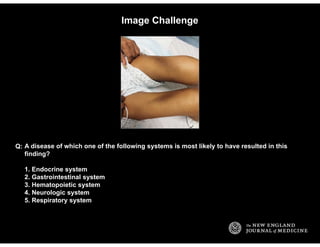
  • 1. Image Challenge A disease of which one of the following systems is most likely to have resulted in this finding? 1. Endocrine system 2. Gastrointestinal system 3. Hematopoietic system 4. Neurologic system 5. Respiratory system Q: Dr.Sherif Badrawy
  • 2. Answer: Image Challenge A disease of which one of the following systems is most likely to have resulted in this finding? Q: 5. Respiratory system Symmetric, slanting regions of hyperpigmentation on both thighs is consistent with Dahl's sign, a result of repeated pressure from the elbows on the epidermis of the thighs in patients spending large amounts of time in the tripod position. As in this patient, this finding provides supporting evidence of advanced chronic obstructive pulmonary disease.
  • 3. Image Challenge What is the most likely diagnosis in this asymptomatic 17-year-old male? 1. Antrochoanal polyp 2. Foreign body 3. Palatine tonsillitis 4. Pharyngeal lymphoma 5. Tonsillar abscess Q:
  • 4. Answer: Image Challenge What is the most likely diagnosis in this asymptomatic 17-year-old male?Q: 1. Antrochoanal polyp Rhinoscopy and computed tomography of the sinuses revealed that the soft, translucent, round mass occupying the left pharyngeal area was an antrochoanal polyp originating from the left maxillary sinus. The diagnosis was confirmed following surgical removal.
  • 5. Image Challenge What is the diagnosis? 1. Diaphragmatic hernia 2. Gastric bezoar 3. Inferior mesenteric artery thrombosis 4. Pancreatic phlegmon 5. Small-bowel volvulus Q:
  • 6. Answer: Image Challenge What is the diagnosis?Q: 5. Small-bowel volvulus The CT reveals small-bowel volvulus with dilated small bowel rotated around its blood supply. There are no signs of free air or fluid and no indication of bowel ischemia on the CT scan. The patient recovered following surgical release of strangulated small bowel that was 1 m from the ligament of Treitz.
  • 7. Image Challenge What is the diagnosis in this patient who developed burning pain, pruritus, and edema of the hands upon their exposure to water and who had this image taken under a Wood's lamp? 1. Acquired acral mycosis 2. Aquagenic keratoderma 3. Dyshidrotic eczema 4. Leprosy 5. Ochronosis Q:
  • 8. Answer: Image Challenge What is the diagnosis in this patient who developed burning pain, pruritus, and edema of the hands upon their exposure to water and who had this image taken under a Wood's lamp? Q: 2. Aquagenic keratoderma Following a biopsy, the patient received a diagnosis of aquagenic keratoderma, an unusual condition characterized by transitory, flat-topped papules and plaques, with hyperwrinkling and eccrine-duct prominence on the palms and fingers induced by exposure to water. Aquagenic keratoderma has been associated with a heterozygous mutation in the cystic fibrosis gene. The patient improved promptly following treatment with subcutaneous injections of botulinum toxin.
  • 9. Image Challenge What is the diagnosis? 1. Aerophagia 2. Congenital diaphragmatic hernia 3. Duodenal atresia 4. Gastroschesis 5. Hypertrophic pyloric stenosis Q:
  • 10. Answer: Image Challenge What is the diagnosis?Q: 3. Duodenal atresia The double-bubble sign comprises distention of both the stomach and proximal duodenum and is pathognomonic of congenital duodenal obstruction. There was no air distal to the level of obstruction in the D2 segment of the duodenum. Laparoscopy showed duodenal atresia type 1; the patient recovered following surgical repair.
  • 11. Image Challenge What is the diagnosis? 1. Angioedema 2. Cardiac tamponade 3. Necrotizing fasciitis 4. Superior vena cava syndrome 5. Toxic epidermal necrolysis Q:
  • 12. Answer: Image Challenge What is the diagnosis?Q: 4. Superior vena cava syndrome The cyanosis that is limited to the head, neck, upper torso, and arms is consistent with superior vena cava syndrome, which was confirmed with venography. The patient recovered following treatment with ultrasound-accelerated thrombolysis.
  • 13. Image Challenge What is the most likely diagnosis in this asymptomatic male? 1. Dercum's disease 2. Familial multiple lipomatosis 3. Madelung's disease 4. Neurofibromatosis, type 1 5. Rheumatoid arthritis Q:
  • 14. Answer: Image Challenge What is the most likely diagnosis in this asymptomatic male?Q: 2. Familial multiple lipomatosis The patient had more than 40 painless, mobile, soft nodules, with a maximum diameter of 10 cm, most of which were located in the arms and legs. Histopathological examination revealed mature adipose tissue, supporting the diagnosis of familial multiple lipomatosis, a rare autosomal dominant disorder, which was present in the patient's father, grandfather, and brothers. The patient's lipid profile was normal. Dercum's disease is a painful syndrome of the adipose tissue associated with obesity; Madelung's disease is characterized by symmetric fat deposits in the head, neck, and upper trunk.
  • 15. Image Challenge What is the most likely diagnosis in this 47-year-old patient with an acute headache? 1. Encephalitis 2. Meningitis 3. Paget's disease 4. Stroke 5. Subdural hemorrhage Q:
  • 16. Answer: Image Challenge What is the most likely diagnosis in this 47-year-old patient with an acute headache?Q: 4. Stroke CT of the brain revealed a linear area of hyperdensity in the first segment of the right middle cerebral artery, consistent with the diagnosis of acute stroke. The linear hyperdensity is suggestive of acute thromboembolic clot within the artery and is an early warning sign of large cerebral infarction, brain edema, and poor functional outcome.
  • 17. Image Challenge What is the most likely diagnosis? 1. Ankyloglossia 2. Atrophic glossitis 3. Creutzfeldt-Jakob disease 4. Hypoglossal nerve palsy 5. Motor neuron disease Q:
  • 18. Answer: Image Challenge What is the most likely diagnosis?Q: 4. Hypoglossal nerve palsy Hypoglossal nerve palsy produces wasting of the ipsilateral side of the tongue, and on attempted protrusion the tongue deviates towards the affected side. This patient's palsy resolved following treatment for bacterial meningitis. Motor neuron disease causes bilateral wasting.
  • 19. Image Challenge What is the diagnosis? 1. Erythema chronicum migrans 2. Granuloma annulare 3. Pityriasis rosea 4. Sarcoidosis 5. Tinea circinata Q:
  • 20. Answer: Image Challenge What is the diagnosis?Q: 5. Tinea circinata A skin-scraping specimen prepared with potassium hydroxide of the violaceous plaque consisting of four concentric rings with intervening areas of normal skin and numerous yellow- white pustules was found to contain multiple hyphae. A diagnosis of tinea circinata, an uncommon morphologic variant of tinea corporis that is caused by the dermatophyte Trichophyton tonsurans, was made.
  • 21. Image Challenge What is the diagnosis in this patient with axillary lymphadenopathy? 1. Bacterial felon 2. Erysipelas 3. Herpetic whitlow 4. Paronychia 5. Scabies Q:
  • 22. Answer: Image Challenge What is the diagnosis in this patient with axillary lymphadenopathy?Q: 3. Herpetic whitlow Direct fluorescence antibody testing of vesicular fluid confirmed the presence of herpes simplex virus type 2, consistent with the diagnosis of herpetic whitlow. The patient responded to treatment with oral acyclovir.
  • 23. Image Challenge What is the diagnosis in this asymptomatic male from North Africa? 1. Cutaneous leishmaniasis 2. Myiasis 3. Paronychia 4. Syphilis 5. Yaws Q:
  • 24. Answer: Image Challenge What is the diagnosis in this asymptomatic male from North Africa?Q: 1. Cutaneous leishmaniasis The verrucous lesion at the base of the thumb does not have the appearance of chronic paronychia; a skin smear revealed amastigote forms of the leishmania parasite.
  • 25. Image Challenge What is the most likely diagnosis in this patient who had suffered a blunt head trauma 2 months earlier? 1. Acute angle-closure glaucoma 2. Carotid cavernous fistula 3. Ocular lymphoma 4. Periorbital cellulitis 5. Thyroid ophthalmopathy Q:
  • 26. Answer: Image Challenge What is the most likely diagnosis in this patient who had suffered a blunt head trauma 2 months earlier? Q: 2. Carotid cavernous fistula Chemosis, proptosis, and an ulcerated cornea that develop after a head trauma are together most consistent with a diagnosis of carotid cavernous fistula. The diagnosis was confirmed with angiography and the patient recovered following endovascular coiling.
  • 27. Image Challenge What is the most likely underlying diagnosis in this 82-year-old patient with diabetes mellitus who had undergone a total hip replacement 10 years previously? 1. Colon cancer 2. Hypogammaglobulinemia 3. Hypophosphatasia 4. Osteosarcoma 5. Tuberculosis Q:
  • 28. Answer: Image Challenge What is the most likely underlying diagnosis in this 82-year-old patient with diabetes mellitus who had undergone a total hip replacement 10 years previously? Q: 1. Colon cancer There is free air extending lateral to the greater trochanter. Intra-articular cultures grew Clostridium septicum, which has a known association with colorectal cancer. Subsequent colonoscopy revealed a fungating colonic adenocarcinoma.
  • 29. Image Challenge What is the most likely diagnosis? 1. Achalasia 2. Diffuse esophageal spasm 3. Esophageal stricture 4. Hiatal hernia 5. Systemic sclerosis Q:
  • 30. Answer: Image Challenge What is the most likely diagnosis?Q: 2. Diffuse esophageal spasm The barium-swallow examination shows diverticula proximal to the marked corkscrew appearance of the distal esophagus, consistent with a diagnosis of diffuse esophageal spasm.
  • 31. Image Challenge What is the diagnosis? 1. Adrenal carcinoma 2. Autosplenectomy 3. Diverticular abscess 4. Emphysematous pyelonephritis 5. Renal vein thrombosis Q:
  • 32. Answer: Image Challenge What is the diagnosis?Q: 4. Emphysematous pyelonephritis Blood tests showed leukocytosis and hyperglycemia. Urinary microscopy revealed pyuria. Unenhanced computed tomography was performed and revealed extensive gas collection in the parenchyma of the left kidney, the perinephric space, and the left renal vein, with corresponding hydronephrosis and hydroureter. These findings and the patient's symptoms suggested emphysematous pyelonephritis.
  • 33. Image Challenge What is the diagnosis in this 52-year-old man with epigastric pain? 1. Bezoar 2. Diverticulitis 3. Intussusception 4. Mesenteric infarction 5. Pseudomembranous colitis Q:
  • 34. Answer: Image Challenge What is the diagnosis in this 52-year-old man with epigastric pain?Q: 3. Intussusception CT of the abdomen revealed dilated small-bowel loops and a "target sign," findings characteristic of intussusception, without evidence of a lead point. Jejunojejunal intussusception caused by an adenocarcinoma was confirmed at laparotomy.
  • 35. Image Challenge This patient would be predicted to have a higher-than-average risk for which one of the following diseases? 1. Cirrhosis 2. Coronary artery disease 3. Gout 4. Hemorrhagic stroke 5. Hypothyroidism Q:
  • 36. Answer: Image Challenge This patient would be predicted to have a higher-than-average risk for which one of the following diseases? Q: 2. Coronary artery disease A diagonal crease in the earlobe that runs backward from the tragus at a 45-degree angle across the lobule to the rear edge of the auricle may be a predictor of coronary artery disease. The finding is thought to indicate premature aging and loss of dermal and vascular elastic fibers, has limited sensitivity, and appears to most useful in persons younger than 60 years of age.
  • 37. Image Challenge What diagnosis is suggested by this fundus photograph from a 5-month-old girl? 1. Exudative retinitis 2. Primary infantile glaucoma 3. Retinopathy of prematurity 4. Retinoblastoma 5. Shaken-baby syndrome Q:
  • 38. Answer: Image Challenge What diagnosis is suggested by this fundus photograph from a 5-month-old girl?Q: 5. Shaken-baby syndrome The image illustrates normal disks and multiple intraretinal and preretinal hemorrhages in the posterior pole. The clinical feature believed to be pathognomonic for the shaken-baby syndrome, the circumpapillary retinal ridge, is present.
  • 39. Image Challenge What is the diagnosis in this patient who presented with a 2-day history of fever, sore throat, arthralgia, and suboccipital lymphadenopathy? 1. Adenovirus infection 2. Infectious mononucleosis 3. Measles 4. Rubella 5. Scarlet fever Q:
  • 40. Answer: Image Challenge What is the diagnosis in this patient who presented with a 2-day history of fever, sore throat, arthralgia, and suboccipital lymphadenopathy? Q: 4. Rubella Testing for rubella IgM antibody was positive, which confirmed the clinical diagnosis of rubella. Rubella is characterized by a maculopapular nonconfluent rash that is pink; it is often associated with suboccipital lymphadenopathy and arthralgia. The rash associated with measles is typically red, and less frequently associated with lymphadenopathy.
  • 41. Image Challenge What diagnosis is suggested by the finding on the sole of this patient's foot? 1. Chemical burn 2. Pemphigus 3. Thrombotic vasculopathy 4. Radiation dermatitis 5. Frostbite Q:
  • 42. Answer: Image Challenge What diagnosis is suggested by the finding on the sole of this patient's foot?Q: 3. Thrombotic vasculopathy The purpuric skin eruption has a netlike arrangement referred to as retiform. Retiform purpura is an indication of an acute thrombotic vasculopathy. If it is acute and rapidly progressive in a febrile patient, it suggests purpura fulminans. Read More: N Engl J Med 2008;358:e1
  • 43. Image Challenge What is the diagnosis? 1. Hand-foot-mouth disease 2. Herpes simplex virus infection 3. Herpes zoster virus infection 4. Folliculitis 5. Scalded skin syndrome Q:
  • 44. Answer: Image Challenge What is the diagnosis?Q: 2. Herpes simplex virus infection There are extensive, friable, hemorrhagic crusts with impetiginization of the chin and around the nares. The appearance is most consistent with herpes simplex virus infection. Herpes simplex infection was confirmed using viral cultures of scrapings from the base of the lesion.
  • 45. Image Challenge This patient developed difficulty swallowing following a dental procedure. What is the diagnosis? 1. Spondylolisthesis 2. Prevertebral air 3. Pharyngeal diverticulum 4. Pharyngeal foreign body 5. Periodontal abscess Q:
  • 46. Answer: Image Challenge This patient developed difficulty swallowing following a dental procedure. What is the diagnosis? Q: 2. Prevertebral air The radiograph shows emphysema with prevertebral air in the cervical soft tissues. A high- speed dental drill was implicated.
  • 47. Image Challenge This finding appeared on microscopical examination of the bronchoalveolar-lavage fluid of a patient with pulmonary nodules and cavitations. What is the diagnosis? 1. Asbestosis 2. Ascariasis 3. Aspergillosis 4. Coal-worker's pneumoconiosis 5. Paragonimiasis Q:
  • 48. Answer: Image Challenge This finding appeared on microscopical examination of the bronchoalveolar-lavage fluid of a patient with pulmonary nodules and cavitations. What is the diagnosis? Q: 5. Paragonimiasis Eggs from the lung fluke paragonimus were discovered on microscopical examination of bronchoalveolar-lavage fluid. Paragonimiasis is acquired by humans through consumption of undercooked freshwater crabs or crayfish. Immature forms migrate through the duodenal wall, peritoneal cavity, and diaphragm and mature within the pulmonary parenchyma. When the encapsulated cyst bursts, eggs are coughed up and swallowed. This patient recovered following treatment with praziquantel.
  • 49. Image Challenge What is the diagnosis? 1. Acromegaly 2. Adrenal insufficiency 3. Histiocytosis 4. Osteopetrosis 5. Paget's disease Q:
  • 50. Answer: Image Challenge What is the diagnosis?Q: 2. Adrenal insufficiency Although rare, calcification and even true ossification of the auricular cartilages have been most typically associated with primary adrenal insufficiency. It is also described in association with mechanical tissue injury, exposure to cold, inflammatory conditions, and other endocrinopathies.
  • 51. Image Challenge What is the diagnosis? 1. Lichen planus 2. Mycosis fungoides 3. Ostraceous psoriasis 4. Paraneoplastic pemphigoid 5. Staphylococcal scalded skin syndrome Q:
  • 52. Answer: Image Challenge What is the diagnosis?Q: 3. Ostraceous psoriasis These sharply demarcated, erythematous, well-defined limpet-like plaques covered with scales and crust are most consistent with ostraceous psoriasis. Read More: N Engl J Med 2010;362:155
  • 53. Image Challenge What is the diagnosis? 1. Spider angioma 2. Hereditary hemorrhagic telangiectasia 3. Pyogenic granuloma 4. Nodular melanoma 5. Cherry hemangioma Q:
  • 54. Answer: Image Challenge What is the diagnosis?Q: 1. Spider angioma Spider angiomas are red vascular lesions with a raised central body and branching spider-like legs. This patient had end-stage liver disease.
  • 55. Image Challenge What is the diagnosis? 1. Basal-cell carcinoma 2. Halo nevus 3. Lichen planus 4. Malignant melanoma 5. Spitz nevus Q:
  • 56. Answer: Image Challenge What is the diagnosis?Q: 2. Halo nevus Halo nevi are benign nevi that develop a border of depigmentation resembling a halo. In addition to the white border, this patient's nevus also has the variegated color, hypertrichosis, small scattered macules, and an irregularly scalloped border that are typical of a congenital melanocytic nevus. The halo phenomenon has been associated with vitiligo and rare spontaneous regression of melanocytic lesions.
  • 57. Image Challenge This patient presented with fatigue, fever, anorexia, and weight loss. What is the most likely diagnosis? 1. Leukemia 2. Scurvy 3. Acquired immunodeficiency syndrome 4. Sarcoidosis 5. Pellagra Q:
  • 58. Answer: Image Challenge This patient presented with fatigue, fever, anorexia, and weight loss. What is the most likely diagnosis? Q: 1. Leukemia Gingival infiltration in a patient with fever, fatigue, and weight loss is most suggestive of acute leukemia, especially monocytic variants of acute myelogenous leukemia. This patient's gingival infiltration resolved after treatment for acute myelomonocytic leukemia. Read More: N Engl J Med 2008;358:274
  • 59. Image Challenge This blood smear was from a child who developed a high fever after returning from a camping trip in eastern California. What is the diagnosis? 1. Babesiosis 2. Brucellosis 3. Leptospirosis 4. Rocky Mountain spotted fever 5. Tickborne relapsing fever Q:
  • 60. Answer: Image Challenge This blood smear was from a child who developed a high fever after returning from a camping trip in eastern California. What is the diagnosis? Q: 5. Tickborne relapsing fever A Borrelia hermsii spirochete is visible on the blood smear. The rodent-associated B. hermsii spirochete is a common cause of tickborne relapsing fever in North America and is transmitted by the night-biting, soft tick Ornithodoros hermsii. The patient defervesced following treatment with doxycycline.
  • 61. Image Challenge What is the diagnosis? 1. Gonococcal arthritis 2. Heberden's node 3. Rheumatoid arthritis 4. Sarcoidosis 5. Tophaceous gout Q:
  • 62. Answer: Image Challenge What is the diagnosis?Q: 5. Tophaceous gout Needle aspiration of the swelling yielded a white viscous fluid with numerous urate crystals identified on polarized microscopy.
  • 63. Image Challenge This patient presented with renal failure. What would be the expected finding on renal biopsy? 1. Cholesterol crystals 2. Crescentic glomerulonephritis 3. Heme pigment 4. Renal cortical necrosis 5. Tubulointerstitial nephritis Q:
  • 64. Answer: Image Challenge This patient presented with renal failure. What would be the expected finding on renal biopsy? Q: 1. Cholesterol crystals This patient presented with renal failure five weeks after coronary-artery bypass grafting. It is likely that atheromatous plaques were disrupted at the time of arterial manipulation, resulting in progressive subacute renal dysfunction, livedo reticularis, and digital cyanosis. Examination of a specimen from a percutaneous kidney biopsy revealed obstructive cholesterol crystals within an arcuate artery, confirming a diagnosis of cholesterol emboli syndrome. She ultimately died of sepsis. Read More: N Engl J Med 2011;364:265
  • 65. Image Challenge What is the diagnosis? 1. Beta-thalassemia 2. Eisenmenger syndrome 3. Metastatic small cell lung carcinoma 4. Sarcoidosis 5. Scleroderma Q:
  • 66. Answer: Image Challenge What is the diagnosis?Q: 1. Beta-thalassemia There is marked medullary expansion of the bony structures suggestive of compensatory extramedullary hematopoiesis. The pulmonary arteries are enlarged suggestive of pulmonary hypertension. Together these findings are most consistent with beta-thalassemia. Pulmonary hypertension in these patients is thought to arise from chronic anemia, hemolysis, and an increased tendency for microscopic thrombi to form within the pulmonary vasculature.
  • 67. Image Challenge Which one of the following biochemical measures would be most likely to be elevated in this patient? 1. Alkaline phosphatase 2. Calcium 3. Ferritin 4. Phosphorus 5. 25-hydroxy-vitamin D Q:
  • 68. Answer: Image Challenge Which one of the following biochemical measures would be most likely to be elevated in this patient? Q: 1. Alkaline phosphatase The patient has genu varum and enlarged wrists consistent with nutritional rickets. Alkaline phosphatase usually is increased markedly over the age-specific reference range in rickets. Serum phosphorus and vitamin D concentrations are usually low; serum calcium concentration is decreased only in hypocalcemic rickets. Ferritin is not usually elevated in these patients.
  • 69. Image Challenge What is the most likely diagnosis? 1. Age-related macular degeneration 2. Melanoma 3. Posterior vitreous detachment 4. Retinitis pigmentosa 5. Toxoplasmosis Q:
  • 70. Answer: Image Challenge What is the most likely diagnosis?Q: 5. Toxoplasmosis The pigmented and sharply demarcated choroidal lesions involving the macular region are most consistent with scarring caused by inactive chorioretinal toxoplasmosis, which can be acquired or congenital. The diagnosis was confirmed with serologic testing.
  • 71. Image Challenge What is the diagnosis? 1. Cutaneous leishmaniasis 2. Cutaneous larva migrans 3. Epidermoid cyst 4. Furuncular myiasis 5. Tungiasis Q:
  • 72. Answer: Image Challenge What is the diagnosis?Q: 4. Furuncular myiasis The image shows a human botfly larva (Dermatobia hominis) emerging from an inflamed nodule on the patient's upper arm. Cutaneous leishmaniasis, cutaneous larva migrans, epidermoid cyst, and tungiasis have distinct appearances. Read More: N Engl J Med 2000;342:937
  • 73. Image Challenge This patient presented with cough. What diagnosis accounts for the combination of findings on the bone scan? 1. Adverse effect of chronic glucocorticoids 2. Mastocytosis 3. Metastatic lung cancer 4. Osteomalacia with fracture 5. Paget's disease Q:
  • 74. Answer: Image Challenge This patient presented with cough. What diagnosis accounts for the combination of findings on the bone scan? Q: 3. Metastatic lung cancer The bone scan shows areas of uptake consistent with metastases as well as diffuse linear uptake in the femoral and tibial bones, consistent with hypertrophic pulmonary osteoarthropathy. Examination of specimens obtained by CT-guided biopsy of an 11-cm right lung mass was consistent with large-cell adenocarcinoma. Hypertrophic pulmonary osteoarthropathy is most frequently associated with lung and gastric carcinomas.
  • 75. Image Challenge Deficiency of which one of the following dietary components is most likely to have caused this rash? 1. Biotin 2. Folate 3. Niacin 4. Riboflavin 5. Vitamin C Q:
  • 76. Answer: Image Challenge Deficiency of which one of the following dietary components is most likely to have caused this rash? Q: 3. Niacin The symmetric, scaly, sunburn-like, hyperpigmented rash extending from the hands to a clearly demarcated border midway up the arm suggests a photosensitive distribution, most consistent with pellagra (niacin deficiency). Deficiency of the other listed dietary components is not typically associated with a photosensitive dermatitis in this area. Read More: N Engl J Med 2011;364:361
  • 77. Image Challenge What is the diagnosis? 1. Cutaneous larva migrans 2. Dirofilariasis 3. Gnathostomiasis 4. Paragonimiasis 5. Toxocariasis Q:
  • 78. Answer: Image Challenge What is the diagnosis?Q: 1. Cutaneous larva migrans The serpiginous, erythematous raised tracts with bulla formation are clinically diagnostic of cutaneous larva migrans. Cutaneous larva migrans is caused by the migration of hookworm larvae in human skin. It is most commonly caused by the hookworm that infects dogs and cats. Read More: N Engl J Med 2010;362:e10
  • 79. Image Challenge This patient presented following a high-speed motor vehicle crash. Which structure has been disrupted? 1. Aorta 2. Diaphragm 3. Esophagus 4. Myocardium 5. Trachea Q:
  • 80. Answer: Image Challenge This patient presented following a high-speed motor vehicle crash. Which structure has been disrupted? Q: 2. Diaphragm The elevated right hemidiaphragm suggests traumatic diaphragmatic rupture. The other listed structures appear to be intact. A computed tomographic scan confirmed the diaphragmatic rupture and showed that the dome of the liver had herniated into the right hemithorax.
  • 81. Image Challenge What diagnosis is most associated with this finding? 1. Congenital adrenal hyperplasia 2. Porphyria cutanea tarda 3. Ovarian teratoma 4. Situs inversus 5. Spinal dysraphism Q:
  • 82. Answer: Image Challenge What diagnosis is most associated with this finding?Q: 5. Spinal dysraphism A patch of hair on the lower back may be a cutaneous marker of spinal dysraphism and warrants consideration of underlying spinal abnormalities to prevent neurologic sequelae.
  • 83. Image Challenge What is the diagnosis? 1. Dental abscess 2. Neurofibromatosis type 1 3. Cleft jaw 4. Hemiatrophy syndrome 5. Mandibular fracture Q:
  • 84. Answer: Image Challenge What is the diagnosis?Q: 5. Mandibular fracture This patient developed a comminuted fracture of the left and right mandible after being struck on his right lower jaw. The open fracture allowed upward displacement of the left half of the mandible. Read More: N Engl J Med 2008;358:512
  • 85. Image Challenge This patient had an elevated lipase level. What is the diagnosis? 1. Adenovirus infection 2. Calciphylaxis 3. Erythema nodosum 4. Panniculitis 5. Tuberculosis Q:
  • 86. Answer: Image Challenge This patient had an elevated lipase level. What is the diagnosis?Q: 4. Panniculitis Pancreatic panniculitis is an uncommon complication of pancreatitis; the diagnosis was confirmed after skin biopsy. The liberation of pancreatic enzymes into the circulation causes fat necrosis in distal panni and the formation of subcutaneous nodules. The nodules regress with improvement in pancreatitis.
  • 87. Image Challenge What is the diagnosis? 1. Diphtheria 2. Secondary syphilis 3. Oral leukoplakia 4. Candida 5. Ludwig's angina Q:
  • 88. Answer: Image Challenge What is the diagnosis?Q: 2. Secondary syphilis These pseudomembranous lesions and erosions of the tongue, the hard and soft palate, and tonsils are consistent with secondary syphilis.
  • 89. Image Challenge What is the diagnosis? 1. Cushing's syndrome 2. Divarication of the rectus abdominis 3. Familial partial lipodystrophy of the Dunnigan type 4. Insulin lipohypertrophy 5. Morgagni hernia Q:
  • 90. Answer: Image Challenge What is the diagnosis?Q: 4. Insulin lipohypertrophy These pendulous subcutaneous periumbilical masses were attributed to 31 years of insulin injection to manage type 1 diabetes. Lipohypertrophy can be associated with glycemic flux and prevented by rotating injection sites.
  • 91. Image Challenge This patient with diffuse, nonencapsulated fatty deposits is most likely to have a history of which one of the following? 1. Alcohol dependency 2. Hypercalcemia 3. Renal insufficiency 4. Rheumatoid arthritis 5. Tuberculosis Q:
  • 92. Answer: Image Challenge This patient with diffuse, nonencapsulated fatty deposits is most likely to have a history of which one of the following? Q: 1. Alcohol dependency Madelungs disease (also known as benign symmetric lipomatosis) is a rare disorder of unknown cause and was the diagnosis in this case. Up to 90% of patients have a history of chronic alcoholism, and there is a strong male predominance. Read More: N Engl J Med 2011;364:465
  • 93. Image Challenge This patient with a history of alcohol abuse developed alopecia with fine, brittle scalp hair, diarrhea, and angular cheilitis. Measurement of which one of the following metals is most likely to be diagnostic? 1. Chromium 2. Copper 3. Manganese 4. Selenium 5. Zinc Q:
  • 94. Answer: Image Challenge This patient with a history of alcohol abuse developed alopecia with fine, brittle scalp hair, diarrhea, and angular cheilitis. Measurement of which one of the following metals is most likely to be diagnostic? Q: 5. Zinc This scaly erythematous eruption that preferentially involved the distal extremities and the perineum is most consistent with acrodermatitis enteropathica, confirmed by measurement of the zinc level. The patient's symptoms resolved after zinc supplementation.
  • 95. Image Challenge What is the diagnosis? 1. Epidural hematoma 2. Glioblastoma multiforme 3. Meningioma 4. Subarachnoid hemorrhage 5. Subdural hematoma Q:
  • 96. Answer: Image Challenge What is the diagnosis?Q: 1. Epidural hematoma Computed tomogram shows a 2.5-cm epidural hematoma in the left parietal region with mass effect, effacement, and left-to-right midline shift. Epidural hematomas have a lens-shaped appearance. Subdural hematomas are typically sickle-shaped.
  • 97. Image Challenge What is the diagnosis? 1. Bowel obstruction 2. Metastatic melanoma 3. Osteitis fibrosa cystica 4. Osteomalacia 5. Paget's disease Q:
  • 98. Answer: Image Challenge What is the diagnosis?Q: 4. Osteomalacia The anteroposterior radiograph of the pelvis reveals an undisplaced transverse fracture of both femora and generalized osteopenia. These features are typical of osteomalacia, a diagnosis that was confirmed in this case by laboratory measurement of vitamin D, calcium, phosphate and alkaline phosphatase levels. Osteitis fibrosa cystica is characterized by radiographic cysts associated with brown tumors.
  • 99. Image Challenge This patient presented with jaw pain and was found to have an elevated alkaline phosphatase and a normal serum creatinine. Which one of the following tests would confirm the diagnosis? 1. Bone scan 2. Insulin-like growth factor-1 level 3. Serum calcium 4. Abdominal ultrasound 5. Testing the function of the facial nerve Q:
  • 100. Answer: Image Challenge This patient presented with jaw pain and was found to have an elevated alkaline phosphatase and a normal serum creatinine. Which one of the following tests would confirm the diagnosis? Q: 1. Bone scan Paget's disease, acromegaly, and renal osteodystrophy are among the causes of jaw enlargement, visible in this image. An elevated alkaline phosphatase makes Paget's disease the most likely diagnosis; the diagnosis can be confirmed with a bone scan. Read More: N Engl J Med 2008;358:625
  • 101. Image Challenge This patient had presented with weight loss. What is the most likely diagnosis? 1. Gastric cancer 2. Lung cancer 3. Melanoma 4. Nasopharyngeal cancer 5. Thyroid cancer Q:
  • 102. Answer: Image Challenge This patient had presented with weight loss. What is the most likely diagnosis?Q: 1. Gastric cancer Left supraclavicular adenopathy can be an indicator of gastric cancer, as in this case. Virchow's node, or Troisier's node, refers to carcinomatous involvement of the supraclavicular nodes at the junction of the thoracic duct and the left subclavian vein. Usually, nodal enlargement is caused by metastatic gastric carcinoma, although supraclavicular nodal involvement can also be seen in other gastrointestinal, thoracic, and pelvic cancers.
  • 103. Image Challenge What is the diagnosis? 1. Tinea barbae 2. Herpes simplex infection 3. Eczema 4. Mycosis fungoides 5. Impetigo Q:
  • 104. Answer: Image Challenge What is the diagnosis?Q: 1. Tinea barbae The combination of alopecia and pustules is most consistent with tinea barbae. Read More: N Engl J Med 1998;338:735
  • 105. Image Challenge This 23-year-old man was involved in a motor vehicle accident. What is the diagnosis? 1. Aortic dissection 2. Cardiac rupture 3. Diaphragmatic rupture 4. Pneumothorax 5. Vertebral fractures Q:
  • 106. Answer: Image Challenge This 23-year-old man was involved in a motor vehicle accident. What is the diagnosis?Q: 4. Pneumothorax The presence of a deep, lucent, right costophrenic angle on supine chest radiography is an indirect sign of a pneumothorax. In addition, a pneumothorax with associated rib fractures and subcutaneous emphysema is evident in the right chest. The patient's endotracheal tube needs to be withdrawn further into the trachea.
  • 107. Image Challenge What is the diagnosis? 1. Bancroftian filariasis 2. Deep-vein thrombosis 3. Erythema ab igne 4. Lipodermatosclerosis 5. Superficial thrombophlebitis Q:
  • 108. Answer: Image Challenge What is the diagnosis?Q: 5. Superficial thrombophlebitis The patient had erythema and tenderness on the medial aspect of the left knee. A palpable, ropelike cord was present from the left medial malleolus to the groin. Duplex Doppler ultrasonography revealed thrombosis of the greater saphenous vein, with no extension into the deep venous system, consistent with superfical thrombophlebitis. Read More: N Engl J Med 2001;344:1214
  • 109. Image Challenge What is the diagnosis? 1. Cutaneous leishmaniasis 2. Leprosy 3. Leukemia cutis 4. Syphilis 5. Yaws Q:
  • 110. Answer: Image Challenge What is the diagnosis?Q: 1. Cutaneous leishmaniasis Cutaneous leishmaniasis typically begins as a painless papule that enlarges to a nodule with a central crust. The papules and nodules enlarge, may develop central ulceration, and take approximately 1 year to heal without treatment. Satellite lesions may also be present. The patient was treated successfully with injections of sodium stibogluconate. Read More: N Engl J Med 2010;362:e15
  • 111. Image Challenge What is the diagnosis? 1. Contact dermatitis 2. Discoid lupus erythematosus 3. Melanoma 4. Nummular eczema 5. Tinea corporis Q:
  • 112. Answer: Image Challenge What is the diagnosis?Q: 4. Nummular eczema The image illustrates discoid (nummular) eczema in an infant. This pattern of eczema is frequently associated with atopic dermatitis and is often confused with ringworm infection.
  • 113. Image Challenge This 43-year-old patient presented with bilateral pain, swelling, and stiffness in the hands and feet. Her chest radiograph was abnormal. What is the most likely diagnosis? 1. Miliary tuberculosis 2. Psoriasis 3. Syphilis 4. Reiter syndrome 5. Sarcoidosis Q:
  • 114. Answer: Image Challenge This 43-year-old patient presented with bilateral pain, swelling, and stiffness in the hands and feet. Her chest radiograph was abnormal. What is the most likely diagnosis? Q: 5. Sarcoidosis The circumscribed, corticated lytic bone lesions on the radiograph are more consistent with sarcoid granuloma than with the other listed choices. Manifestations of sarcoidosis involving the bones and joints can occur early or late in the illness. Read More: N Engl J Med 2008;358:e7
  • 115. Image Challenge These oral ulcers were painless. What is the most likely diagnosis? 1. Chancroid 2. Herpes simplex 3. Iron deficiency 4. Squamous cell cancer 5. Syphilis Q:
  • 116. Answer: Image Challenge These oral ulcers were painless. What is the most likely diagnosis?Q: 5. Syphilis The ulcers have clean, smooth bases and slightly elevated, indurated borders. This type of painless ulcer is typical of a syphilitic chancre. The patient reported having had unprotected orogenital sex with his girlfriend about 2 weeks before the onset of the ulcers. Rapid plasma reagin testing was positive with a titer of at least 1:32. In addition, a Treponema pallidum particle agglutination assay was strongly positive, and a fluorescent treponemal antibody absorption test was positive for both IgG and IgM antibodies. The patient's girlfriend also had positive results on serologic analyses for syphilis, and both were treated successfully with intramuscular penicillin. Chancroid and herpetic ulcers tend to be painful. Cancers do not typically involve both upper and lower lips simultaneously.
  • 117. Image Challenge What is the diagnosis? 1. Arcus juvenilus 2. Calcific band keratopathy 3. Herpetic keratitis 4. Kayser-Fleischer ring 5. Vernal conjunctivitis Q:
  • 118. Answer: Image Challenge What is the diagnosis?Q: 5. Vernal conjunctivitis The diagnosis of vernal conjunctivitis is typically based on the clinical findings of giant cobblestone papillae on the tarsal conjunctiva or at the limbus as in this case. This problem typically affects young males and often resolves at puberty. Both eyes are generally involved, and patients often report ocular burning or itching, tearing, and photophobia. Treatment consists of saline eyedrops, topical antihistamines, and nonsteroidal antiinflammatory drugs.
  • 119. Image Challenge What is the most likely diagnosis? 1. Acromegaly 2. Cystic fibrosis 3. Eisenmenger's syndrome 4. Squamous-cell lung cancer 5. Ulcerative colitis Q:
  • 120. Answer: Image Challenge What is the most likely diagnosis?Q: 3. Eisenmenger's syndrome When Eisenmengers syndrome occurs in concert with a patent ductus arteriosus, deoxygenated blood from the right ventricle is delivered to the aorta distal to the left subclavian artery. The upper extremities are thus spared the effects of the shunt, whereas the lower extremities are not, resulting in differential clubbing and cyanosis. The other diagnoses are not typically associated with differential clubbing. Read More: N Engl J Med 2011;364:666
  • 121. Image Challenge What is the diagnosis? 1. Gastric outlet obstruction 2. Hirschsprung disease 3. Ileal intussusception 4. Ulcerative colitis 5. Zollinger-Ellison syndrome Q:
  • 122. Answer: Image Challenge What is the diagnosis?Q: 4. Ulcerative colitis The abdominal radiograph shows a severely dilated transverse colon and free air. The findings are typical of fulminant ulcerative colitis. The patient recovered after a total abdominal colectomy with end ileostomy. Read More: N Engl J Med 2010;362:635
  • 123. Image Challenge This 22-year-old man presented with a 1-month history of severe pubic itch that was worst at night. What is the most appropriate topical treatment? 1. Hydrocortisone 2. Hydroxyzine 3. Mupirocin 4. Permethrin 5. Selenium sulfide Q:
  • 124. Answer: Image Challenge This 22-year-old man presented with a 1-month history of severe pubic itch that was worst at night. What is the most appropriate topical treatment? Q: 4. Permethrin The visible nits are consistent with pubic pediculosis. The recommended treatments include permethrin or pyrethrin lotions. Alternative regimens to treat lice include topical malathion or oral ivermectin. Patients with pediculosis pubis should be evaluated for other sexually transmitted diseases. Read More: N Engl J Med 2009;360:e11
  • 125. Image Challenge What diagnosis explains the loss of visual acuity in this woman who is at 36 weeks of gestation? 1. Central serous chorioretinitis 2. Diabetes mellitus 3. Glaucoma 4. Graves' disease 5. Preeclampsia Q:
  • 126. Answer: Image Challenge What diagnosis explains the loss of visual acuity in this woman who is at 36 weeks of gestation? Q: 5. Preeclampsia The findings are indicative of grade 4 hypertensive retinopathy, with widespread hemorrhages, cotton-wool spots, hard exudates in a star shape in the macular region, and swelling of the optic disks. The blood pressure was 220/140 mm Hg, and severe preeclampsia was diagnosed. The exudates resolved spontaneously in the months following stabilization and delivery of an underweight baby boy.
  • 127. Image Challenge This patient presented with transient, painless visual obscuration in the left eye. What is the diagnosis? 1. Papilledema 2. Hypertensive retinopathy 3. Cholesterol emboli 4. Temporal arteritis 5. Diabetic retinopathy Q:
  • 128. Answer: Image Challenge This patient presented with transient, painless visual obscuration in the left eye. What is the diagnosis? Q: 3. Cholesterol emboli The retinal photograph demonstrates multiple, tiny refractile retinal arteriolar cholesterol emboli and a saddle embolus superior to the optic nerve. Read More: N Engl J Med 2008;358:826
  • 129. Image Challenge What is the diagnosis? 1. Aphthous stomatitis 2. Bullous pemphigoid 3. Chickenpox 4. Herpes zoster 5. Paraneoplastic pemphigus Q:
  • 130. Answer: Image Challenge What is the diagnosis?Q: 4. Herpes zoster The multiple erythematous ulcers, some covered with a whitish membrane, on the hard and soft palates are most consistent with a diagnosis of herpes zoster. The lesions healed completely after 7 days of treatment with acyclovir.
  • 131. Image Challenge What is the most likely underlying diagnosis? 1. Cirrhosis 2. Chronic renal failure 3. Hypothyroidism 4. Myeloma 5. Sickle cell disease Q:
  • 132. Answer: Image Challenge What is the most likely underlying diagnosis?Q: 1. Cirrhosis This lesion has the characteristic appearance of a spider angioma. Spider angiomas are suggestive of liver disease; this patient was diagnosed with cirrhosis.
  • 133. Image Challenge What is the diagnosis? 1. Anterior uveitis 2. Carotid cavernous sinus fistula 3. Graves' disease 4. Orbital lymphoma 5. Scleral rupture Q:
  • 134. Answer: Image Challenge What is the diagnosis?Q: 2. Carotid cavernous sinus fistula The patient had inferior chemosis and conjunctival injection. Contrast-enhanced computed tomography of the orbit showed a dilated left superior ophthalmic vein, and angiography confirmed the presence of a carotid cavernous sinus fistula. The patient had complete resolution following embolization of the fistula. Read More: N Engl J Med 2011;364:e15
  • 135. Image Challenge This 12-year-old presented with decreased appetite and vomiting. What is the most likely diagnosis? 1. Endometrioma 2. Hernia 3. Keloid 4. Intraabdominal cancer 5. Urachal duct cyst Q:
  • 136. Answer: Image Challenge This 12-year-old presented with decreased appetite and vomiting. What is the most likely diagnosis? Q: 4. Intraabdominal cancer Sister Mary Joseph's nodule is a lymphatic metastasis to the umbilical region. This 12-year- old girl had presented with decreased appetite, vomiting, and weight loss. Computed tomographic scanning showed a large pelvic mass probably originating from the ovary, omental and hepatic metastases, ascites, and a mass through the umbilicus. The appearance is not typical of the other listed diagnoses. Read More: N Engl J Med 2005;352:1913
  • 137. Image Challenge What clinical presentation would be expected in this patient? 1. Asymmetrical mydriasis 2. Ataxic hemiparesis 3. Hypothermia 4. Quadriplegia 5. Upward gaze palsy Q:
  • 138. Answer: Image Challenge What clinical presentation would be expected in this patient?Q: 4. Quadriplegia The most common presentation of a pontine hemorrhage is quadriplegia. Small, reactive pupils are characteristic of pontine hemorrhages. Hemiparesis would be expected if the hemorrhage were asymmetrical. Hypothermia is unusual. Upward gaze palsy occurs with midbrain involvement.
  • 139. Image Challenge What is the most likely diagnosis in this patient who underwent fine-needle aspiration after reporting several weeks of submandibular pain? 1. Adenoid cystic carcinoma 2. Cat scratch disease 3. Infectious mononucleosis 4. Sclerosing sialadenitis 5. Systemic lupus erythematosus Q:
  • 140. Answer: Image Challenge What is the most likely diagnosis in this patient who underwent fine-needle aspiration after reporting several weeks of submandibular pain? Q: 4. Sclerosing sialadenitis Chronic sclerosing sialadenitis is an inflammatory fibrosing condition of the salivary glands that can be bilateral and manifests as firm swelling of the involved gland. Carcinomas do not tend to be bilateral. The other listed conditions are not typically painful and do not disproportionally involve the submandibular glands.
  • 141. Image Challenge What is the most likely diagnosis? 1. Chronic venous insufficiency 2. Reiter syndrome 3. Gunshot wound 4. Chronic renal failure 5. Pseudohypoparathyroidism Q:
  • 142. Answer: Image Challenge What is the most likely diagnosis?Q: 1. Chronic venous insufficiency Chronic venous insufficiency may be accompanied by subcutaneous calcifications. These calcifications are often discovered by chance on plain radiographs. Read More: N Engl J Med 2008;358:e10
  • 143. Image Challenge What is the diagnosis? 1. Parulis 2. Pyogenic granuloma 3. Peripheral ossifying fibroma 4. Retrocuspid papillae 5. Torus mandibularis Q:
  • 144. Answer: Image Challenge What is the diagnosis?Q: 5. Torus mandibularis Four hard, sessile nodules were noted on the lingual surface, with normal overlying mucosa. No notable abnormalities were identified on panoramic radiographs. Exostoses are localized, benign bony protrusions. The most common oral exostoses are torus palatinus and torus mandibularis, which do not have cartilage involvement, owing to their anatomical location.
  • 145. Image Challenge Which one of the following organs is enlarged? 1. Colon 2. Ovary 3. Stomach 4. Spleen 5. Gall bladder Q:
  • 146. Answer: Image Challenge Which one of the following organs is enlarged?Q: 3. Stomach Massive dilation of the stomach with distal gas is most consistent with gastric outlet obstruction. Read More: N Engl J Med 2007;356:942
  • 147. Image Challenge This patient had diabetes. What is the diagnosis? 1. Diabetic bullae 2. Eruptive xanthomas 3. Lichen amyloidosis 4. Necrobiosis lipoidica 5. Pustular psoriasis Q:
  • 148. Answer: Image Challenge This patient had diabetes. What is the diagnosis?Q: 2. Eruptive xanthomas The lesions were reddish yellow, pruritic, and painful and were present on the backs of both legs and on the buttocks and knees. A blood specimen was lipemic and the triglyceride level was 8168 mg per deciliter. Histologic analysis of a lesion-biopsy specimen confirmed the suspicion that the lesions were eruptive xanthomas.
  • 149. Image Challenge What laboratory test is most appropriate for this patient? 1. Alpha-fetoprotein 2. Beta2-microglobulin 3. Calcitonin 4. Insulin-like growth factor-1 5. Red-cell transketolase Q:
  • 150. Answer: Image Challenge What laboratory test is most appropriate for this patient?Q: 3. Calcitonin This patient had multiple flesh-colored papules on the eyelids, lips, and tongue. This phenotype is suggestive of multiple endocrine neoplasia (MEN) type 2B (MEN-2B), an autosomal dominant condition characterized by medullary thyroid cancer among other features. Serum calcitonin can be used to screen for medullary thyroid cancer. Read More: N Engl J Med 2011;364:870
  • 151. Image Challenge What is the diagnosis in this baby girl who was delivered at 30 weeks' gestation? 1. Congenital cytomegalovirus infection 2. Ecthyma gangrenosum 3. Homozygous protein C deficiency 4. Klippel-Trenaunay syndrome 5. Langerhans cell histiocytosis Q:
  • 152. Answer: Image Challenge What is the diagnosis in this baby girl who was delivered at 30 weeks' gestation?Q: 1. Congenital cytomegalovirus infection The girl had anemia and thrombocytopenia. The skin findings were due to congenital cytomegalovirus infection, with dermal hematopoiesis. Similar findings can occur as a result of severe anemia, congenital rubella, and parvovirus infection. The child did not survive.
  • 153. Image Challenge Serum levels of which one of the following laboratory tests would be expected to be most abnormal in this patient? 1. 17-hydroxyprogesterone 2. Angiotensin-converting enzyme 3. Anti-tissue transglutaminase antibody 4. Prolactin 5. Vitamin B6 Q:
  • 154. Answer: Image Challenge Serum levels of which one of the following laboratory tests would be expected to be most abnormal in this patient? Q: 2. Angiotensin-converting enzyme Lupus pernio is a manifestation of sarcoidosis that involves the nasal bridge and cheeks. Serum levels of the angiotensin-converting enzyme are elevated in the majority of patients with untreated sarcoidosis. Read More: N Engl J Med 2007;357:2153
  • 155. Image Challenge What maternal diagnosis is most likely to expain the development of this rash 2 hours after delivery of this child with trisomy 21? 1. Measles 2. Polyarteritis nodosa 3. Rubella 4. Streptococcal A infection 5. Systemic lupus erythematosus Q:
  • 156. Answer: Image Challenge What maternal diagnosis is most likely to expain the development of this rash 2 hours after delivery of this child with trisomy 21? Q: 5. Systemic lupus erythematosus Maternal antibodies crossing the placenta can lead to the clinical manifestations of neonatal lupus, which was diagnosed in this case. The rash resolved over the subsequent 6 months.
  • 157. Image Challenge What is the most likely diagnosis? 1. Cisplatin overdose 2. Lesch-Nyhan syndrome 3. Rhabdomyolysis 4. Primary hyperparathyroidism 5. Ethylene glycol poisoning Q:
  • 158. Answer: Image Challenge What is the most likely diagnosis?Q: 5. Ethylene glycol poisoning This urine sediment contains calcium oxalate crystals of two types. The crystals shaped like envelopes with diagonally crossing lines are octahedrons of calcium oxalate dihydrate. The needle-shaped crystals are calcium oxalate monohydrate. Calcium oxalate monohydrate crystals are rarely seen in the urinary sediment but are typical of ethylene glycol ingestion, and therefore when seen, they strongly suggest the diagnosis. Read More: N Engl J Med 2006;354:1065
  • 159. Image Challenge This specimen was resected from a child with intestinal obstruction. What is the most likely diagnosis? 1. Ascariasis 2. Kala-azar 3. Meckel's diverticulum 4. Strongyloidiasis 5. Trichobezoar Q:
  • 160. Answer: Image Challenge This specimen was resected from a child with intestinal obstruction. What is the most likely diagnosis? Q: 1. Ascariasis A mass of Ascaris lumbricoides in the terminal ileum was found to be causing intestinal obstruction in this child from Kenya. The child recovered uneventfully after surgery. In the absence of obstruction, peritonitis, or bowel strangulation, nonsurgical management can include albendazole, ivermectin, or mebendazole.
  • 161. Image Challenge This patient was hypothyroid. What is the diagnosis? 1. Branchial cleft cyst 2. Ectopic thyroid 3. Laryngocele 4. Lipoma 5. Papillary thyroid cancer Q:
  • 162. Answer: Image Challenge This patient was hypothyroid. What is the diagnosis?Q: 2. Ectopic thyroid An ultrasonogram showed a solid midline mass. A technetium-99 thyroid scan showed uptake in the region of the mass but no uptake in the area of the thyroid gland. A fine-needle aspiration biopsy did not reveal any evidence of a malignant condition. The diagnosis was an ectopic thyroid gland in a thyroglossal duct cyst.
  • 163. Image Challenge What is the diagnosis? 1. Adrenal adenoma 2. Gastric bezoar 3. Pancreatic pseudocyst 4. Pulmonary echinococcosis 5. Splenic cyst Q:
  • 164. Answer: Image Challenge What is the diagnosis?Q: 5. Splenic cyst The plain abdominal film shows a large circular calcified lesion in the left upper abdomen, most consistent with a calcified splenic cyst. Computed tomography demonstrated a large, well-defined, cystic mass with mural calcification in the spleen. The patient had been involved in a motor vehicle accident 20 years earlier.
  • 165. Image Challenge What is the diagnosis? 1. Dermatobia hominis 2. Pediculus humanus capitis 3. Pediculus humanus corporis 4. Phthirus pubis 5. Sarcoptes scabiei Q:
  • 166. Answer: Image Challenge What is the diagnosis?Q: 4. Phthirus pubis Dermoscopy reveals the typical broad body and the large middle and hind legs of Phthirus pubis, the pubic louse. Though most commonly found in pubic hair, the trunk, limbs, and eyelashes can be colonized in heavy infestations.
  • 167. Image Challenge Which one of the following conditions is the most likely to be responsible for this clinical picture? 1. Excessive fluoride supplementation 2. Hyperbilirubinemia 3. Treatment with tetracycline 4. Trichophyton rubrum infection 5. Pseudomonas aeruginosa infection Q:
  • 168. Answer: Image Challenge Which one of the following conditions is the most likely to be responsible for this clinical picture? Q: 5. Pseudomonas aeruginosa infection Green nails, a form of chromonychia, may be caused by bacterial infection with P. aeruginosa. This syndrome is typically seen in patients with nail disease such as onycholysis, onychotillomania, or paronychia, particularly in those whose abnormal nails have been exposed to moist environments. The green color is caused by the fluorescent siderophore pyoverdin, produced by P. aeruginosa.
  • 169. Image Challenge Treatment with which one of the following medications is associated with this clinical finding? 1. Erlotinib 2. Leflunomide 3. Methotrexate 4. Pegvisomant 5. Psoralen Q:
  • 170. Answer: Image Challenge Treatment with which one of the following medications is associated with this clinical finding? Q: 1. Erlotinib Erlotinib, a tyrosine kinase inhibitor of the epidermal growth factor receptor, induces characteristic hair alterations. Trichomegaly, curling, elongation, and trichorrhexis are typical; these reverse after discontinuation of therapy. The pictured changes are not typical of leflunomide, methotrexate, pegvisomant, or psoralen. Read More: N Engl J Med 2008;358:1175
  • 171. Image Challenge What is the most likely diagnosis for this finding detected during esophageal endoscopy? 1. Adenocarcinoma 2. Candidiasis 3. Dieulafoy's lesion 4. Schatzki ring 5. Systemic sclerosis Q:
  • 172. Answer: Image Challenge What is the most likely diagnosis for this finding detected during esophageal endoscopy?Q: 1. Adenocarcinoma This nodular abnormality was found to be an early adenocarcinoma in a patient with Barrett's esophagus.
  • 173. Image Challenge What is the diagnosis? 1. Addison's disease 2. Amalgam tattoo 3. Blue nevus 4. Hemangioma 5. Melanoma Q:
  • 174. Answer: Image Challenge What is the diagnosis?Q: 5. Melanoma Histopathological examination of the slightly elevated, bluish-brown lesions with irregular boundaries revealed an infiltrating lentiginous melanoma. Primary melanoma of the oral cavity accounts for 0.5% of all oral cancers and has a poor prognosis. The patient underwent partial maxillectomy and had no signs of recurrence 6 months after surgery.
  • 175. Image Challenge This patient's serum was found to be discolored four hours following a surgical procedure. What is the most likely cause? 1. Fluorescent dye 2. Methemoglobinemia 3. Propofol 4. Pseudomonal sepsis 5. Ethylene glycol Q:
  • 176. Answer: Image Challenge This patient's serum was found to be discolored four hours following a surgical procedure. What is the most likely cause? Q: 1. Fluorescent dye The patient had undergone intraoperative angiography with a fluorescent dye and a Wood's lamp was used to evaluate mesenteric-vessel viability. Read More: N Engl J Med 2007;356:e10
  • 177. Image Challenge What is the diagnosis? 1. Coarctation of the aorta 2. Left atrial enlargement 3. Phrenic nerve palsy 4. Pulmonary embolism 5. Sarcoidosis Q:
  • 178. Answer: Image Challenge What is the diagnosis?Q: 4. Pulmonary embolism The chest radiograph demonstrates a Westermark sign with a focal area of oligemia in the right middle zone and cutoff of the pulmonary artery in the upper lobe of the right lung. Computed tomographic pulmonary angiography confirmed the presence of a thrombus in the right pulmonary artery with an occlusive thrombus in the pulmonary arteries of the right upper and middle lobes. The patient made a good recovery.
  • 179. Image Challenge What is the diagnosis? 1. Neurofibromatosis 2. Rhinophyma 3. Rosacea 4. Systemic lupus erythematosus 5. Tuberous sclerosis Q:
  • 180. Answer: Image Challenge What is the diagnosis?Q: 5. Tuberous sclerosis The yellow-orange papules on the patient's nose and cheeks are characteristic of tuberous sclerosis, an autosomal dominant disorder in which mutations in tumor suppressor gene TSC1 or TSC2 result in the formation of benign hamartomas throughout the body. Almost all patients with this condition have at least one characteristic dermatologic feature. Read More: N Engl J Med 2011;364:1061
  • 181. Image Challenge A woman underwent colonoscopy after presenting with colicky abdominal pain and loose stool. What type of worm is causing this presentation? 1. Ascaris lumbricoides 2. Diphyllobothrium latum 3. Necator americanus 4. Trichinella spiralis 5. Trichuris trichiura Q:
  • 182. Answer: Image Challenge A woman underwent colonoscopy after presenting with colicky abdominal pain and loose stool. What type of worm is causing this presentation? Q: 2. Diphyllobothrium latum Colonoscopy revealed a long, moving tapeworm, Diphyllobothrium latum, located in the terminal ileum and extending to the sigmoid colon. D. latum is a fish tapeworm that can infect humans after they consume infected undercooked or raw fish. She was treated with a single dose of praziquantel.
  • 183. Image Challenge Where is the abnormality on this chest radiograph? 1. Left lower lobe 2. Left upper lobe 3. Right lower lobe 4. Right upper lobe 5. Superior mediastinum Q:
  • 184. Answer: Image Challenge Where is the abnormality on this chest radiograph?Q: 2. Left upper lobe A pulmonary mass is visible in the left upper lung.
  • 185. Image Challenge What is the most likely diagnosis? 1. Paget's disease 2. Meningioma 3. Neurocysticercosis 4. Pneumocephalus 5. Hyperparathyroidism Q:
  • 186. Answer: Image Challenge What is the most likely diagnosis?Q: 4. Pneumocephalus The cranial radiograph shows air in the left temporal region without evidence of fracture. Pneumocephalus can occur after neurosurgical procedures, head and facial trauma, or ear infection and can even occur spontaneously. The finding is not typical of a brown tumor, Paget's disease, neurocysticercosis, or meningioma. Read More: N Engl J Med 2008;358:e13
  • 187. Image Challenge What is the diagnosis? 1. Acromegaly 2. Ankylosing spondylitis 3. Fluorosis 4. Mastocytosis 5. Multiple myeloma Q:
  • 188. Answer: Image Challenge What is the diagnosis?Q: 3. Fluorosis Radiography of the spine reveals a rugger-jersey appearance (striated pattern of increased density in the upper and lower zones of the vertebrae), suggesting skeletal fluorosis. The patient's serum fluoride concentration was confirmed to be abnormally high. Skeletal fluorosis is endemic in areas with high concentrations of fluoride in the drinking water, but it is rare in other parts of the world. The patient's symptoms improved after cessation of excessive tea consumption.
  • 189. Image Challenge What is the diagnosis? 1. Carpal tunnel syndrome 2. Rheumatoid Arthritis 3. Scleroderma 4. Diabetic peripheral neuropathy 5. Dupuyten's contracture Q:
  • 190. Answer: Image Challenge What is the diagnosis?Q: 5. Dupuyten's contracture The pictured flexion contractures involving bilateral third digits and the right fifth digit are most consistent with Dupuytren's contracture. Read More: N Engl J Med 2007;356:e11
  • 191. Image Challenge What is the diagnosis? 1. Arcus senilis 2. Galactosialidosis 3. Hemochromatosis 4. Keratoconus 5. Wilson's disease Q:
  • 192. Answer: Image Challenge What is the diagnosis?Q: 5. Wilson's disease This 18-year-old patient had a postural tremor of her arms and legs, mild dysphagia and dysarthria, and bradykinesia. Measurement of urinary copper and ceruloplasmin confirmed the diagnosis of Wilson's disease. The rings resolved following chelation therapy.
  • 193. Image Challenge Which vector was responsible for infecting this 14-year-old immigrant from Cameroon? 1. Chrysops fly 2. Mosquito 3. Reduviid bug 4. Sand fly 5. Tsetse fly Q:
  • 194. Answer: Image Challenge Which vector was responsible for infecting this 14-year-old immigrant from Cameroon?Q: 1. Chrysops fly Microfilaria of Loa loa are transmitted by the hematophagous Chrysops fly. This patient had 17% eosinophilia and high levels of parasites in the bloodstream. She recovered following treatment with diethylcarbamazine. Read More: N Engl J Med 2006;355:e6
  • 195. Image Challenge What is the diagnosis? 1. Chemical burn 2. Cicatricial pemphigoid 3. Epidermomycosis 4. Herpes zoster 5. Squamous cell carcinoma Q:
  • 196. Answer: Image Challenge What is the diagnosis?Q: 4. Herpes zoster Vesicular, purulent, and crusted lesions consistent with herpes zoster are evident in the ophthalmic distribution of the trigeminal nerve. The patient completed a 10-day course of oral acyclovir, and clindamycin was added to the regimen for suspected bacterial superinfection. Read More: N Engl J Med 2010;362:1128
  • 197. Image Challenge What is the diagnosis? 1. Coarctation of the aorta 2. Lung cancer 3. Pneumothorax 4. Rib fracture 5. Substernal goiter Q:
  • 198. Answer: Image Challenge What is the diagnosis?Q: 5. Substernal goiter The chest radiograph demonstrates tracheal deviation. Ultrasonography of the neck revealed a large goiter with the right lobe extending into the anterior superior mediastinum.
  • 199. Image Challenge What is the diagnosis? 1. Small-bowel obstruction 2. Echinococcosis 3. Mesenteric ischemia 4. Pancreatic pseudocysts 5. Cecal volvulus Q:
  • 200. Answer: Image Challenge What is the diagnosis?Q: 1. Small-bowel obstruction The computed tomogram reveals small-bowel obstruction by a left-sided luminal mass. The mass has a hyperdense periphery and an aerated core. Read More: N Engl J Med 2008;358:1381
  • 201. Image Challenge What is the most likely diagnosis for this boy who was born at 36 weeks gestation weighing 1800 g? 1. Congenital parvovirus B19 infection 2. Fetal alcohol syndrome 3. Neonatal thyrotoxicosis 4. Nesidioblastosis 5. Williams syndrome Q:
  • 202. Answer: Image Challenge What is the most likely diagnosis for this boy who was born at 36 weeks gestation weighing 1800 g? Q: 3. Neonatal thyrotoxicosis The upper eyelids are retracted and subcutaneous fat is virtually absent, findings that are most consistent with a diagnosis of neonatal thyrotoxicosis. Mother and child recovered with treatment.
  • 203. Image Challenge What is the diagnosis? 1. Bulimia nervosa 2. Cocaine abuse 3. Lichen planus 4. Ludwig's angina 5. Osteonecrosis Q:
  • 204. Answer: Image Challenge What is the diagnosis?Q: 1. Bulimia nervosa The combination of a deep ulcer of the posterior palate with severe tooth erosion is most consistent with a diagnosis of bulimia nervosa. The patient reported binge eating and self- induced vomiting several times daily.
  • 205. Image Challenge What is the diagnosis? 1. Chalazion 2. Papilloma 3. Pterygium 4. Pinguecula 5. Coloboma Q:
  • 206. Answer: Image Challenge What is the diagnosis?Q: 2. Papilloma This 9-year-old boy was diagnosed with conjunctival viral papilloma. Read More: N Engl J Med 2007;356:1352
  • 207. Image Challenge What is the diagnosis? 1. Beta-galactosidase deficiency 2. Fordyce's angiokeratomas 3. Radiation dermatitis 4. Scabies 5. Varicocele Q:
  • 208. Answer: Image Challenge What is the diagnosis?Q: 2. Fordyce's angiokeratomas Angiokeratomas of the scrotum (Fordyce's angiokeratomas) often arise in the second or third decade but are most commonly diagnosed in elderly men. No treatment is necessary.
  • 209. Image Challenge Ultraviolet light was shone on this patient's rash. What is the diagnosis? 1. Erythrasma 2. Intertrigo 3. Pityriasis rosea 4. Psoriasis 5. Tinea versicolor Q:
  • 210. Answer: Image Challenge Ultraviolet light was shone on this patient's rash. What is the diagnosis?Q: 1. Erythrasma Under examination with ultraviolet A light from a Woods lamp, the rash exhibited coral-red fluorescence, a finding pathognomonic for erythrasma. This common disorder results from overgrowth of Corynebacterium minutissimum. The coral-red fluorescence helps to distinguish this disorder from dermatophytoses, candidal intertrigo, and psoriasis. Read More: N Engl J Med 2011;364:e25
  • 211. Image Challenge What is the diagnosis? 1. Bullous pemphigoid 2. Dermatitis herpetiformis 3. Impetigo 4. Syphilis 5. Varicella Q:
  • 212. Answer: Image Challenge What is the diagnosis?Q: 5. Varicella This polymorphic rash with vesicles, pustules, and crusty lesions is most consistent with varicella infection. Read More: N Engl J Med 2010;362:1227
  • 213. Image Challenge The appearance of this Tunisian woman's ear is most consistent with which one of the following infectious diseases? 1. Leprosy 2. Leishmaniasis 3. Syphilis 4. Tuberculosis 5. Yaws Q:
  • 214. Answer: Image Challenge The appearance of this Tunisian woman's ear is most consistent with which one of the following infectious diseases? Q: 2. Leishmaniasis Cutaneous leishmaniasis is endemic in Tunisia, particularly in the central and northern parts of the country. Lupoid leishmaniasis is an uncommon form of chronic cutaneous leishmaniasis associated with Leishmania tropica. A polymerase-chain-reaction assay performed on a biopsy specimen from this patient was positive for leishmania.
  • 215. Image Challenge What is the diagnosis? 1. Behçet's syndrome 2. Lichen simplex chronicus 3. Condyloma acuminatum 4. Lichen sclerosus 5. Vestibular papillomatosis Q:
  • 216. Answer: Image Challenge What is the diagnosis?Q: 5. Vestibular papillomatosis These shiny, soft, linearly arrayed papules are the typical presentation of vestibular papillomatosis, a variant of vestibular mucosa commonly mistaken for genital warts. Read More: N Engl J Med 2008;358:1495
  • 217. Image Challenge What is the diagnosis in this patient who presented with sudden painful vision impairment after vigorous exercise? 1. Central retinal artery occlusion 2. Corneal ulcer 3. Dislocation of the lens 4. Episcleritis 5. Retinal detachment Q:
  • 218. Answer: Image Challenge What is the diagnosis in this patient who presented with sudden painful vision impairment after vigorous exercise? Q: 3. Dislocation of the lens There is anterior dislocation of the lens. Rupture of the zonular fibers may result from ocular trauma or other conditions. The patient underwent surgical extraction of the dislocated lens, anterior vitrectomy, and implantation of an iris-fixated intraocular lens.
  • 219. Image Challenge What is the diagnosis? 1. Carcinoid syndrome 2. Mastocytosis 3. Normal pregnancy 4. Radial-artery occlusion 5. Raynaud's phenomenon Q:
  • 220. Answer: Image Challenge What is the diagnosis?Q: 5. Raynaud's phenomenon Raynaud's phenomenon is characterized by exaggerated vasoconstrictive color changes (pallor and cyanosis) in the fingers, usually due to exposure to cold. The phenomenon is considered primary if there is no evidence of an underlying medical illness. Secondary Raynaud's phenomenon occurs in association with another condition, such as systemic lupus erythematosus, scleroderma, or a vascular occlusive disease. The patient was given a recommendation to keep her hands warm to avoid further attacks.
  • 221. Image Challenge What is the diagnosis? 1. Subcutaneous metastases 2. Filariasis 3. Caput Medusae 4. Neurofibromatosis 5. Hepatocellular carcinoma Q:
  • 222. Answer: Image Challenge What is the diagnosis?Q: 3. Caput Medusae These enlarged veins on his abdomen are consistent with caput medusae. Read More: N Engl J Med 1999;341:419
  • 223. Image Challenge Which coronary artery is occluded? 1. Left anterior descending 2. Left circumflex 3. Left diagonal branch 4. Left main stem 5. Right Q:
  • 224. Answer: Image Challenge Which coronary artery is occluded?Q: 4. Left main stem The 12-lead electrocardiogram shows ST-segment elevation in leads V1, V2, V3, I, aVL, and aVR and ST-segment depression in leads V4, V5, V6, II, III, and aVF, findings suggestive of occlusion of the left main stem of the coronary artery. The patient recovered following revascularization.
  • 225. Image Challenge What is the diagnosis? 1. Intestinal malrotation 2. Intestinal perforation 3. Lynch syndrome 4. Mesenteric ischemia 5. Portal vein thrombosis Q:
  • 226. Answer: Image Challenge What is the diagnosis?Q: 4. Mesenteric ischemia Abdominal computed tomography with contrast material shows occlusion of the main trunk of the superior mesenteric artery with mesenteric venous gas and pneumatosis intestinalis. The patient did not survive. Read More: N Engl J Med 2011;364:1349
  • 227. Image Challenge This 4-year-old boy presented with a 5-day history of mild fever and malaise and a 3-day history of rash. What is the diagnosis? 1. Erythema infectiosum 2. Hand, foot, and mouth disease 3. Kawasaki disease 4. Measles 5. Pityriasis rosea Q:
  • 228. Answer: Image Challenge This 4-year-old boy presented with a 5-day history of mild fever and malaise and a 3-day history of rash. What is the diagnosis? Q: 2. Hand, foot, and mouth disease This clinical picture is highly characteristic of hand, foot, and mouth disease, a self-limiting viral disease that is usually caused by coxsackievirus A16 or enterovirus 71. Typical skin lesions are elliptical vesicles surrounded by an erythematous halo. The patient was treated supportively at home without medication and recovered.
  • 229. Image Challenge What is the diagnosis? 1. Carcinoid syndrome 2. Dermatomyositis 3. Endocarditis 4. Lichen planus 5. Porphyria Q:
  • 230. Answer: Image Challenge What is the diagnosis?Q: 2. Dermatomyositis Dilated and tortuous blood vessels with areas of atrophy, telangiectases, and bushy loop formation along the fingernail bed are most consistent with dermatomyositis. Periungual telangiectases also occur in patients with scleroderma and systemic lupus erythematosus.
  • 231. Image Challenge A patient with this tomogram would be most likely to present with which one of the following signs? 1. Uniocular blindness 2. Hemiplegia 3. Alexia without agraphia 4. Hemiballismus 5. Internuclear ophthalmoplegia Q:
  • 232. Answer: Image Challenge A patient with this tomogram would be most likely to present with which one of the following signs? Q: 2. Hemiplegia The tomogram shows a calcified object in the proximal right middle cerebral artery. Occlusion of the middle cerebral artery would be most likely to be associated with contralateral hemiparesis, as in this case. The other listed choices represent stroke syndromes that most typically involve other vascular territories.
  • 233. Image Challenge What is the diagnosis? 1. Empyema 2. Lymphangioleiomyomatosis 3. Paraesophageal hernia 4. Pericardial effusion 5. Plombage Q:
  • 234. Answer: Image Challenge What is the diagnosis?Q: 3. Paraesophageal hernia Posteroanterior chest radiography revealed a large intrathoracic gastric bubble, and a barium- contrast study of the upper gastrointestinal tract confirmed the presence of a large paraesophageal hernia. An exploratory laparotomy revealed a large paraesophageal hernia (i.e., type II hiatal hernia), without evidence of gastric strangulation. The crura were repaired, and a Nissen fundoplication with anterior gastropexy was performed. The patient recovered from surgery uneventfully.
  • 235. Image Challenge What is the diagnosis? 1. Adrenal cancer 2. Echinococcal infection 3. Meckel's diverticulitis 4. Pneumatosis intestinalis 5. Trichobezoar Q:
  • 236. Answer: Image Challenge What is the diagnosis?Q: 4. Pneumatosis intestinalis Pneumatosis intestinalis is diagnosed by the presence of air pockets in the intestinal wall. In certain cases, pneumatosis intestinalis may be considered a surrogate marker for intestinal ischemia and impending perforation. However, the condition may also occur in a benign context and is no longer considered a disease but rather a sign, and its significance needs to be considered in accordance with each patient's clinical context.
  • 237. Image Challenge This 9-kg liver was removed at the time of liver transplantation. What is the diagnosis? 1. Macronodular cirrhosis 2. Hepatocellular carcinoma 3. Echinococcosis 4. Polycystic liver disease 5. Trophoblastic tumor Q:
  • 238. Answer: Image Challenge This 9-kg liver was removed at the time of liver transplantation. What is the diagnosis?Q: 4. Polycystic liver disease The cystic changes are most consistent with polycystic liver disease. Polycystic kidney disease was also diagnosed in this patient. Read More: N Engl J Med 2007;356:1560
  • 239. Image Challenge What is the diagnosis? 1. Granulomatosis with polyangiitis (Wegener's) 2. Lepromatous leprosy 3. Neurofibromatosis type 1 4. Sarcoidosis 5. Tertiary syphilis Q:
  • 240. Answer: Image Challenge What is the diagnosis?Q: 2. Lepromatous leprosy The patient's face had multiple nodular lesions that coalesced into plaques, especially on the forehead, ears, nose, and lips. Screening for IgM antibodies to phenolic glycolipid I, which is specific for Mycobacterium leprae, was highly positive. The patient underwent multidrug therapy, with the addition of prednisone, for 5 months. After 9 months of multidrug treatment, the skin infiltration and weakness in the left eyelid had diminished.
  • 241. Image Challenge What is the diagnosis? 1. Dermatitis herpetiformis 2. Impetigo 3. Measles 4. Secondary syphilis 5. Varicella Q:
  • 242. Answer: Image Challenge What is the diagnosis?Q: 5. Varicella This unvaccinated adolescent had fever and a classic, extensive varicella rash characterized by pruritic vesicular lesions with erythematous bases. Read More: N Engl J Med 2005;352:439
  • 243. Image Challenge This patient was trying to look right when the image was taken. What is the diagnosis? 1. Internuclear ophthalmoplegia 2. Left fourth cranial nerve palsy 3. Left sixth cranial nerve palsy 4. Right fourth cranial nerve palsy 5. Right sixth cranial nerve palsy Q:
  • 244. Answer: Image Challenge This patient was trying to look right when the image was taken. What is the diagnosis?Q: 5. Right sixth cranial nerve palsy The neurologic examination reveals an inability to abduct the right eye with horizontal gaze to the right, a finding that is consistent with an isolated right abducens nerve palsy. Read More: N Engl J Med 2010;362:e52
  • 245. Image Challenge What term is used to describe this finding? 1. Arc eye 2. Asthenopia 3. Choroideremia 4. Coloboma 5. Corectopia Q:
  • 246. Answer: Image Challenge What term is used to describe this finding?Q: 4. Coloboma Colobomas are the result of abnormal closure of the optic fissure. They may occur anywhere along the optic fissure and can affect the iris, choroid, or macula. Isolated iris colobomas are asymptomatic, but those involving the macula or the optic disk can result in severe visual impairment. Typical iris colobomas occur in the inferonasal quadrant.
  • 247. Image Challenge This rash appeared following treatment for leukemia. What is the diagnosis? 1. Cryoglobulinemia 2. Leukemia cutis 3. Herpes zoster 4. Graft-versus-host disease 5. Urticaria pigmentosa Q:
  • 248. Answer: Image Challenge This rash appeared following treatment for leukemia. What is the diagnosis?Q: 4. Graft-versus-host disease There is hyperpigmentation and hypopigmentation of the skin, cutaneous atrophy, telangiectasia, and ulcerations. This is most consistent with graft-versus-host disease of the skin. Read More: N Engl J Med 2002;347:36
  • 249. Image Challenge What is the diagnosis? 1. Blastomycosis 2. Celiac disease 3. Down's syndrome 4. Hyperparathyroidism 5. IgA nephropathy Q:
  • 250. Answer: Image Challenge What is the diagnosis?Q: 2. Celiac disease This patient was diagnosed with celiac disease. Folate malabsorption is a suggested mechanism of cerebral calcification in patients with celiac disease, because cerebral calcification can be seen with other conditions related to folate deficiency, such as treatment wtih methotrexate. The patient's condition improved following treatment with a gluten-free diet. The other diagnoses are not associated with cerebral calcification in this pattern.
  • 251. Image Challenge What is the diagnosis? 1. Amelanotic melanoma 2. Angioma 3. Dermal nevus 4. Pyogenic granuloma 5. Wart Q:
  • 252. Answer: Image Challenge What is the diagnosis?Q: 1. Amelanotic melanoma The red nodule had a blue-gray pigmented area at the 2 o'clock position. Excision confirmed a diagnosis of melanoma; the patient was found to have diffuse metastases. Amelanotic melanoma may appear to be similar to many common benign skin lesions (e.g., pyogenic granulomas, angiomas, and dermal nevi) and is therefore often erroneously treated with the use of diathermy or laser vaporization.
  • 253. Image Challenge What is the diagnosis? 1. Intracranial hemorrhage 2. Osteoma 3. Neurocysticercosis 4. Arachnoid cyst 5. Meningioma Q:
  • 254. Answer: Image Challenge What is the diagnosis?Q: 5. Meningioma This well-circumscribed and highly calcified extra-axial mass is most consistent with a meningioma. Read More: N Engl J Med 2007;356:e14
  • 255. Image Challenge What is the diagnosis? 1. Cryptorchidism 2. Femoral-artery pseudoaneurysm 3. Iliopectineal bursitis 4. Inguinal hernia 5. Metastatic inguinal lymphadenopathy Q:
  • 256. Answer: Image Challenge What is the diagnosis?Q: 5. Metastatic inguinal lymphadenopathy The findings are most consistent with metastatic inguinal lymphadenopathy, a common site of metastasis for tumors located in the lower extremities, pelvis, and back. Enlarged lymph nodes are not always visible, but should be palpable. This patient died 3 months after presentation, following a diagnosis of metastatic malignant melanoma that had arisen on the back.
  • 257. Image Challenge What is the most likely diagnosis? 1. Calciphylaxis 2. Factor V Leiden 3. Protein C deficiency 4. Scleroderma 5. Waldenstrom's macroglobulinemia Q:
  • 258. Answer: Image Challenge What is the most likely diagnosis?Q: 5. Waldenstrom's macroglobulinemia The patient was diagnosed with Waldenstrom's macroglobulinemia after he presented with progressive ischemic injury of the fingers and toes, probably related to hyperviscosity and cryoglobulinemic vasculitis. The other listed diagnoses do not typically present with this appearance. The patient required digital amputation. Read More: N Engl J Med 2011;364:e34
  • 259. Image Challenge What is the diagnosis? 1. Duodenal perforation 2. Emphysematous pyelonephritis 3. Perinephric cyst 4. Situs inversus 5. Ureterocele Q:
  • 260. Answer: Image Challenge What is the diagnosis?Q: 1. Duodenal perforation The computed tomography shows air surrounding the right kidney and extending to the retroperitoneum. A duodenal ulcer with retroperitoneal perforation was diagnosed. The normal appearance of the kidney excludes emphysematous pyelonephritis. Read More: N Engl J Med 2006;354:e9
  • 261. Image Challenge What is the diagnosis? 1. Duodenal perforation 2. Emphysematous pyelonephritis 3. Perinephric cyst 4. Situs inversus 5. Ureterocele Q:
  • 262. Answer: Image Challenge What is the diagnosis?Q: 1. Duodenal perforation The computed tomography shows air surrounding the right kidney and extending to the retroperitoneum. A duodenal ulcer with retroperitoneal perforation was diagnosed. The normal appearance of the kidney excludes emphysematous pyelonephritis. Read More: N Engl J Med 2006;354:e9
  • 263. Image Challenge What is the diagnosis? 1. Epidermolysis bullosa 2. Hereditary hemorrhagic telangiectasia 3. Neurofibromatosis 4. Peutz-Jeghers syndrome 5. Scleroderma Q:
  • 264. Answer: Image Challenge What is the diagnosis?Q: 2. Hereditary hemorrhagic telangiectasia Telangiectasias of the tongue are most consistent with hereditary hemorrhagic telangiectasia. Peutz-Jeghers syndrome does not typically involve the tongue. Read More: N Engl J Med 2009;360:1769
  • 265. Image Challenge What is the diagnosis? 1. Babesiosis 2. Iron deficiency anemia 3. Hereditary spherocytosis 4. Malaria 5. Sideroblastic anemia Q:
  • 266. Answer: Image Challenge What is the diagnosis?Q: 1. Babesiosis The peripheral-blood smear shows numerous intracellular organisms in red blood cells. Ring forms are seen, as well as rare tetrads. These so-called Maltese cross formations are essentially pathognomonic of babesiosis, since they are not seen in malaria, the primary consideration in the differential diagnosis. Read More: N Engl J Med 2008;358:e19
  • 267. Image Challenge What is the diagnosis? 1. Blepharitis 2. Entropion 3. Eyelid wart 4. Keratoconjunctivitis sicca 5. Vernal keratoconjunctivitis Q:
  • 268. Answer: Image Challenge What is the diagnosis?Q: 5. Vernal keratoconjunctivitis There are large, flat-topped, confluent cobblestone papillae in the upper palpebral conjunctiva in the right eye. Tarsal conjunctiva shows loss of architecture, scarring, and hyperemia. These findings are most consistent with a diagnosis of vernal keratoconjunctivitis. The patient responded well to a supratarsal injection of triamcinolone acetonide.
  • 269. Image Challenge What diagnosis explains these lesions that developed after acupuncture to the area? 1. Herpetic whitlow 2. Nummular eczema 3. Psoriasis 4. Ringworm 5. Scabies Q:
  • 270. Answer: Image Challenge What diagnosis explains these lesions that developed after acupuncture to the area?Q: 3. Psoriasis There are multiple discrete, erythematous, scaly, indurated papules on the lower back and buttocks. Needling of the skin can produce trauma that is sufficient to cause Koebner's phenomenon and subsequent psoriatic flare. Clobetasol cream was prescribed. Irritation and infection are rare complications of acupuncture when performed to established standards.
  • 271. Image Challenge What diagnosis explains these lesions that developed after acupuncture to the area? 1. Herpetic whitlow 2. Nummular eczema 3. Psoriasis 4. Ringworm 5. Scabies Q:
  • 272. Answer: Image Challenge What diagnosis explains these lesions that developed after acupuncture to the area?Q: 3. Psoriasis There are multiple discrete, erythematous, scaly, indurated papules on the lower back and buttocks. Needling of the skin can produce trauma that is sufficient to cause Koebner's phenomenon and subsequent psoriatic flare. Clobetasol cream was prescribed. Irritation and infection are rare complications of acupuncture when performed to established standards.
  • 273. Image Challenge What is the diagnosis? 1. Syphilis 2. Phenytoin exposure 3. Marfan syndrome 4. Torus palatinus 5. Kaposi sarcoma Q:
  • 274. Answer: Image Challenge What is the diagnosis?Q: 4. Torus palatinus The appearance of this mass is most consistent with torus palatinus. Read More: N Engl J Med 2007;356:1759
  • 275. Image Challenge Which of these medications, used to treat this patient's glioblastoma multiforme, is most likely to have contributed to this complication? 1. Acetazolamide 2. Dexamethasone 3. Levetiracetam 4. Temozolomide 5. Topotecan Q:
  • 276. Answer: Image Challenge Which of these medications, used to treat this patient's glioblastoma multiforme, is most likely to have contributed to this complication? Q: 2. Dexamethasone The patient was found to have pneumoperitoneum, with gas extending from the infradiaphragmatic region to the inferior margin of the liver, outlining the gallbladder. The findings are highly suggestive of bowel perforation; dexamethasone increases the risk for this complication. Comfort care was pursued in accordance with the patient's wishes and she died shortly thereafter.
  • 277. Image Challenge What is the diagnosis? 1. Erythema marginatum 2. Erythema multiforme 3. Pityriasis rosea 4. Tinea versicolor 5. Tuberculoid leprosy Q:
  • 278. Answer: Image Challenge What is the diagnosis?Q: 5. Tuberculoid leprosy Examination of this patient revealed multiple large, annular, hypopigmented, atrophic macules with well-defined, erythematous, raised borders. The lesions were hairless, hypohidrotic, and anesthetic. There was no peripheral-nerve enlargement. Histopathological analysis of a skin- biopsy specimen revealed well-developed epithelioid granulomas, lymphocytes, and Langerhans giant cells. On the basis of clinical and histological findings, the condition was diagnosed as tuberculoid leprosy. Read More: N Engl J Med 2011;364:1657
  • 279. Image Challenge What is the most likely diagnosis? 1. End-stage renal disease 2. Graves' disease 3. Iron deficiency 4. Pseudohypoparathyroidism 5. Sarcoidosis Q:
  • 280. Answer: Image Challenge What is the most likely diagnosis?Q: 3. Iron deficiency There is pallor of the nail beds, as well as koilonychia (spoon-shaped nails). Though koilonychia can be present in other diseases, together these findings are most suggestive of iron-deficiency anemia. Read More: N Engl J Med 2010;362:e59
  • 281. Image Challenge What is the diagnosis? 1. Hypercholesterolemia 2. Leukemia 3. Lichen planus 4. Pompe's disease 5. Psoriasis Q:
  • 282. Answer: Image Challenge What is the diagnosis?Q: 1. Hypercholesterolemia The image reveals diffuse, flat xanthomas and larger tuberous xanthomas on the buttocks of this 18-year-old. These findings are consistent with familial hypercholesterolemia which was confirmed on evaluation of her lipid profile and review of her family history.
  • 283. Image Challenge What is the diagnosis? 1. Central retinal artery occlusion 2. Diabetic papillopathy 3. Ocular toxoplasmosis 4. Optic neuritis 5. Malignant hypertension Q:
  • 284. Answer: Image Challenge What is the diagnosis?Q: 5. Malignant hypertension The fundus photograph shows disk edema, cottonwool spots, a swollen optic nerve, and retinal hemorrhages. Together, these findings suggest a diagnosis of malignant hypertension. Read More: N Engl J Med 2008;358:1951
  • 285. Image Challenge What is the cause of this new skin lesion in this patient whose blood CD4+ cell count was 20 per cubic millimeter (1%) following treatment for chronic lymphocytic leukemia? 1. Bacillary angiomatosis 2. Bullous impetigo 3. Cryptococcosis 4. Kaposi's sarcoma 5. Verruca vulgaris Q:
  • 286. Answer: Image Challenge What is the cause of this new skin lesion in this patient whose blood CD4+ cell count was 20 per cubic millimeter (1%) following treatment for chronic lymphocytic leukemia? Q: 3. Cryptococcosis Biopsy of the umbilicated facial papule revealed numerous encapsulated yeast forms; Cryptococcus neoformans was cultured. The facial and other lesions gradually disappeared following treatment with amphotericin B, flucytosine, and ultimately fluconazole.
  • 287. Image Challenge In addition to neurofibromatosis, what other examination finding would you expect for this patient? 1. Aortic regurgitation 2. Chvostek sign 3. Lisch nodules 4. Thyroid bruit 5. Web neck Q:
  • 288. Answer: Image Challenge In addition to neurofibromatosis, what other examination finding would you expect for this patient? Q: 3. Lisch nodules There is hypertrophy of the right lower extremity and an irregular peduncular mass with overlying hyperpigmentation and hypertrichosis involving the right trunk, right buttock, and right leg. Histopathological examination was consistent with a soft-tissue neurofibroma. Lisch nodules were seen on ophthalmologic examination, and neurofibromatosis type 1 was diagnosed.
  • 289. Image Challenge In what vessel is this patient's dialysis catheter? 1. Left axillary vein 2. Left internal mammary vein 3. Left-sided superior vena cava 4. Azygous vein 5. Highest intercostal vein Q:
  • 290. Answer: Image Challenge In what vessel is this patient's dialysis catheter?Q: 3. Left-sided superior vena cava This left internal jugular dialysis catheter is in a left-sided superior vena cava that drained into the right atrium via a coronary sinus. Read More: N Engl J Med 2007;356:1870
  • 291. Image Challenge What is the most likely infecting organism in this patient with prostatitis? 1. Blastomyces dermatitidis 2. Escherichia coli 3. Histoplasma capsulatum 4. Pseudomonas aeruginosa 5. Staphylococcus aureus Q:
  • 292. Answer: Image Challenge What is the most likely infecting organism in this patient with prostatitis?Q: 1. Blastomyces dermatitidis The appearance of these skin lesions is most typical of infection with Blastomyces dermatitidis, Coccidioides immitis, and Cryptococcus neoformans. The genitourinary tract is the second most common nonpulmonary site of involvement after the skin for these infections. The lesions are not typical of infection with the other listed infections.
  • 293. Image Challenge What is the diagnosis? 1. Addison's disease 2. Alkaptonuria 3. Hematoma 4. Melanoma 5. Zidovudine exposure Q:
  • 294. Answer: Image Challenge What is the diagnosis?Q: 4. Melanoma Subungual melanoma, a variant of acral lentiginous melanoma, arises from the nail matrix, most commonly in the great toe or thumb. Hutchinsons nail sign is an important clinical clue to subungual melanoma and is characterized by extension of brown or black pigment from the nail bed, matrix, and nail plate to the adjacent cuticle and proximal or lateral nail folds. The patient underwent amputation of the great toe, and he remains healthy 8 years later. Read More: N Engl J Med 2011;364:e38
  • 295. Image Challenge What is the diagnosis? 1. Cerebral aneurysm 2. Chiari I malformation 3. Neurofibromatosis 4. Paget disease 5. Subdural hematoma Q:
  • 296. Answer: Image Challenge What is the diagnosis?Q: 2. Chiari I malformation The T1-weighted sagittal image shows a tonsillar herniation (type I Chiari malformation) more than 5 mm below the level of the foramen magnum. Read More: N Engl J Med 2007;357:1821
  • 297. Image Challenge What is the diagnosis? 1. Coxsackievirus infection 2. Gorlin's syndrome 3. Herpes zoster 4. Orbital cellulitis 5. Superficial pyoderma Q:
  • 298. Answer: Image Challenge What is the diagnosis?Q: 3. Herpes zoster The image illustrates the classical appearance of acute herpes zoster involving the first division of the trigeminal nerve.
  • 299. Image Challenge Which structure is most dilated? 1. Aorta 2. Left atrium 3. Left ventricle 4. Right atrium 5. Right ventricle Q:
  • 300. Answer: Image Challenge Which structure is most dilated?Q: 2. Left atrium The chest radiograph reveals cardiomegaly, with splaying of the carina and an elevated left main bronchus. These findings are most suggestive of an enlarged left atrium. Read More: N Engl J Med 2008;358:2050
  • 301. Image Challenge What is the diagnosis in this patient who had been involved in a motorcycle accident? 1. Aortic dissection 2. Cardiac tamponade 3. Diffuse pulmonary hemorrhage 4. Tension pneumothorax 5. Traumatic diaphragmatic hernia Q:
  • 302. Answer: Image Challenge What is the diagnosis in this patient who had been involved in a motorcycle accident?Q: 4. Tension pneumothorax The chest radiograph reveals a 90-degree rightward rotation of the cardiac silhouette with left tension pneumothorax, pulmonary contusion, and multiple rib fractures. The mediastinal shift that was caused by the tension pneumothorax resolved following pleural drainage.
  • 303. Image Challenge What is the diagnosis? 1. Achalasia 2. Esophageal cancer 3. Esophageal web 4. Gastroesophageal reflux disease 5. Nutcracker esophagus Q:
  • 304. Answer: Image Challenge What is the diagnosis?Q: 5. Nutcracker esophagus The barium swallow shows a typical corkscrew appearance consistent with nutcracker esophagus. High-resolution manometry of the esophagus confirmed a hypertensive peristaltic disorder of the esophagus.
  • 305. Image Challenge What is the diagnosis? 1. Central retinal vein occlusion 2. Profilerative diabetic retinopathy 3. Hypertensive retinopathy 4. Chorioretinitis 5. Papilledema Q:
  • 306. Answer: Image Challenge What is the diagnosis?Q: 2. Profilerative diabetic retinopathy Severe bilateral proliferative diabetic retinopathy with significant optic-disk neovascularization is visible. Read More: N Engl J Med 2007;356:1979
  • 307. Image Challenge What is the diagnosis? 1. Digoxin poisoning 2. Hyperkalemia 3. Intra-aortic balloon pump 4. Pericardial effusion 5. Right fascicular block Q:
  • 308. Answer: Image Challenge What is the diagnosis?Q: 2. Hyperkalemia The electrocardiogram shows a regular rhythm, with a widened QRS complex in a sine-wave configuration, and there no discernible P waves. The T waves were fused with the widened QRS complexes to form the sine-wave pattern (sinoventricular rhythm). The patients serum potassium level was 9.1 mmol per liter. His condition stabilized after the administration of calcium chloride, bicarbonate, glucose, and insulin therapy, which was followed by hemodialysis.
  • 309. Image Challenge A prolapse such as this may be effectively reduced by applying what common household substance to the tissue? 1. Baking powder 2. Flour 3. Salt 4. Sugar 5. Tea Q:
  • 310. Answer: Image Challenge A prolapse such as this may be effectively reduced by applying what common household substance to the tissue? Q: 4. Sugar Plain granulated sugar can be applied to the mucosa of the prolapsed structure (such as the ileum in this case) to promote the osmotic shift of fluid out of the edematous tissue. In this case, within 2 minutes, the edema had diminished sufficiently to allow spontaneous reduction of the prolapse. Read More: N Engl J Med 2011;364:1855
  • 311. Image Challenge What is the most likely diagnosis? 1. Cutaneous larva migrans 2. Glucagonoma 3. Lung cancer 4. Systemic lupus erythematosus 5. Ulcerative colitis Q: