SlideShare a Scribd company logo
1 of 388
Download to read offline
A 38 year old lady presents with a recent episode of renal colic. As part of her
investigations the following results are obtained:
Corrected Calcium 3.84 mmol/l
PTH 88pg/ml (increased)
Her serum urea and electrolytes are normal.
What is the most likely diagnosis?
A. Carcinoma of the bronchus
B. Secondary hyperparathyroidism
C. Primary hyperparathyroidism
D. Tertiary hyperparathyroidism
E. Carcinoma of the breast
Theme from September 2012 exam
Theme from September 2011 exam
In this situation the most likely diagnosis is primary hyperparathyroidism. The
question mentions that serum urea and electrolytes are normal, which makes tertiary
hyperparathyroidism unlikely.
Primary hyperparathyroidism
In exams primary hyperparathyroidism is stereotypically seen in elderly females with
an unquenchable thirst and an inappropriately normal or raised parathyroid hormone
level. It is most commonly due to a solitary adenoma
Causes of primary hyperparathyroidism
 80%: solitary adenoma
 15%: hyperplasia
 4%: multiple adenoma
 1%: carcinoma
Features - 'bones, stones, abdominal groans and psychic moans'
 Polydipsia, polyuria
 Peptic ulceration/constipation/pancreatitis
 Bone pain/fracture
 Renal stones
 Depression
 Hypertension
Associations
 Hypertension
 Multiple endocrine neoplasia: MEN I and II
Investigations
 Raised calcium, low phosphate
 PTH may be raised or normal
 Technetium-MIBI subtraction scan
Treatment
 Parathyroidectomy, if imaging suggests target gland then a focused approach
may be used
 Theme: Head and neck lumps
A. Branchial cyst
B. Cystic hygroma
C. Carotid body tumour
D. Lymphadenopathy
E. Adenolymphoma of the parotid
F. Pleomorphic adenoma of the parotid
G. Submandibular tumour
H. Thyroglossal cyst
I. Thoracic outlet syndrome
J. Submandibular gland calculus

Please select the most likely lesion to account for the clinical scenario given.
Each option may be used once, more than once or not at all.
2. A 60 year old Tibetan immigrant is referred to the surgical clinic with a painless
neck swelling. On examination it is located on the left side immediately anterior
to the sternocleidomastoid muscle. There are no other abnormalities to find on
examination.
You answered Branchial cyst
The correct answer is Carotid body tumour
Carotid body tumours typically present as painless masses. They may compress
the vagus or hypoglossal nerves with symptoms attributable to these structures.
Over 90% occur spontaneously and are more common in people living at high
altitude. In familial cases up to 30% may be bilateral. Treatment is with
excision.
3. A 40 year old women presents as an emergency with a painful mass underneath
her right mandible. The mass has appeared over the previous week with the pain
worsening as the lump has increased in size. On examination there is a 4cm mass
underneath her mandible, there is no associated lymphadenopathy.
Submandibular gland calculus
The sub mandibular gland is the most common site for salivary calculi. Patients
will usually complain of pain, which is worse on eating. When the lesion is
located distally the duct may be laid open and the stone excised. Otherwise the
gland will require removal.
4. A 73 year old male smoker is referred to the clinic by his GP. On examination he
has a 3cm soft mass immediately anterior to his ear. It has been present for the
past five years and is otherwise associated with no symptoms.
You answered Pleomorphic adenoma of the parotid
The correct answer is Adenolymphoma of the parotid
Warthins tumours (a.k.a. adenolymphoma) are commoner in older men
(especially smokers). They are the second commonest benign tumour of the
parotid gland, they may be bilateral. They are soft and slow growing and
relatively easy to excise. Pleomorphic adenomas typically present in females
aged between 40 - 60 years.

 Neck lumps

The table below gives characteristic exam question features for conditions
causing neck lumps:
Reactive
lymphadenopathy
By far the most common cause of neck swellings. There may
be a history of local infection or a generalised viral illness
Lymphoma Rubbery, painless lymphadenopathy
The phenomenon of pain whilst drinking alcohol is very
uncommon
There may be associated night sweats and splenomegaly
Thyroid swelling May be hypo-, eu- or hyperthyroid symptomatically
Moves upwards on swallowing
Thyroglossal cyst More common in patients < 20 years old
Usually midline, between the isthmus of the thyroid and the
hyoid bone
Moves upwards with protrusion of the tongue
May be painful if infected
Pharyngeal pouch More common in older men
Represents a posteromedial herniation between
thyropharyngeus and cricopharyngeus muscles
Usually not seen, but if large then a midline lump in the neck
that gurgles on palpation
Typical symptoms are dysphagia, regurgitation, aspiration
and chronic cough
Cystic hygroma A congenital lymphatic lesion (lymphangioma) typically
found in the neck, classically on the left side
Most are evident at birth, around 90% present before 2 years
of age
Branchial cyst An oval, mobile cystic mass that develops between the
sternocleidomastoid muscle and the pharynx
Develop due to failure of obliteration of the second branchial
cleft in embryonic development
Usually present in early adulthood
Cervical rib More common in adult females
Around 10% develop thoracic outlet syndrome
Carotid aneurysm Pulsatile lateral neck mass which doesn't move on swallowing
A 12 year old child is admitted with a 12 hour history of colicky right upper quadrant
pain. On examination the child is afebrile and is jaundiced. The abdomen is soft and
non tender at the time of examination. What is the most likely cause?
A. Infectious hepatitis
B. Acute cholecystitis
C. Cholangitis
D. Hereditary spherocytosis
E. Gilberts syndrome
Theme from September 2012 Exam
The child is most likely to have hereditary spherocytosis. In these individuals there
may be disease flares precipitated by acute illness. They form small pigment stones.
These may cause biliary colic and some may require cholecystectomy.
Hereditary Spherocytosis
Most common disorder of the red cell membrane, it has an incidence of 1 in 5000.
The abnormally shaped erythrocytes are prone to splenic sequestration and
destruction. This can result in hyperbilirubinaemia, jaundice and splenomegaly. In
older patients an intercurrent illness may increase the rate of red cell destruction
resulting in more acute symptoms.
Severe cases may benefit from splenectomy.
A 2 day old baby is noted to have voiding difficulties and on closer inspection is
noted to have hypospadias. Which of the following abnormalities is most commonly
associated with the condition?
A. Cryptorchidism
B. Diaphragmatic hernia
C. Ventricular - septal defect
D. Bronchogenic cyst
E. Atrial septal defect
Theme from January 2012 Exam
Hypospadias most commonly occurs as an isolated disorder. Associated urological
abnormalities may be seen in up to 40% of infants, of these cryptorchidism is the most
frequent (10%).
Hypospadias
The urethral meatus opens on the ventral surface of the penis. There is also a ventral
deficiency of the foreskin. The uretral meatus may open more proximally in the more
severe variants. However, 75% of the openings are distally located. The incidence is 1
in 300 male births.
Features include:
 Absent frenular artery
 Ventrally opened glans
 Skin tethering to hypoplastic urethra
 Splayed columns of spongiosum tissue distal to the meatus
 Deficiency of the foreskin ventrally
Management:
 No routine cultural circumcisions
 Urethroplasty
 Penile reconstruction
The foreskin is often utilised in the reconstructive process. In boys with very distal
disease no treatment may be needed.
Theme: Liver lesions
A. Cystadenoma
B. Hyatid cyst
C. Amoebic abscess
D. Mesenchymal hamartoma
E. Liver cell adenoma
F. Cavernous haemangioma
Please select the most likely lesion for the scenario given. Each option may be used
once, more than once or not at all.
7. A 38 year old lady presents with right upper quadrant pain and nausea. She is
otherwise well and her only medical therapy is the oral contraceptive pill which
she has taken for many years with no ill effects. Her liver function tests are
normal. An ultrasound examination demonstrates a hyperechoic well defined
lesion in the left lobe of the liver which measures 14 cm in diameter.
Cavernous haemangioma
Cavernous haemangioma often presents with vague symptoms and signs. They
may grow to considerable size. Liver function tests are usually normal. The
lesions are typically well defined and hyperechoic on ultrasound. A causative
link between OCP use and haemangiomata has yet to be established, but is
possible.
8. A 37 year old lady presents with right upper quadrant pain and nausea. She is
otherwise well and her only medical therapy is the oral contraceptive pill which
she has taken for many years with no ill effects. Her liver function tests and
serum alpha feto protein are normal. An ultrasound examination demonstrates a
4cm non encapsulated lesion in the right lobe of the liver which has a mixed
echoity and heterogeneous texture.
Liver cell adenoma
Liver cell adenomas are linked to OCP use and 90% of patients with liver cell
adenomas have used the OCP. Liver function tests are often normal. The lesions
will typically have a mixed echoity and heterogeneous texture.
9. A 38 year old shepherd presents to the clinic with a 3 month history of malaise
and right upper quadrant pain. On examination he is mildly jaundiced. His liver
function tests demonstrate a mild elevation in bilirubin and transaminases, his
full blood count shows an elevated eosinophil level. An abdominal x-ray is
performed by the senior house officer and demonstrates a calcified lesion in the
right upper quadrant of the abdomen.
Hyatid cyst
Similar theme in September 2011 Exam
Hyatid disease is more common in those who work with sheep or dogs. Liver
function tests may be abnormal and an eosinophilia is often present. Plain
radiographs may reveal a calcified cyst wall.
Benign liver lesions
Haemangioma  Most common benign tumours of mesenchymal origin
 Incidence in autopsy series is 8%
 Cavernous haemangiomas may be enormous
 Clinically they are reddish purple hypervascular lesions
 Lesions are normally separated from normal liver by ring of
fibrous tissue
 On ultrasound they are typically hyperechoic
Liver cell
adenoma
 90% develop in women in their third to fifth decade
 Linked to use of oral contraceptive pill
 Lesions are usually solitary
 They are usually sharply demarcated from normal liver
although they usually lack a fibrous capsule
 On ultrasound the appearances are of mixed echoity and
heterogeneous texture. On CT most lesions are hypodense
when imaged prior to administration of IV contrast agents
 In patients with haemorrhage or symptoms removal of the
adenoma may be required
Mesenchymal
hamartomas
Congential and benign, usually present in infants. May compress
normal liver
Liver abscess  Biliary sepsis is a major predisposing factor
 Structures drained by the portal venous system form the
second largest source
 Common symptoms include fever, right upper quadrant
pain. Jaundice may be seen in 50%
 Ultrasound will usually show a fluid filled cavity,
hyperechoic walls may be seen in chronic abscesses
Amoebic abscess  Liver abscess is the most common extra intestinal
manifestation of amoebiasis
 Between 75 and 90% lesions occur in the right lobe
 Presenting complaints typically include fever and right
upper quadrant pain
 Ultrasonography will usually show a fluid filled structure
with poorly defined boundaries
 Aspiration yield sterile odourless fluid which has an
anchovy paste consistency
 Treatment is with metronidazole
Hyatid cysts  Seen in cases of Echinococcus infection
 Typically an intense fibrotic reaction occurs around sites of
infection
 The cyst has no epithelial lining
 Cysts are commonly unilocular and may grow to 20cm in
size. The cyst wall is thick and has an external laminated
hilar membrane and an internal enucleated germinal layer
 Typically presents with malaise and right upper quadrant
pain. Secondary bacterial infection occurs in 10%.
 Liver function tests are usually abnormal and eosinophilia is
present in 33% cases
 Ultrasound may show septa and hyatid sand or daughter
cysts.
 Percutaneous aspiration is contra indicated
 Treatment is by sterilisation of the cyst with mebendazole
and may be followed by surgical resection. Hypertonic
swabs are packed around the cysts during surgery
Polycystic liver
disease
 Usually occurs in association with polycystic kidney disease
 Autosomal dominant disorder
 Symptoms may occur as a result of capsular stretch
Cystadenoma  Rare lesions with malignant potential
 Usually solitary multiloculated lesions
 Liver function tests usually normal
 Ultrasonography typically shows a large anechoic, fluid
filled area with irregular margins. Internal echos may result
from septa
 Surgical resection is indicated in all cases
A 72 year old man presents with symptoms and signs of benign prostatic hyperplasia.
Which of the following structures is most likely to be enlarged on digital rectal
examination?
A. Posterior lobe of the prostate
B. Median lobe of the prostate
C. Right lateral lobe of the prostate
D. Left lateral lobe of the prostate
E. Anterior lobe of the prostate
Carcinoma of the prostate typically occurs in the posterior lobe. The median lobe is
usually enlarged in BPH. The anterior lobe has little in the way of glandular tissue and
is seldom enlarged.
Benign Prostatic Hyperplasia
 Prostatic enlargement occurs in many elderly men
 >90% of men aged over 80 will have at least microscopic evidence of benign
prostatic hyperplasia
Pathology
As part of the hyperplastic process increase in both stromal and glandular components
are seen. The changes are most notable in the central and periurethral region of the
gland.
Image showing enlarged prostate removed by transvesical prostatectomy with
massive enlargement of the median lobe
Image sourced from Wikipedia
Presentation
The vast majority of men will present with lower urinary tract symptoms. These will
typically be:
 Poor flow
 Nocturia
 Hesitancy
 Incomplete and double voiding
 Terminal dribbling
 Urgency
 Incontinence
Investigation
 Digital rectal examination to assess prostatic size and morphology.
 Urine dipstick for infections and haematuria.
 Uroflowmetry (a flow rate of >15ml/second helps to exclude BOO)
 Bladder pressure studies may help identify detrusor failure and whilst may not
form part of first line investigations should be included in those with atypical
symptoms and prior to redo surgery.
 Bladder scanning to demonstrate residual volumes. USS if high pressure
chronic retention.
Management
 Lifestyle changes such as stopping smoking and altering fluid intake may help
those with mild symptoms.
 Medical therapy includes alpha blockers and 5 alpha reductase inhibitors. The
former work quickly on receptor zones located at the bladder neck.
Cardiovascular side effects are well documented. The latter work on
testosterone metabolising enzymes. Although they have a slower onset of
action, the 5 alpha reductase inhibitors may prevent acute urinary retention.
 Surgical therapy includes transurethral resection of the prostate and is the
treatment of choice in those with severe symptoms and those who fail to
respond to medical therapy. More tailored bladder neck incision procedures
may be considered in those with small prostates. Retrograde ejaculation may
occur following surgery. The change in the type of irrigation solutions used
has helped to minimise the TURP syndrome of electrolyte disturbances.
A 58 year old man has been suffering from mechanical back pain for several years.
One morning he awakes from sleep and feels a sudden onset of pain in his back
radiating down his left leg. Which of the following events is most likely to account for
his symptoms?
A. Prolapse of inner annulus fibrosus
B. Prolapse of outer annulus fibrosus
C. Prolapse of nucleus pulposus
D. Rupture of the ligamentum flavum
E. None of the above
Theme from 2009 Exam
Theme from September 2012 Exam
The symptoms would be most likely the result of intervertebral disk prolapse. In disk
prolapse the nucleus pulposus is the structure which usually herniates.
Intervertebral discs
 Consist of an outer annulus fibrosus and an inner nucleus pulposus.
 The anulus fibrosus consists of several layers of fibrocartilage.
 The nucleus pulposus contains loose fibres suspended in a mucoprotein gel
with the consistency of jelly. The nucleus of the disc acts as a shock absorber.
 Pressure on the disc causes posterior protrusion of the nucleus pulposus. Most
commonly in the lumbrosacral and lower cervical areas.
 The discs are separated by hyaline cartilage.
 There is one disc between each pair of vertebrae, except for C1/2 and the
sacrococcygeal vertebrae.
heme: Paediatric neck masses
A. Cystic hygroma
B. Thyroglossal cyst
C. Rhabdomyosarcoma
D. Branchial cyst
E. Dermoid cyst
Please select the most likely underlying diagnosis for the situation that is described.
Each option may be used once, more than once, or not at all.
12. A 2 year old boy is brought to the clinic by his mother who has noticed that he
has developed a small mass. On examination a small smooth cyst is identified
which is located above the hyoid bone. On ultrasound the lesion appears to be a
heterogenous and multiloculated mass.
You answered Thyroglossal cyst
The correct answer is Dermoid cyst
Dermoid cysts are usually multiloculated and heterogeneous. Most are located
above the hyoid and their appearances on imaging differentiate them from
thyroglossal cysts.
13. A 22 month old baby is brought to the clinic by her mother who is concerned
that she has developed a swelling in her neck. On examination she has a soft,
lesion located in the posterior triangle that transilluminates.
Cystic hygroma
Cystic hygromas are soft and transilluminate. Most are located in the posterior
triangle.
14. A 3 year old boy is brought to the clinic by his mother who has noticed a mass
in his neck. On examination he has a smooth mass located on the lateral aspect
of his anterior triangle, near to the angle of the mandible. On ultrasound it has a
fluid filled, anechoic, appearance.
You answered Dermoid cyst
The correct answer is Branchial cyst
Branchial cysts are usually located laterally and derived from the second
branchial cleft. Unless infection has occurred they will usually have an
anechoic appearance on ultrasound.
Neck Masses in Children
Thyroglossal cyst  Located in the anterior triangle, usually in the midline
and below the hyoid (65% cases)
 Derived from remnants of the thyroglossal duct
 Thin walled and anechoic on USS (echogenicity
suggests infection of cyst)
Branchial cyst  Six branchial arches separated by branchial clefts
 Incomplete obliteration of the branchial apparatus may
result in cysts, sinuses or fistulae
 75% of branchial cysts originate from the second
branchial cleft
 Usually located anterior to the sternocleidomastoid
near the angle of the mandible
 Unless infected the fluid of the cyst has a similar
consistency to water and is anechoic on USS
Dermoids  Derived from pleuripotent stem cells and are located
in the midline
 Most commonly in a suprahyoid location
 They have heterogeneous appearances on imaging and
contain variable amounts of calcium and fat
Thyroid gland  True thyroid lesions are rare in children and usually
represent thyroglossal cysts or tumours like lymphoma
Lymphatic
malformations
 Usually located posterior to the sternocleidomastoid
 Cystic hygroma result from occlusion of lymphatic
channels
 The painless, fluid filled, lesions usually present prior
to the age of 2
 They are often closely linked to surrounding structures
and surgical removal is difficult
 They are typically hypoechoic on USS
Infantile
haemangioma
 May present in either triangle of the neck
 Grow rapidly initially and then will often
spontaneously regress
 Plain x-rays will show a mass lesion, usually
containing calcified phleboliths
 As involution occurs the fat content of the lesions
increases
Lymphadenopathy  Located in either triangle of the neck
 May be reactive or neoplastic
 Generalised lymphadenopathy usually secondary to
infection in children (very common)
An unusually tall 43 year old lady presents to the surgical clinic with bilateral inguinal
hernias. She develops chest pain and collapses. As part of her investigations a chest x-
ray shows evidence of mediastinal widening. What is the most likely underlying
diagnosis?
A. Pulmonary embolus
B. Aortic dissection
C. Tietze syndrome
D. Boerhaaves syndrome
E. Myocardial infarct
Marfans syndrome may present with a variety of connective tissue disorders such as
bilateral inguinal hernia. They are at high risk of aortic dissection, as in this case.
Aortic dissection
 More common than rupture of the abdominal aorta
 33% of patients die within the first 24 hours, and 50% die within 48 hours if
no treatment received
 Associated with hypertension
 Features of aortic dissection: tear in the intimal layer, followed by formation
and propagation of a subintimal hematoma. Cystic medial necrosis (Marfan's)
 Most common site of dissection: 90% occurring within 10 centimetres of the
aortic valve
Stanford Classification
Type Location Treatment
A Ascending aorta/ aortic root Surgery- aortic root replacement
B Descending aorta Medical therapy with antihypertensives
DeBakey classification
Type Site affected
I Ascending aorta, aortic arch, descending aorta
II Ascending aorta only
III Descending aorta distal to left subclavian artery
Clinical features
 Tearing, sudden onset chest pain (painless 10%)
 Hypertension or Hypotension
 A blood pressure difference greater than 20 mm Hg
 Neurologic deficits (20%)
Investigations
 CXR: widened mediastinum, abnormal aortic knob, ring sign, deviation
trachea/oesophagus
 CT (spiral)
 MRI
 Angiography (95% of patients diagnosed)
Management
 Beta-blockers: aim HR 60-80 bpm and systolic BP 100-120 mm Hg.
 Urgent surgical intervention: type A dissections. This will usually involve
aortic root replacement.
 A 72 year old man has just undergone an emergency repair for a ruptured
abdominal aortic aneurysm. Pre operatively he was taking aspirin, clopidogrel
and warfarin. Intra operatively he received 5000 units of unfractionated
heparin prior to application of the aortic cross clamp. His blood results on
admission to the critical care unit are as follows:
Full blood count
Hb 8 g/dl
Platelets 40 * 109
/l
WBC 7.1 * 109
/l
 His fibrin degradation products are measured and found to be markedly
elevated. Which of the following accounts for these results?
A. Anastomotic leak
B. Disseminated intravascular coagulation
C. Heparin induced thrombocytopenia
D. Adverse effect of warfarin
E. Adverse effects of antiplatelet agents

Theme from April 2012 Exam
The combination of low platelet counts and raised FDP in this setting maked
DIC the most likely diagnosis.
 Disseminated intravascular coagulation - Diagnosis

Under homeostatic conditions, coagulation and fibrinolysis are coupled. The
activation of the coagulation cascade yields thrombin that converts fibrinogen
to fibrin; the stable fibrin clot being the final product of hemostasis. The
fibrinolytic system breaks down fibrinogen and fibrin. Activation of the
fibrinolytic system generates plasmin (in the presence of thrombin), which is
responsible for the lysis of fibrin clots. The breakdown of fibrinogen and
fibrin results in polypeptides (fibrin degradation products). In a state of
homeostasis, the presence of plasmin is critical, as it is the central proteolytic
enzyme of coagulation and is also necessary for fibrinolysis.
In DIC, the processes of coagulation and fibrinolysis are dysregulated, and the
result is widespread clotting with resultant bleeding. Regardless of the
triggering event of DIC, once initiated, the pathophysiology of DIC is similar
in all conditions. One critical mediator of DIC is the release of a
transmembrane glycoprotein (tissue factor =TF). TF is present on the surface
of many cell types (including endothelial cells, macrophages, and monocytes)
and is not normally in contact with the general circulation, but is exposed to
the circulation after vascular damage. For example, TF is released in response
to exposure to cytokines (particularly interleukin 1), tumor necrosis factor, and
endotoxin. This plays a major role in the development of DIC in septic
conditions. TF is also abundant in tissues of the lungs, brain, and placenta.
This helps to explain why DIC readily develops in patients with extensive
trauma. Upon activation, TF binds with coagulation factors that then triggers
the extrinsic pathway (via Factor VII) which subsequently triggers the intrinsic
pathway (XII to XI to IX) of coagulation.
Diagnosis
Fibrin degradation products are often raised.
Disorder Prothrombin time APTT Bleeding
time
Platelet
count
Warfarin
administration
Prolonged Normal Normal Normal
Aspirin
administration
Normal Normal Prolonged Normal
Heparin Often normal (may be
prolonged)
Prolonged Normal Normal
DIC Prolonged Prolonged Prolonged Low
A 53 year old man from Hong Kong presents with symptoms of fatigue, weight loss
and recurrent epistaxis. Clinical examination reveals left sided cervical
lymphadenopathy and oropharyngeal examination reveals an ulcerated mass in the
naso pharynx. Which of the following viral agents is most commonly implicated in
the development of this condition?
A. Cytomegalovirus
B. Epstein Barr virus
C. Coxsackie virus
D. Herpes simplex virus
E. None of the above
The clinical scenario is most typical for nasopharyngeal carcinoma. An association
with previous Epstein Barr Virus is well established. Infection with the other viruses
listed is not a recognised risk factor for the development of the condition.
Nasopharyngeal carcinoma
 Squamous cell carcinoma of the nasopharynx
 Rare in most parts of the world, apart from individuals from Southern China
 Associated with Epstein Barr virus infection
Presenting features
Systemic Local
Cervical lymphadenopathy Otalgia
Unilateral serous otitis media
Nasal obstruction, discharge and/ or epistaxis
Cranial nerve palsies e.g. III-VI
Imaging
Combined CT and MRI.
Treatment
Radiotherapy is first line therapy.
An 18 year old male presents with lethargy, night sweats and on examination is found
to have left supraclavicular lymphadenopathy. A surgical registrar performs a left
supraclavicular lymph node biopsy. The pathologist identifies Reed- Sternberg cells
on the subsequent histology sections, what is the most likely diagnosis?
A. Metastatic gastric cancer
B. Hodgkins lymphoma
C. Non Hodgkins lymphoma
D. Tuberculosis
E. None of the above
Reed-Sternberg cells are characteristic histological cell type found in Hodgkins
disease.
Lymphadenopathy
 Lymphadenopathy in the neck, axillae, groins and abdomen
 Need to note: solitary/multiple, defined/indistinct, hard/rubbery/soft,
tender/painless
Causes of lymphadenopathy
Mnemonic: Hodgkins disease
H aematological: Hodgkins lymphoma, NHL, Leukaemia
O ncological: metastases
D ermatopathic lympadenitis
G aucher's disease
K awasaki disease
I nfections: TB, glandular fever, Syphilis
N iemann Pick disease
S erum sickness
D rug reaction (phenytoin)
I mmunological (SLE)
S arcoidosis
E ndocrinological (Hyperthyroidism)
A ngioimmunoplastic lymphadenopathy
S LE
E osinophilic granulomatosis
Which of the following lesions is least likely to occur in the presence of severe
atrophic gastritis?
A. Duodenal ulcer
B. Gastric cancer
C. Gastric polyp
D. Iron deficiency anaemia
E. Pernicious anaemia
Due the absence of acid a duodenal ulcer is unlikely to occur.
Gastric cancer
Overview
There are 700,000 new cases of gastric cancer worldwide each year. It is most
common in Japan and less common in western countries. It is more common in men
and incidence rises with increasing age. The exact cause of many sporadic cancer is
not known, however, familial cases do occur in HNPCC families. In addition,
smoking and smoked or preserved foods increase the risk. Japanese migrants retain
their increased risk (decreased in subsequent generations). The distribution of the
disease in western countries is changing towards a more proximal location (perhaps
due to rising obesity).
Pathology
There is some evidence of support a stepwise progression of the disease through
intestinal metaplasia progressing to atrophic gastritis and subsequent dysplasia,
through to cancer. The favoured staging system is TNM. The risk of lymph node
involvement is related to size and depth of invasion; early cancers confined to
submucosa have a 20% incidence of lymph node metastasis. Tumours of the gastro-
oesophageal junction are classified as below:
Type
1
True oesophageal cancers and may be associated with Barrett's oesophagus.
Type
2
Carcinoma of the cardia, arising from cardiac type epithelium
or short segments with intestinal metaplasia at the oesophagogastric junction.
Type
3
Sub cardial cancers that spread across the junction. Involve similar nodal
stations to gastric cancer.
Groups for close endoscopic monitoring
 Intestinal metaplasia of columnar type
 Atrophic gastritis
 Low to medium grade dysplasia
 Patients who have previously undergone resections for benign peptic ulcer
disease (except highly selective vagotomy).
Referral to endoscopy
Patients of any age with
dyspepsia and any of the
following
Patients without
dyspepsia
Worsening dyspepsia
Chronic gastrointestinal
bleeding
Dysphagia Barretts oesophagus
Dysphagia Unexplained
abdominal pain or
weight loss
Intestinal metaplasia
Weight loss Vomiting Dysplasia
Iron deficiency anaemia Upper abdominal mass Atrophic gastritis
Upper abdominal mass Jaundice Patient aged over 55 years
with unexplained or persistent
dyspepsia
Upper GI endoscopy performed for dyspepsia. The addition of dye spraying (as
shown in the bottom right) may facilitate identification of smaller tumours
Image sourced from Wikipedia
Staging
 CT scanning of the chest abdomen and pelvis is the routine first line staging
investigation in most centres.
 Laparoscopy to identify occult peritoneal disease
 PET CT (particularly for junctional tumours)
Treatment
 Proximally sited disease greater than 5-10cm from the OG junction may be
treated by sub total gastrectomy
 Total gastrectomy if tumour is <5cm from OG junction
 For type 2 junctional tumours (extending into oesophagus)
oesophagogastrectomy is usual
 Endoscopic sub mucosal resection may play a role in early gastric cancer
confined to the mucosa and perhaps the sub mucosa (this is debated)
 Lymphadenectomy should be performed. A D2 lymphadenectomy is widely
advocated by the Japanese, the survival advantages of extended
lymphadenectomy have been debated. However, the overall recommendation
is that a D2 nodal dissection be undertaken.
 Most patients will receive chemotherapy either pre or post operatively.
Prognosis
UK Data
Disease extent Percentage 5 year survival
All RO resections 54%
Early gastric cancer 91%
Stage 1 87%
Stage 2 65%
Stage 3 18%
Operative procedure
Total Gastrectomy , lymphadenectomy and Roux en Y anastomosis
General anaesthesia
Prophylactic intravenous antibiotics
Incision: Rooftop.
Perform a thorough laparotomy to identify any occult disease.
Mobilise the left lobe of the liver off the diaphragm and place a large pack over it.
Insert a large self retaining retractor e.g. omnitract or Balfour (take time with this, the
set up should be perfect). Pack the small bowel away.
Begin by mobilising the omentum off the transverse colon.
Proceed to detach the short gastric vessels.
Mobilise the pylorus and divide it at least 2cm distally using a linear cutter stapling
device.
Continue the dissection into the lesser sac taking the lesser omentum and left gastric
artery flush at its origin.
The lymph nodes should be removed en bloc with the specimen where possible.
Place 2 stay sutures either side of the distal oesophagus. Ask the anaesthetist to pull
back on the nasogastric tube. Divide the distal oesophagus and remove the stomach.
The oesphago jejunal anastomosis should be constructed. Identify the DJ flexure and
bring a loop of jejunum up to the oesophagus (to check it will reach). Divide the
jejunum at this point. Bring the divided jejunum either retrocolic or antecolic to the
oesophagus. Anastamose the oesophagus to the jejunum, using either interrupted 3/0
vicryl or a stapling device. Then create the remainder of the Roux en Y reconstruction
distally.
Place a jejunostomy feeding tube.
Wash out the abdomen and insert drains (usually the anastomosis and duodenal
stump). Help the anaesthetist insert the nasogastric tube (carefully!)
Close the abdomen and skin.
Enteral feeding may commence on the first post-operative day. However, most
surgeons will leave patients on free NG drainage for several days and keep them nil
by mouth.
A 28 year old man develops an acute paronychia and subsequent spreading sepsis.
The tissue exudate has a higher protein content than normal tissue because?
A. Breakdown of tissue cells release protein
B. Capillary walls are more permeable
C. Increased blood flow transports more protein into the area
D. Intracapillary pressure is raised
E. Plasma cells release gamma globulin
The increased permeability allows the exudation of plasma proteins.
Acute inflammation
Inflammation is the reaction of the tissue elements to injury. Vascular changes occur,
resulting in the generation of a protein rich exudate. So long as the injury does not
totally destroy the existing tissue architecture, the episode may resolve with
restoration of original tissue architecture.
Vascular changes
 Vasodilation occurs and persists throughout the inflammatory phase.
 Inflammatory cells exit the circulation at the site of injury.
 The equilibrium that balances Starlings forces within capillary beds is
disrupted and a protein rich exudate will form as the vessel walls also become
more permeable to proteins.
 The high fibrinogen content of the fluid may form a fibrin clot. This has
several important immunomodulatory functions.
Sequelae
Resolution  Typically occurs with minimal initial injury
 Stimulus removed and normal tissue architecture
results
Organisation  Delayed removed of exudate
 Tissues undergo organisation and usually fibrosis
Suppuration  Typically formation of an abscess or an empyema
 Sequestration of large quantities of dead
neutrophils
Progression to chronic
inflammation
 Coupled inflammatory and reparative activities
 Usually occurs when initial infection or
suppuration has been inadequately managed
Causes
 Microbacterial infections e.g. Viruses, exotoxins or endotoxins released by
bacteria
 Chemical agents
 Physical agents e.g. Trauma
 Hypersensitivity reactions
 Tissue necrosis
Presence of neutrophil polymorphs is a histological diagnostic feature of acute
inflammation
As a busy surgical trainee on the colorectal unit you are given the unenviable task of
reviewing the unit's histopathology results for colonic polyps. Which of the polyp
types described below has the greatest risk of malignancy?
A. Hyperplastic polyp
B. Tubular adenoma
C. Villous adenoma
D. Hamartomatous polyp
E. Serrated polyp
Villous adenomas carry the highest risk of malignant transformation. Hyperplastic
polyps carry little in the way of increased risk. Although, patients with
hamartomatous polyp syndromes may have a high risk of malignancy, the polyps
themselves have little malignant potential.
Colonic polyps
Colonic Polyps
May occur in isolation of greater numbers as part of the polyposis syndromes. In FAP
greater than 100 polyps are typically present. The risk of malignancy in association
with adenomas is related to size and is the order of 10% in a 1cm adenoma. Isolated
adenomas seldom give risk of symptoms (unless large and distal). Distally sited
villous lesions may produce mucous and if very large electrolyte disturbances may
occur.
Follow up of colonic polyps
Low risk
 1 or 2 adenomas <1cm. No follow up or re-colonoscopy at 5 years.
Moderate risk
 3 or 4 small adenomas or 1 adenoma >1cm. Re-scope at 3 years.
High risk
 >5 small adenomas or >3 with 1 of them >1cm. Re scope at 1 year.
From Atkins and Saunders Gut 2002 51 (suppl V:V6-V9). It is important to stratify
patients appropriately and ensure that a complete colonoscopy with good views was
performed.
Segmental resection or complete colectomy should be considered when:
1. Incomplete excision of malignant polyp
2. Malignant sessile polyp
3. Malignant pedunculated polyp with submucosal invasion
4. Polyps with poorly differentiated carcinoma
5. Familial polyposis coli
-Screening from teenager up to 40 years by 2 yearly sigmoidoscopy/colonoscopy
-Panproctocolectomy and Ileostomy or Restorative Panproctocolectomy.
Rectal polypoidal lesions may be amenable to trans anal endoscopic microsurgery.
A 23 year old man presents to the surgical clinic with an inguinal hernia. On
examination he has a small direct hernia. However, you also notice that he has
pigmented spots around his mouth, on his palms and soles. In his history he
underwent a reduction of an intussusception aged 12 years. Which of the following
lesions is most likely to be identified if a colonoscopy were performed?
A. Hamartomas
B. Tubulovillous adenoma
C. Colorectal cancer
D. Crohns disease
E. Hyperplastic polyps
Theme from April 2012 Exam
He is most likely to have Peutz-Jeghers syndrome which is associated with
Hamartomas.
Peutz-Jeghers syndrome
Peutz-Jeghers syndrome is an autosomal dominant condition characterised by
numerous benign hamartomatous polyps in the gastrointestinal tract. It is also
associated with pigmented freckles on the lips, face, palms and soles. Around 50% of
patients will have died from a gastrointestinal tract cancer by the age of 60 years.
Genetics
 Autosomal dominant
 Responsible gene encodes serine threonine kinase LKB1 or STK11
Features
 Hamartomatous polyps in GI tract (mainly small bowel)
 Pigmented lesions on lips, oral mucosa, face, palms and soles
 Intestinal obstruction e.g. intussusception (which may lead to diagnosis)
 Gastrointestinal bleeding
Management
 Conservative unless complications develop
 A 56 year old surgeon has been successfully operating for many years. Over
the past few weeks she has begun to notice that her hands are becoming
blistering and weepy. A latex allergy is diagnosed. Which of the following
pathological processes accounts for this scenario?
A. Type 1 hypersensitivity reaction
B. Type 2 hypersensitivity reaction
C. Type 4 hypersensitivity reaction
D. Type 3 hypersensitivity reaction
E. None of the above
Hypersensitivity
reactions: ACID
type 1 --Anaphylactic
type 2 --Cytotoxic
type 3 --Immune complex
type 4 --Delayed
hypersensitivity

Theme from 2012 Exam
Contact dermatitis of a chronic nature is an example of a type 4
hypersensitivity reaction. Type 4 hypersensitivity reactions are cell mediated
rather than antibody mediated.
 Hypersensitivity reactions

The Gell and Coombs classification divides hypersensitivity reactions into 4
types
Type I Type II Type III Type IV
Description Anaphylactic Cytotoxic Immune
complex
Delayed type
Mediator IgE IgG, IgM IgG, IgM T-cells
Antigen Exogenous Cell surface Soluble Tissues
Response
time
Minutes Hours Hours 2-3 days
Examples Asthma
Hay fever
Autoimmune
haemolytic anaemia
Pemphigus
Goodpasture's
Serum
sickness
SLE
Aspergillosis
Graft versus host
disease
Contact
dermatitis

A 56 year old motorcyclist is involved in a road traffic accident and sustains a
displaced femoral shaft fracture. Not other injuries are identified on the primary or
secondary surveys. The fracture is treated with closed, antegrade intramedullary
nailing. The following day the patient becomes increasingly agitated and confused.
On examination he is pyrexial, hypoxic SaO2 90% on 6 litres O2, tachycardic and
normotensive. Systemic examination demonstrates a non blanching petechial rash
present over the torso. What is the most likely explanation for this?
A. Pulmonary embolism with paradoxical embolus
B. Fat embolism
C. Meningococcal sepsis
D. Alcohol withdrawl
E. Chronic sub dural haematoma
This man has a recent injury and physical signs that would be concordant with fat
embolism syndrome. Meningococcal sepsis is not usually associated with hypoxia
initially. Pulmonary emboli are not typically associated with pyrexia.
Fat embolism
Diagnosis and clinical features
System Feature
Cardiothoracic  Early persistent tachycardia
 Tachypnoea, dyspnoea, hypoxia usually 72 hours following
injury
 Pyrexia
Dermatological  Red/ brown impalpable petechial rash (usually only in 25-
50%)
 Subconjunctival and oral haemorrhage/ petechiae
CNS  Confusion and agitation
 Retinal haemorrhages and intra-arterial fat globules on
fundoscopy
Imaging
 May be normal
 Fat emboli tend to lodge distally and therefore CTPA may not show any
vascular occlusion, a ground glass appearance may be seen at the periphery
Treatment
 Prompt fixation of long bone fractures
 Some debate regarding benefit Vs. risk of medullary reaming in femoral shaft/
tibial fractures in terms of increasing risk (probably does not).
 DVT prophylaxis
 General supportive care
Which of these tumour markers is most helpful in identifying an individual with
hepatocellular carcinoma?
A. Serum AFP
B. Serum CA19-9
C. CEA
D. Beta HCG
E. CA125
Theme from September 2011 Exam
Hepatocellular carcinoma is commonly diagnosed with imaging and an elevated alpha
fetoprotein. Biopsy may seed the tumour and should be avoided. Up to 80% of
hepatocellular carcinoma arise in cirrhotic livers.
Liver tumours
Primary liver tumours
The most common primary tumours are cholangiocarcinoma and hepatocellular
carcinoma. Overall metastatic disease accounts for 95% of all liver malignancies
making the primary liver tumours comparatively rare.
Primary liver tumours include:
 Cholangiocarcinoma
 Hepatocellular carcinoma
 Hepatoblastoma
 Sarcomas (Rare)
 Lymphomas
 Carcinoids (most often secondary although primary may occur)
Hepatocellular carcinoma
These account for the bulk of primary liver tumours (75% cases). Its worldwide
incidence reflects its propensity to occur on a background of chronic inflammatory
activity. Most cases arise in cirrhotic livers or those with chronic hepatitis B infection,
especially where viral replication is actively occurring. In the UK it accounts for less
than 5% of all cancers, although in parts of Asia its incidence is 100 per 100,000.
The majority of patients (80%) present with existing liver cirrhosis, with a mass
discovered on screening ultrasound.
Diagnosis
 CT/ MRI (usually both) are the imaging modalities of choice
 a-fetoprotein is elevated in almost all cases
 Biopsy should be avoided as it seeds tumours cells through a resection plane.
 In cases of diagnostic doubt serial CT and aFP measurements are the preferred
strategy.
Treatment
 Patients should be staged with liver MRI and chest, abdomen and pelvic CT
scan.
 The testis should be examined in males (testicular tumours may cause raised
AFP). PET CT may be used to identify occult nodal disease.
 Surgical resection is the mainstay of treatment in operable cases. In patients
with a small primary tumour in a cirrhotic liver whose primary disease process
is controlled, consideration may be given to primary whole liver resection and
transplantation.
 Liver resections are an option but since most cases occur in an already
diseased liver the operative risks and post-operative hepatic dysfunction are
far greater than is seen following metastectomy.
 These tumours are not particularly chemo or radiosensitive however, both may
be used in a palliative setting. Tumour ablation is a more popular strategy.
Survival
Poor, overall survival is 15% at 5 years.
Cholangiocarcinoma
This is the second most common type of primary liver malignancy. As its name
suggests these tumours arise in the bile ducts. Up to 80% of tumours arise in the extra
hepatic biliary tree. Most patients present with jaundice and by this stage the majority
will have disease that is not resectable.
Primary scelerosing cholangitis is the main risk factor. In deprived countries typhoid
and liver flukes are also major risk factors.
Diagnosis
 Patients will typically have an obstructive picture on liver function tests.
 CA 19-9, CEA and CA 125 are often elevated
 CT/ MRI and MRCP are the imaging methods of choice.
Treatment
 Surgical resection offers the best chance of cure. Local invasion of peri hilar
tumours is a particular problem and this coupled with lobar atrophy will often
contra indicate surgical resection.
 Palliation of jaundice is important, although metallic stents should be avoided
in those considered for resection.
Survival
Is poor, approximately 15% 5 year survival.
A 39 year old man has suffered from terminal ileal Crohns disease for the past 20
years. Which condition is he least likely to develop?
A. Gallstones
B. Malabsorption
C. Pyoderma gangrenosum
D. Amyloidosis
E. Feltys syndrome
Felteys syndrome:
 Rheumatoid disease
 Splenomegaly
 Neutropenia
Feltys syndrome is associated with rheumatoid disease. Individuals with long standing
crohns disease are at risk of gallstones because of impairment of the enterohepatic
recycling of bile salts. Formation of entero-enteric fistulation may produce
malabsorption. Amyloidosis may complicate chronic inflammatory states.
Crohns disease
Crohns disease is a chronic transmural inflammation of a segment(s) of the
gastrointestinal tract and may be associated with extra intestinal manifestations.
Frequent disease patterns observed include ileal, ileocolic and colonic disease. Peri-
anal disease may occur in association with any of these. The disease is often
discontinuous in its distribution. Inflammation may cause ulceration, fissures, fistulas
and fibrosis with stricturing. Histology reveals a chronic inflammatory infiltrate that is
usually patchy and transmural.
Ulcerative colitis Vs Crohns
Crohn's disease Ulcerative colitis
Distribution Mouth to anus Rectum and colon
Macroscopic
changes
Cobblestone appearance, apthoid
ulceration
Contact bleeding
Depth of
disease
Transmural inflammation Superficial inflammation
Distribution
pattern
Patchy Continuous
Histological
features
Granulomas (non caseating epithelioid
cell aggregates with Langhans' giant
cells)
Crypt abscesses,
Inflammatory cells in the
lamina propria
Extraintestinal manifestations of Crohns
Related to disease extent Unrelated to disease extent
Aphthous ulcers (10%) Sacroiliiitis (10-15%)
Erythema nodosum (5-10%) Ankylosing spondylitis (1-2%)
Pyoderma gangrenosum (0.5%) Primary sclerosing cholangitis (Rare)
Acute arthropathy (6-12%) Gallstones (up to 30%)
Ocular complications (up to 10%) Renal calculi (up to 10%)
Theme: Renal stones
A. Calcium oxalate
B. Uric acid
C. Cystine
D. Struvite
E. Calcium phosphate
Please select the most likely stone type for each of the following urinary tract stone
scenarios. Each option may be used once, more than once or not at all.
28. A 73 year old lady is undergoing chemotherapy for treatment of acute
leukaemia. She develops symptoms of renal colic. Her urine tests positive for
blood. A KUB x-ray shows no evidence of stones.
Uric acid
Chemotherapy and cell death can increase uric acid levels. In this acute setting
the uric acid stones are unlikely to be coated with calcium and will therefore be
radiolucent.
29. A 16 year old boy presents with renal colic. His parents both have a similar
history of the condition. His urine tests positive for blood. A KUB style x-ray
shows a relatively radiodense stone in the region of the mid ureter.
Cystine
Cystine stones are associated with an inherited metabolic disorder.
30. A 43 year old lady with episodes of recurrent urinary tract sepsis presents with
a staghorn calculus of the left kidney. Her urinary pH is 7.3. A KUB x-ray
shows a faint outline of the calculus.
Struvite
Theme from April 2012 Exam
Chronic infection with urease producing enzymes can produce an alkaline urine
with formation of struvate stone.
Renal stones
Type of
stones
Features Percentage of
all calculi
Calcium
oxalate
Hypercalciuria is a major risk factor (various causes)
Hyperoxaluria may also increase risk
Hypocitraturia increases risk because citrate forms
complexes with calcium making it more soluble
Stones are radio-opaque (though less than calcium
phosphate stones)
Hyperuricosuria may cause uric acid stones to which
calcium oxalate binds
85%
Cystine Inherited recessive disorder of transmembrane cystine
transport leading to decreased absorption of cystine
from intestine and renal tubule
Multiple stones may form
Relatively radiodense because they contain sulphur
1%
Uric acid Uric acid is a product of purine metabolism
May precipitate when urinary pH low
May be caused by diseases with extensive tissue
breakdown e.g. malignancy
More common in children with inborn errors of
metabolism
Radiolucent
5-10%
Calcium
phosphate
May occur in renal tubular acidosis, high urinary pH
increases supersaturation of urine with calcium and
phosphate
Renal tubular acidosis types 1 and 3 increase risk of
stone formation (types 2 and 4 do not)
Radio-opaque stones (composition similar to bone)
10%
Struvite Stones formed from magnesium, ammonium and
phosphate
Occur as a result of urease producing bacteria (and are
thus associated with chronic infections)
Under the alkaline conditions produced, the crystals
can precipitate
Slightly radio-opaque
2-20%
Effect of urinary pH on stone formation
Urine pH will show individual variation (from pH 5-7). Post prandially the pH falls as
purine metabolism will produce uric acid. Then the urine becomes more alkaline
(alkaline tide). When the stone is not available for analysis the pH of urine may help
to determine which stone was present.
Stone type Urine acidity Mean urine pH
Calcium phosphate Normal- alkaline >5.5
Calcium oxalate Variable 6
Uric acid Acid 5.5
Struvate Alkaline >7.2
Cystine Normal 6.5
A 64 year old man presents to the clinic with
right upper quadrant discomfort. He has never attended the hospital previously and is
usually well. He has just retired from full time employment as a machinist in a PVC
factory. CT scanning shows a large irregular tumour in the right lobe of his liver.
Which of the following lesions is the most likely?
A. Liposarcoma
B. Angiosarcoma
C. Hamartoma
D. Hyatid liver disease
E. Benign angioma
Angiosarcoma of the liver is a rare tumour. However, it is linked to working with
vinyl chloride, as in this case. Although modern factories minimise the exposure to
this agent, this has not always been the case.
Occupational cancers
Occupational cancers accounted for 5.3% cancer deaths in 2005.
In men the main cancers include:
 Mesothelioma
 Bladder cancer
 Non melanoma skin cancer
 Lung cancer
 Sino nasal cancer
Occupations with high levels of occupational tumours include:
 Construction industry
 Working with coal tar and pitch
 Mining
 Metalworkers
 Working with asbestos (accounts for 98% of all mesotheliomas)
 Working in rubber industry
Shift work has been linked to breast cancer in women (Health and safety executive
report RR595).
The latency between exposure and disease is typically 15 years for solid tumours and
20 for leukaemia.
Many occupational cancers are otherwise rare. For example sino nasal cancer is an
uncommon tumour, 50% will be SCC. They are linked to conditions such as wood
dust exposure and unlike lung cancer is not strongly linked to cigarette smoking.
Another typical occupational tumour is angiosarcoma of the liver which is linked to
working with vinyl chloride. Again in the non occupational context this is an
extremely rare sporadic tumour.
A 64 year old man presents to the clinic with right upper quadrant discomfort. He has
never attended the hospital previously and is usually well. He has just retired from full
time employment as a machinist in a PVC factory. CT scanning shows a large
irregular tumour in the right lobe of his liver. Which of the following lesions is the
most likely?
A. Liposarcoma
B. Angiosarcoma
C. Hamartoma
D. Hyatid liver disease
E. Benign angioma
Angiosarcoma of the liver is a rare tumour. However, it is linked to working with
vinyl chloride, as in this case. Although modern factories minimise the exposure to
this agent, this has not always been the case.
Occupational cancers
Occupational cancers accounted for 5.3% cancer deaths in 2005.
In men the main cancers include:
 Mesothelioma
 Bladder cancer
 Non melanoma skin cancer
 Lung cancer
 Sino nasal cancer
Occupations with high levels of occupational tumours include:
 Construction industry
 Working with coal tar and pitch
 Mining
 Metalworkers
 Working with asbestos (accounts for 98% of all mesotheliomas)
 Working in rubber industry
Shift work has been linked to breast cancer in women (Health and safety executive
report RR595).
The latency between exposure and disease is typically 15 years for solid tumours and
20 for leukaemia.
Many occupational cancers are otherwise rare. For example sino nasal cancer is an
uncommon tumour, 50% will be SCC. They are linked to conditions such as wood
dust exposure and unlike lung cancer is not strongly linked to cigarette smoking.
Another typical occupational tumour is angiosarcoma of the liver which is linked to
working with vinyl chloride. Again in the non occupational context this is an
extremely rare sporadic tumour.
A 32 year old man is involved in a house fire and sustains extensive partial thickness
burns to his torso and thigh. Two weeks post operatively he develops oedema of both
lower legs. The most likely cause of this is:
A. Iliofemoral deep vein thrombosis
B. Venous obstruction due to scarring
C. Hypoalbuminaemia
D. Excessive administration of intravenous fluids
E. None of the above
Theme from 2009 Exam
Loss of plasma proteins is the most common cause of oedema developing in this time
frame.
Burns pathology
Extensive burns
 Haemolysis due to damage of erythrocytes by heat and microangiopathy
 Loss of capillary membrane integrity causing plasma leakage into interstitial
space
 Extravasation of fluids from the burn site causing hypovolaemic shock (up to
48h after injury)- decreased blood volume and increased haematocrit
 Protein loss
 Secondary infection e.g. Staphylococcus aureus
 ARDS
 Risk of Curlings ulcer (acute peptic stress ulcers)
 Danger of full thickness circumferential burns in an extremity as these may
develop compartment syndrome
Healing
 Superficial burns: keratinocytes migrate to form a new layer over the burn site
 Full thickness burns: dermal scarring. Usually need keratinocytes from skin
grafts to provide optimal coverage.
What is the diagnostic marker for carcinoid syndrome?
A. B-HCG
B. Histamine
C. Chromogranin A
D. 5-Hydroxyindoleacetic acid
E. 5-Hydroxytryptamine
Urinary measurement of 5- HIAA is an important part of clinical follow up.
Carcinoid syndrome
 Carcinoid tumours secrete serotonin
 Originate in neuroendocrine cells mainly in the intestine (midgut-distal
ileum/appendix)
 Can occur in the rectum, bronchi
 Hormonal symptoms mainly occur when disease spreads outside the bowel
Clinical features
- Onset: years
- Flushing face
- Palpitations
- Tricuspid stenosis causing dyspnoea
- Asthma
- Severe diarrhoea (secretory, persists despite fasting)
Investigation
- 5-HIAA in a 24-hour urine collection
- Scintigraphy
- CT scan
Treatment
 Octreotide
 Surgical removal
A 42 year old man from Southern India presents with chronic swelling of both lower
legs, they are brawny and indurated with marked skin tophic changes. Which of the
following organisms is the most likely origin of this disease process?
A. Loa loa
B. Wuchereria bancrofti
C. Trypanosoma cruzi
D. Trypanosoma gambiense
E. None of the above
W. Bancrofti is the commonest cause of filariasis leading to lymphatic obstruction.
Infection with Loa loa typically occurs in the African sub continent and usually
results in generalised sub cutaneous infections without lymphatic obstruction.
Trypanosomal infections would not produce this clinical picture.
Wuchereria bancrofti
 Parasitic filarial nematode
 Accounts for 90% of cases of filariasis
 Usually diagnosed by blood smears
 Usually transmitted by mosquitos
 Treatment is with diethylcarbamazine
A 45 year old lady has recently undergone a thyroidectomy for treatment of medullary
thyroid cancer. Which of the following tumour markers is used clinically to screen for
recurrence?
A. Free T3
B. Thyroglobulin
C. Calcitonin
D. Free T4
E. Thyroid stimulating hormone
Theme from 2011 Exam
Calcitonin is clinically utilised to screen for medullary thyroid cancer recurrence.
Thyroid function testing does not form part of either diagnosis or follow up from a
malignancy perspective. However, routine assessment of TSH may be needed in
patients on thyroxine.
Thyroid malignancy
Papillary carcinoma
 Commonest sub-type
 Accurately diagnosed on fine needle aspiration cytology
 Histologically they may demonstrate psammoma bodies (areas of
calcification) and so called 'orphan Annie' nuclei
 They typically metastasise via the lymphatics and thus laterally located
apparently ectopic thyroid tissue is usually a metastasis from a well
differentiated papillary carcinoma.
Follicular carcinoma
 Are less common than papillary lesions
 Like papillary tumours they may present as a discrete nodule. Although they
appear to be well encapsulated macroscopically there invasion on microscopic
evaluation.
 Lymph node metastases are uncommon and these tumours tend to spread
haematogenously. This translates into a higher mortality rate.
 Follicular lesions cannot be accurately diagnosed on fine needle aspiration
cytology and thus all follicular FNA's will require at least a hemi
thyroidectomy.
Anaplastic carcinoma
 Less common and tend to occur in elderly females
 Disease is usually advanced at presentation and often only palliative
decompression and radiotherapy can be offered.
Medullary carcinoma
 These are tumours of the parafollicular cells ( C Cells) and are of neural crest
origin.
 The serum calcitonin may be elevated which is of use when monitoring for
recurrence.
 They may be familial and occur as part of the MEN -2A disease spectrum.
 Spread may be either lymphatic or haematogenous and as these tumours are
not derived primarily from thyroid cells they are not responsive to radioiodine.
Lymphoma
 These respond well to radiotherapy
 Radical surgery is unnecessary once the disease has been diagnosed on biopsy
material. Such biopsy material is not generated by an FNA and thus a core
biopsy has to be obtained (with care!).
A 22 year old man is kicked in the head during a rugby match. He is temporarily
concussed, but then regains consciousness. Half an hour later he develops slurred
speech, ataxia and loses consciousnesses. On arrival in hospital he is intubated and
ventilated. A CT Scan is performed which shows an extradural haematoma. What is
the most likely cause?
A. Basilar artery laceration
B. Middle meningeal artery laceration
C. Laceration of the sigmoid sinus
D. Laceration of the anterior cerebral artery
E. Laceration of the middle cerebral artery
Theme based on September 2011 Exam
The most likely vessel from those in the list to cause an acute extra dural haemorrhage
is the middle meningeal artery. The anterior and middle cerebral arteries may cause
acute sub dural haemorrhage. Acute sub dural haemorrhages usually take slightly
longer to evolve than acute extra dural haemorrhages.
Middle meningeal artery
 Middle meningeal artery is typically the third branch of the first part of the
maxillary artery, one of the two terminal branches of the external carotid
artery. After branching off the maxillary artery in the infratemporal fossa, it
runs through the foramen spinosum to supply the dura mater (the outermost
meninges) .
 The middle meningeal artery is the largest of the three (paired) arteries which
supply the meninges, the others being the anterior meningeal artery and the
posterior meningeal artery.
 The middle meningeal artery runs beneath the pterion. It is vulnerable to
injury at this point, where the skull is thin. Rupture of the artery may give rise
to an extra dural hematoma.
 In the dry cranium, the middle meningeal, which runs within the dura mater
surrounding the brain, makes a deep indention in the calvarium.
 The middle meningeal artery is intimately associated with the
auriculotemporal nerve which wraps around the artery making the two easily
identifiable in the dissection of human cadavers and also easily damaged in
surgery.
Which of the following is not characteristic of a granuloma?
A. Altered macrophages
B. Fused macrophages
C. Epithelioid cells
D. Mixture of chronic inflammatory cells
E. Polymorphnuclear leucocytes, cellular debris and fibrin
These are typical components of an abscess cavity. Polymorphonuclear leucocytes
may be found in a granuloma if there is a focus of suppuration.
Chronic inflammation
Overview
Chronic inflammation may occur secondary to acute inflammation.In most cases
chronic inflammation occurs as a primary process. These may be broadly viewed as
being one of three main processes:
 Persisting infection with certain organisms such as Mycobacterium
tuberculosis which results in delayed type hypersensitivity reactions and
inflammation.
 Prolonged exposure to non-biodegradable substances such as silica or suture
materials which may induce an inflammatory response.
 Autoimmune conditions involving antibodies formed against host antigens.
Acute vs. Chronic inflammation
Acute inflammation Chronic inflammation
Changes to existing vascular structure and
increased permeability of endothelial cells
Angiogenesis predominates
Infiltration of neutrophils Macrophages, plasma cells and
lymphocytes predominate
Process may resolve with: Healing by fibrosis is the main
result
 Suppuration
 Complete resolution
 Abscess formation
 Progression to chronic inflammation
 Healing by fibrosis
Granulomatous inflammation
A granuloma consists of a microscopic aggregation of macrophages (with epithelial
type arrangement =epitheliod). Large giant cells may be found at the periphery of
granulomas.
Mediators
Growth factors released by activated macrophages include agents such as interferon
and fibroblast growth factor (plus many more). Some of these such as interferons may
have systemic features resulting in systemic symptoms and signs, which may be
present in individuals with long standing chronic inflammation.
The finding of granulomas is pathognomonic of chronic inflammation, as illustrated
in this biopsy from a patient with colonic Crohns disease
Image sourced from Wikipedia
A 42 year old man presents with a painless lump in the left testicle that he noticed on
self examination. Clinically there is a firm nodule in the left testicle, ultrasound
appearances show an irregular mass lesion. His serum AFP and HCG levels are both
within normal limits. What is the most likely diagnosis?
A. Yolk sack tumour
B. Seminoma
C. Testicular teratoma
D. Epididymo-orchitis
E. Adenomatoid tumour
Seminomas typically have normal AFP and HCG. These are usually raised in
teratomas and yolk sac tumours
This mans age, presenting symptoms and normal tumour markers make a seminoma
the most likely diagnosis. Epididymo-orchitis does not produce irregular mass lesions
which are painless.
Testicular disorders
Testicular cancer
Testicular cancer is the most common malignancy in men aged 20-30 years. Around
95% of cases of testicular cancer are germ-cell tumours. Germ cell tumours may
essentially be divided into:
Tumour type Key features Tumour
markers
Pathology
Seminoma  Commonest
subtype (50%)
 Average age at
diagnosis = 40
 Even advanced
disease
associated with
5 year survival
of 73%
 AFP usually
normal
 HCG
elevated in 10%
seminomas
 Lactate
dehydrogenase;
elevated in 10-
20% seminomas
(but also in
many other
conditions)
Sheet like
lobular
patterns of
cells with
substantial
fibrous
component.
Fibrous septa
contain
lymphocytic
inclusions and
granulomas
may be seen.
Non seminomatous germ
cell tumours (42%)
 Teratoma
 Yolk sac tumour
 Choriocarcinoma
 Mixed germ cell
tumours (10%)
 Younger age at
presentation =20-30
years
 Advanced disease
carries worse prognosis
(48% at 5 years)
 Retroperitoneal
lymph node dissection
may be needed for
residual disease after
chemotherapy
 AFP elevated
in up to 70% of
cases
 HCG
elevated in up
to 40% of cases
 Other
markers rarely
helpful
Heterogenous
texture with
occasional
ectopic tissue
such as hair
Image demonstrating a classical seminoma, these tumours are typically more uniform
than teratomas
Image sourced from Wikipedia
Risk factors for testicular cancer
 Cryptorchidism
 Infertility
 Family history
 Klinefelter's syndrome
 Mumps orchitis
Features
 A painless lump is the most common presenting symptom
 Pain may also be present in a minority of men
 Other possible features include hydrocele, gynaecomastia
Diagnosis
 Ultrasound is first-line
 CT scanning of the chest/ abdomen and pelvis is used for staging
 Tumour markers (see above) should be measured
Management
 Orchidectomy (Inguinal approach)
 Chemotherapy and radiotherapy may be given depending on staging
 Abdominal lesions >1cm following chemotherapy may require retroperitoneal
lymph node dissection.
Prognosis is generally excellent
 5 year survival for seminomas is around 95% if Stage I
 5 year survival for teratomas is around 85% if Stage I
Benign disease
Epididymo-orchitis
Acute epididymitis is an acute inflammation of the epididymis, often involving the
testis and usually caused by bacterial infection.
 Infection spreads from the urethra or bladder. In men <35 years, gonorrhoea or
chlamydia are the usual infections.
 Amiodarone is a recognised non infective cause of epididymitis, which
resolves on stopping the drug.
 Tenderness is usually confined to the epididymis, which may facilitate
differentiating it from torsion where pain usually affects the entire testis.
Testicular torsion
 Twist of the spermatic cord resulting in testicular ischaemia and necrosis.
 Most common in males aged between 10 and 30 (peak incidence 13-15 years)
 Pain is usually severe and of sudden onset.
 Cremasteric reflex is lost and elevation of the testis does not ease the pain.
 Treatment is with surgical exploration. If a torted testis is identified then both
testis should be fixed as the condition of bell clapper testis is often bilateral.
A baby is born by normal vaginal delivery at 39 weeks gestation. Initially all appears
well and then the clinical staff become concerned because the baby develops recurrent
episodes of cyanosis. These are worse during feeding and improve dramatically when
the baby cries. The most likely underlying diagnosis is:
A. Choanal atresia
B. Oesophageal reflux
C. Tetralogy of Fallot
D. Oesophageal atresia
E. Congenital diaphragmatic hernia
Question theme from 2011 exam
In Choanal atresia the episodes of cyanosis are usually worst during feeding.
Improvement may be seen when the baby cries as the oropharyngeal airway is used.
Choanal atresia
 Congenital disorder with an incidence of 1 in 7000 births.
 Posterior nasal airway occluded by soft tissue or bone.
 Associated with other congenital malformations e.g. coloboma
 Babies with unilateral disease may go unnoticed.
 Babies with bilateral disease will present early in life as they are obligate nasal
breathers.
 Treatment is with fenestration procedures designed to restore patency.
A 28 year old lady presents with a pigmented lesion on her calf. Excisional biopsy
confirms a diagnosis of melanoma measuring 1cm in diameter with a Breslow
thickness of 0.5mm. The lesion is close <1 mm to all resection margins. Which of the
following surgical resection margins is acceptable for this lesion?
A. 5 cm
B. 1 cm
C. 0.5 cm
D. 2 cm
E. 3 cm
Malignant melanoma
The main diagnostic features (major
criteria):
Secondary features (minor
criteria)
 Change in size
 Change in shape
 Change in colour
 Diameter >6mm
 Inflammation
 Oozing or bleeding
 Altered sensation
Treatment
 Suspicious lesions should undergo excision biopsy. The lesion should be
removed in completely as incision biopsy can make subsequent
histopathological assessment difficult.
 Once the diagnosis is confirmed the pathology report should be reviewed to
determine whether further re-exicision of margins is required (see below):
Margins of excision-Related to Breslow thickness
Lesions 0-1mm thick 1cm
Lesions 1-2mm thick 1- 2cm (Depending upon site and pathological features)
Lesions 2-4mm thick 2-3 cm (Depending upon site and pathological features)
Lesions >4 mm thick 3cm
Marsden J et al Revised UK guidelines for management of Melanoma. Br J Dermatol
2010 163:238-256.
Further treatments such as sentinel lymph node mapping, isolated limb perfusion and
block dissection of regional lymph node groups should be selectively applied.
A 20 year old man is involved in a road traffic accident. Following the incident he is
unable to extend his wrist. However, this improves over the following weeks. Which
type of injury is he most likely to have sustained?
A. Radial nerve neurotmesis
B. Radial nerve neuropraxia
C. Axillary nerve axonotmesis
D. Ulnar nerve neuropraxia
E. Ulnar nerve axonotmesis
Theme from April 2011 Exam
Transient loss of function makes neuropraxia the most likely injury. The wrist
extensors are innervated by the radial nerve making this the most likely site of injury.
Neuropraxia
 Nerve intact but electrical conduction is affected
 Myelin sheath integrity is preserved
 Full recovery
 Autonomic function preserved
 Wallerian degeneration does not occur
 A 53 year old lady has undergone a bilateral breast augmentation procedure
many years previously. The implants are tense and uncomfortable and are
removed. During their removal the surgeon encounters a dense membrane
surrounding the implants, it has a coarse granular appearance. The tissue is
sent for histology and it demonstrates fibrosis with the presence of
calcification. The underlying process responsible for these changes is:
A. Hyperplasia
B. Dysplasia
C. Metastatic calcification
D. Dystrophic calcification
E. Necrosis

Breast implants often become surrounded by a pseudocapsule and this may
secondarily then be subjected to a process of dystrophic calcification.
 Pathological calcification

Dystrophic
calcification
Deposition of calcium deposits in tissues that have undergone,
degeneration, damage or disease in the presence of normal serum
calcium levels
Metastatic
calcification
Deposition of calcium deposits in tissues that are otherwise normal
in the presence of increased serum calcium levels
A 4 year old girl presents with symptoms of right sided loin pain, lethargy and
haematuria. On examination she is pyrexial and has a large mass in the right upper
quadrant. The most likely underlying diagnosis is:
A. Perinephric abscess
B. Nephroblastoma
C. Renal cortical adenoma
D. Grawitz tumour
E. Squamous cell carcinoma of the kidney
In a child of this age, with the symptoms described a nephroblastoma is the most
likely diagnosis. A perinephric abscess is most unlikely. If an abscess were to occur it
would be confined to Gertotas fascia in the first instance, and hence anterior extension
would be unlikely.
Nephroblastoma
Nephroblastoma (Wilms tumours)
 Usually present in first 4 years of life
 May often present as a mass associated with haematuria (pyrexia may occur in
50%)
 Often metastasise early (usually to lung)
 Treated by nephrectomy
 Younger children have better prognosis (<1 year of age =80% overall 5 year
survival)
Theme: Thyroid neoplasms
A. Follicular carcinoma
B. Anaplastic carcinoma
C. Medullary carcinoma
D. Papillary carcinoma
E. Lymphoma
F. Hashimotos thyroiditis
G. Graves disease
For the following histological descriptions please select the most likely underlying
thyroid neoplasm. Each option may be used once, more than once or not at all.
45. A 22 year old female undergoes a thyroidectomy. The resected specimen
shows a non encapsulated tumour with papillary projections and pale empty
nuclei.
Papillary carcinoma
Theme from April 2012
The presence of papillary structures together with the cytoplasmic features
described is strongly suggestive of papillary carcinoma. They are seldom
encapsulated.
46. A thyroidectomy specimen from a 43 year old lady shows a mass with
prominent oxyphil cells and scanty thyroid colloid.
You answered Medullary carcinoma
The correct answer is Follicular carcinoma
Hurthle cell tumours are a variant of follicular neoplasms in which oxyphil
cells predominate. They have a poorer prognosis than conventional follicular
neoplasms
47. A 32 year old lady undergoes a thyroidectomy for a mild goitre. The resected
specimen shows an intense lymphocytic infiltrate with acinar destruction and
fibrosis.
You answered Lymphoma
The correct answer is Hashimotos thyroiditis
Lymphocytic infiltrates and fibrosis are typically seen in Hashimotos
thyroiditis. In Lymphoma only dense lymphatic type tissue is usually present.
Thyroid neoplasms
Lesion Common features
Follicular
adenoma
 Usually present as a solitary thyroid nodule
 Malignancy can only be excluded on formal histological
assessment
Papillary
carcinoma
 Usually contain a mixture of papillary and colloidal filled
follicles
 Histologically tumour has papillary projections and pale empty
nuclei
 Seldom encapsulated
 Lymph node metastasis predominate
 Haematogenous metastasis rare
 Account for 60% of thyroid cancers
Follicular
carcinoma
 May appear macroscopically encapsulated, microscopically
capsular invasion is seen. Without this finding the lesion is a
follicular adenoma.
 Vascular invasion predominates
 Multifocal disease rare
 Account for 20% of all thyroid cancers
Anaplastic
carcinoma
 Most common in elderly females
 Local invasion is a common feature
 Account for 10% of thyroid cancers
 Treatment is by resection where possible, palliation may be
achieved through isthmusectomy and radiotherapy.
Chemotherapy is ineffective.
Medullary
carcinoma
 Tumours of the parafollicular cells (C Cells)
 C cells derived from neural crest and not thyroid tissue
 Serum calcitonin levels often raised
 Familial genetic disease accounts for up to 20% cases
 Both lymphatic and haematogenous metastasis are recognised,
nodal disease is associated with a very poor prognosis.
A 43 year old lady with hypertension is suspected of having a phaeochromocytoma.
Which of the following investigations is most likely to be beneficial in this situation?
A. Dexamethasone suppression test
B. Urinary 5-Hydroxyindoleacetic Acid (5-HIAA)
C. Histamine provocation test
D. Tyramine provocation test
E. Urinary vanillymandelic acid measurements
Theme from September 2011 Exam
Theme from September 2012 Exam
Urinary VMA measurements are not completely specific but constitute first line
assessment. Stimulation tests of any sort are not justified in first line assessments.
Phaeochromocytoma and adrenal lesions
Phaeochromocytoma
Neuroendocrine tumour of the chromaffin cells of the adrenal medulla. Hypertension
and hyperglycaemia are often found.
 10% of cases are bilateral.
 10% occur in children.
 11% are malignant (higher when tumour is located outside the adrenal).
 10% will not be hypertensive.
Familial cases are usually linked to the Multiple endocrine neoplasia syndromes
(considered under its own heading).
Most tumours are unilateral (often right sided) and smaller than 10cm.
Diagnosis
Urine analysis of vanillymandelic acid (VMA) is often used (false positives may
occur e.g. in patients eating vanilla ice cream!)
Blood testing for plasma metanephrine levels.
CT and MRI scanning are both used to localise the lesion.
Treatment
Patients require medical therapy first. An irreversible alpha adrenoreceptor blocker
should be given, although minority may prefer reversible bockade(1). Labetolol may
be co-administered for cardiac chronotropic control. Isolated beta blockade should not
be considered as it will lead to unopposed alpha activity.
These patients are often volume depleted and will often require moderate volumes of
intra venous normal saline perioperatively.
Once medically optimised the phaeochromocytoma should be removed. Most
adrenalectomies can now be performed using a laparoscopic approach(2). The
adrenals are highly vascular structures and removal can be complicated by
catastrophic haemorrhage in the hands of the inexperienced. This is particularly true
of right sided resections where the IVC is perilously close. Should the IVC be
damaged a laparotomy will be necessary and the defect enclosed within a Satinsky
style vascular clamp and the defect closed with prolene sutures. Attempting to
interfere with the IVC using any instruments other than vascular clamps will result in
vessel trauma and make a bad situation much worse.
Incidental adrenal lesions
Adrenal lesions may be identified on CT scanning performed for other reasons(3).
Factors suggesting benign disease on CT include(4):
 Size less than 3cm
 Homogeneous texture
 Lipid rich tissue
 Thin wall to lesion
All patients with incidental lesions should be managed jointly with an endocrinologist
and full work up as described above. Patients with functioning lesions or those with
adverse radiological features (Particularly size >3cm) should proceed to surgery.
References
1. Weingarten TN, Cata JP, O'Hara JF, Prybilla DJ, Pike TL, Thompson GB, et al.
Comparison of two preoperative medical management strategies for laparoscopic
resection of pheochromocytoma. Urology. 2010 Aug;76(2):508 e6-11.
2. Nguyen PH, Keller JE, Novitsky YW, Heniford BT, Kercher KW. Laparoscopic
approach to adrenalectomy: review of perioperative outcomes in a single center. Am
Surg. 2011 May;77(5):592-6.
3. Ng VW, Ma RC, So WY, Choi KC, Kong AP, Cockram CS, et al. Evaluation of
functional and malignant adrenal incidentalomas. Arch Intern Med. 2010 Dec
13;170(22):2017-20.
4. Muth A, Hammarstedt L, Hellstrom M, Sigurjonsdottir HA, Almqvist E, Wangberg
B. Cohort study of patients with adrenal lesions discovered incidentally. Br J Surg.
2011 May 27.
A 46 year old lady presents with symptoms of diarrhoea, weight loss of 10 Kg and a
skin rash of erythematous blisters involving the abdomen and buttocks. The blisters
have an irregular border and both intact and ruptured vesicles. What is the most likely
diagnosis?
A. Colonic adenocarcinoma
B. Pancreatic adenocarcinoma
C. Tropical sprue
D. Glucagonoma
E. Insulinoma
Theme from September 2011 Exam
Theme from September 2012 Exam
Glucagonoma is strongly associated with necrolytic migratory erythema.
Glucagonoma
 Rare pancreatic tumours arising from the alpha cells of the pancreas.
 Glucagon levels markedly elevated.
 Symptoms include diarrhoea, weight loss and necrolytic migratory erythema.
 A serum level of glucagon >1000pg/ml usually suggests the diagnosis,
imaging with CT scanning is also required.
 Treatment is with surgical resection.
A 56 year old man presents with symptoms of neuropathic facial pain and some
weakness of the muscles of facial expression on the right side. On examination he has
a hard mass approximately 6cm anterior to the right external auditory meatus. What is
the most likely diagnosis?
A. Pleomorphic adenoma
B. Adenocarcinoma
C. Mucoepidermoid carcinoma
D. Adenoid cystic carcinoma
E. Lymphoma
Theme from September 2011 Exam
The patient is most likely to have a malignant lesion within the parotid. Of the
malignancies listed; adenoid cystic carcinoma has the greatest tendency to perineural
invasion.
Parotid gland malignancy
 Most parotid neoplasms (80%) are benign lesions
 Most commonly present with painless mass in cheek region
 Up to 30% may present with pain, when this is associated with a discrete mass
lesion in the parotid it usually indicates perineural invasion.
 Perineural invasion is very unlikely to occur in association with benign lesions
 80% of patients with facial nerve weakness caused by parotid malignancies
will have nodal metastasis and a 5 year survival of 25%
Types of malignancy
Mucoepidermoid
carcinoma
30% of all parotid malignancies
Usually low potential for local invasiveness and metastasis
(depends mainly on grade)
Adenoid cystic
carcinoma
Unpredictable growth patter
Tendency for perineural spread
Nerve growth may display skip lesions resulting in incomplete
excision
Distant metastasis more common (visceral rather than nodal
spread)
5 year survival 35%
Mixed tumours Often a malignancy occurring in a previously benign parotid
lesion
Acinic cell carcinoma Intermediate grade malignancy
May show perineural invasion
Low potential for distant metastasis
5 year survival 80%
Adenocarcinoma Develops from secretory portion of gland
Risk of regional nodal and distant metastasis
5 year survival depends upon stage at presentation, may be up
to 75% with small lesions with no nodal involvement
Lymphoma Large rubbery lesion, may occur in association with Warthins
tumours
Diagnosis should be based on regional nodal biopsy rather
than parotid resection Treatment is with chemotherapy (and
radiotherapy)
A 20 year old African lady undergoes an open appendicectomy. She is reviewed for
an unrelated problem 8 months later. On abdominal inspection the wound site is
covered by shiny dark protuberant scar tissue that projects beyond the limits of the
skin incision. Which of the following is the most likely underlying process?
A. Hypertrophic scar
B. Keloid scar
C. Marjolins ulcer
D. Repeated episodes of wound sepsis
E. Mycosis fungoides
Keloid scars extend beyond the limits of the incision. Mycosis fungoides is a
cutaneous T cell lymphoma.
Wound healing
Surgical wounds are either incisional or excisional and either clean, clean
contaminated or dirty. Although the stages of wound healing are broadly similar their
contributions will vary according to the wound type.
The main stages of wound healing include:
Haemostasis
 Vasospasm in adjacent vessels, platelet plug formation and generation of
fibrin rich clot.
Inflammation
 Neutrophils migrate into wound (function impaired in diabetes).
 Growth factors released, including basic fibroblast growth factor and vascular
endothelial growth factor.
 Fibroblasts replicate within the adjacent matrix and migrate into wound.
 Macrophages and fibroblasts couple matrix regeneration and clot substitution.
Regeneration
 Platelet derived growth factor and transformation growth factors stimulate
fibroblasts and epithelial cells.
 Fibroblasts produce a collagen network.
 Angiogenesis occurs and wound resembles granulation tissue.
Remodeling
 Longest phase of the healing process and may last up to one year (or longer).
 During this phase fibroblasts become differentiated (myofibroblasts) and these
facilitate wound contraction.
 Collagen fibres are remodeled.
 Microvessels regress leaving a pale scar.
The above description represents an idealised scenario. A number of diseases may
distort this process. It is obvious that one of the key events is the establishing well
vascularised tissue. At a local level angiogenesis occurs, but if arterial inflow and
venous return are compromised then healing may be impaired, or simply nor occur at
all. The results of vascular compromise are all too evidence in those with peripheral
vascular disease or those poorly constructed bowel anastomoses.
Conditions such as jaundice will impair fibroblast synthetic function and overall
immunity with a detrimental effect in most parts of healing.
Problems with scars:
Hypertrophic scars
Excessive amounts of collagen within a scar. Nodules may be present histologically
containing randomly arranged fibrils within and parallel fibres on the surface. The
tissue itself is confined to the extent of the wound itself and is usually the result of a
full thickness dermal injury. They may go on to develop contractures.
Image of hypertrophic scarring. Note that it remains confined to the boundaries of the
original wound:
Image sourced from Wikipedia
Keloid scars
Excessive amounts of collagen within a scar. Typically a keloid scar will pass beyond
the boundaries of the original injury. They do not contain nodules and may occur
following even trivial injury. They do not regress over time and may recur following
removal.
Image of a keloid scar. Note the extension beyond the boundaries of the original
incision:
Image sourced from Wikipedia
Drugs which impair wound healing:
 Non steroidal anti inflammatory drugs
 Steroids
 Immunosupressive agents
 Anti neoplastic drugs
Closure
Delayed primary closure is the anatomically precise closure that is delayed for a few
days but before granulation tissue becomes macroscopically evident.
Secondary closure refers to either spontaneous closure or to surgical closure after
granulation tissue has formed.
The pathogenicity of the tubercle bacillus is due to which of the following?
A. Necrosis caused by expanding granulomas
B. Ability to multiply within fibroblasts
C. Delayed hypersensitivity reaction against bacteria
D. Effect of antibody response
E. Direct toxic effect on host cells
Mycobacteria stimulate a specific T cell response of cell mediated immunity. This is
effective in reducing the infection, the delayed hypersensitivity also damages tissues.
Necrosis occurs in TB but is usually within the granuloma.
Tuberculosis pathology
 Is a form of primary chronic inflammation, caused by the inability of
macrophages to kill the Mycobacterium tuberculosis.
 The macrophages often migrate to regional lymph nodes, the lung lesion plus
affected lymph nodes is referred to as a Ghon complex.
 This leads to the formation of a granuloma which is a collection of epithelioid
histiocytes.
 There is the presence of caseous necrosis in the centre.
 The inflammatory response is mediated by a type 4 hypersensitivity reaction.
 In healthy individuals the disease may be contained, in the
immunocompromised disseminated (miliary TB) may occur.
Diagnosis
 Waxy membrane of mycobacteria prevents binding with normal stains. Ziehl -
Neelsen staining is typically used.
 Culture based methods take far longer.
Image showing acid- alcohol fast mycobacteria stained using the Ziehl- Neelsen
method
Image sourced from Wikipedia
A 45 year old women with a thyroid carcinoma undergoes a total thyroidectomy. The
post operative histology report shows a final diagnosis of medullary type thyroid
cancer. Which of the tests below is most likely to be of clinical use in screening for
disease recurrence?
A. Serum CA 19-9 Levels
B. Serum thyroglobulin levels
C. Serum PTH levels
D. Serum calcitonin levels
E. Serum TSH levels
Theme from September 2012 Exam
Medullary thyroid cancers often secrete calcitonin and monitoring the serum levels of
this hormone is useful in detecting sub clinical recurrence.
Thyroid neoplasms
Lesion Common features
Follicular
adenoma
 Usually present as a solitary thyroid nodule
 Malignancy can only be excluded on formal histological
assessment
Papillary
carcinoma
 Usually contain a mixture of papillary and colloidal filled
follicles
 Histologically tumour has papillary projections and pale empty
nuclei
 Seldom encapsulated
 Lymph node metastasis predominate
 Haematogenous metastasis rare
 Account for 60% of thyroid cancers
Follicular
carcinoma
 May appear macroscopically encapsulated, microscopically
capsular invasion is seen. Without this finding the lesion is a
follicular adenoma.
 Vascular invasion predominates
 Multifocal disease rare
 Account for 20% of all thyroid cancers
Anaplastic
carcinoma
 Most common in elderly females
 Local invasion is a common feature
 Account for 10% of thyroid cancers
 Treatment is by resection where possible, palliation may be
achieved through isthmusectomy and radiotherapy.
Chemotherapy is ineffective.
Medullary
carcinoma
 Tumours of the parafollicular cells (C Cells)
 C cells derived from neural crest and not thyroid tissue
 Serum calcitonin levels often raised
 Familial genetic disease accounts for up to 20% cases
 Both lymphatic and haematogenous metastasis are recognised,
nodal disease is associated with a very poor prognosis.
A 15 year old boy undergoes an emergency splenectomy for trauma. He makes a full
recovery and is discharged home. Eight weeks post operatively the general
practitioner performs a full blood count with a blood film. Which of the following is
most likely to be present?
A. Myofibroblasts
B. Howell-Jolly bodies
C. Multinucleate giant cells
D. Reed Sternberg Cells
E. None of the above
Post splenectomy blood
film features:
Howell- Jolly bodies
MRCS preparation emrcs questions Pathology

More Related Content

What's hot

Complication of laparoscopy
Complication of laparoscopyComplication of laparoscopy
Complication of laparoscopyTariq Mohammed
 
Surgery Questions
Surgery QuestionsSurgery Questions
Surgery Questionsaxix
 
5 scanning for dvt
5 scanning for dvt5 scanning for dvt
5 scanning for dvtnswhems
 
Natural Orifice Transluminal Endoscopic Surgery"NOTES"
Natural Orifice Transluminal Endoscopic Surgery"NOTES"Natural Orifice Transluminal Endoscopic Surgery"NOTES"
Natural Orifice Transluminal Endoscopic Surgery"NOTES"Hisham Ahmed,M.D,PhD,MRCS
 
Day care surgery by manjusb
Day care surgery by manjusbDay care surgery by manjusb
Day care surgery by manjusbmanjusb61
 
Colposcopy case studies2
Colposcopy case studies2Colposcopy case studies2
Colposcopy case studies2Tariq Mohammed
 
obstetrics and gynaecology Picture test for medical students
obstetrics and gynaecology Picture test for medical studentsobstetrics and gynaecology Picture test for medical students
obstetrics and gynaecology Picture test for medical studentsAloy Okechukwu Ugwu
 
Post operative care General chhabi
Post operative  care  General chhabi Post operative  care  General chhabi
Post operative care General chhabi chhabilal bastola
 
100 picture osce in obstetrics and gynaecology
100 picture osce in obstetrics and gynaecology100 picture osce in obstetrics and gynaecology
100 picture osce in obstetrics and gynaecologyAloy Okechukwu Ugwu
 
Progressive dyspnea in cancer patients
Progressive dyspnea in cancer patientsProgressive dyspnea in cancer patients
Progressive dyspnea in cancer patientsSamir Elkafrawy
 
Osce picture test in obstetrics and gynaecology
Osce picture test in obstetrics and gynaecology Osce picture test in obstetrics and gynaecology
Osce picture test in obstetrics and gynaecology Aloy Okechukwu Ugwu
 
Medical student surgery osce
Medical student surgery osceMedical student surgery osce
Medical student surgery osceBashir BnYunus
 
Obstetrics and gynaecology picture test osce 2
Obstetrics and gynaecology picture test osce 2Obstetrics and gynaecology picture test osce 2
Obstetrics and gynaecology picture test osce 2Aloy Okechukwu Ugwu
 
Picture test IN OBSTETRICS AND GYNAECOLOGY
Picture test IN OBSTETRICS AND GYNAECOLOGYPicture test IN OBSTETRICS AND GYNAECOLOGY
Picture test IN OBSTETRICS AND GYNAECOLOGYAloy Okechukwu Ugwu
 

What's hot (20)

Complication of laparoscopy
Complication of laparoscopyComplication of laparoscopy
Complication of laparoscopy
 
Surgery Questions
Surgery QuestionsSurgery Questions
Surgery Questions
 
5 scanning for dvt
5 scanning for dvt5 scanning for dvt
5 scanning for dvt
 
Natural Orifice Transluminal Endoscopic Surgery"NOTES"
Natural Orifice Transluminal Endoscopic Surgery"NOTES"Natural Orifice Transluminal Endoscopic Surgery"NOTES"
Natural Orifice Transluminal Endoscopic Surgery"NOTES"
 
Day care surgery by manjusb
Day care surgery by manjusbDay care surgery by manjusb
Day care surgery by manjusb
 
Obstetrics picture test
Obstetrics picture testObstetrics picture test
Obstetrics picture test
 
Colposcopy case studies2
Colposcopy case studies2Colposcopy case studies2
Colposcopy case studies2
 
obstetrics and gynaecology Picture test for medical students
obstetrics and gynaecology Picture test for medical studentsobstetrics and gynaecology Picture test for medical students
obstetrics and gynaecology Picture test for medical students
 
Post operative care General chhabi
Post operative  care  General chhabi Post operative  care  General chhabi
Post operative care General chhabi
 
100 picture osce in obstetrics and gynaecology
100 picture osce in obstetrics and gynaecology100 picture osce in obstetrics and gynaecology
100 picture osce in obstetrics and gynaecology
 
Empty scrotum
Empty scrotum Empty scrotum
Empty scrotum
 
Trauma & Pregnancy
Trauma & PregnancyTrauma & Pregnancy
Trauma & Pregnancy
 
Radiation therapy in gynecologic cancer 17-03-15
Radiation therapy in gynecologic cancer 17-03-15Radiation therapy in gynecologic cancer 17-03-15
Radiation therapy in gynecologic cancer 17-03-15
 
Progressive dyspnea in cancer patients
Progressive dyspnea in cancer patientsProgressive dyspnea in cancer patients
Progressive dyspnea in cancer patients
 
Osce picture test in obstetrics and gynaecology
Osce picture test in obstetrics and gynaecology Osce picture test in obstetrics and gynaecology
Osce picture test in obstetrics and gynaecology
 
Medical student surgery osce
Medical student surgery osceMedical student surgery osce
Medical student surgery osce
 
Obstetrics and gynaecology picture test osce 2
Obstetrics and gynaecology picture test osce 2Obstetrics and gynaecology picture test osce 2
Obstetrics and gynaecology picture test osce 2
 
Circumcision
CircumcisionCircumcision
Circumcision
 
Picture test IN OBSTETRICS AND GYNAECOLOGY
Picture test IN OBSTETRICS AND GYNAECOLOGYPicture test IN OBSTETRICS AND GYNAECOLOGY
Picture test IN OBSTETRICS AND GYNAECOLOGY
 
Gynae mcq
Gynae mcqGynae mcq
Gynae mcq
 

Viewers also liked

Intercollegiate MRCS Examiners Newsletter Volume 2
Intercollegiate MRCS Examiners Newsletter Volume 2Intercollegiate MRCS Examiners Newsletter Volume 2
Intercollegiate MRCS Examiners Newsletter Volume 2meducationdotnet
 
Tips for orthopedics exam
Tips for orthopedics examTips for orthopedics exam
Tips for orthopedics examKareem Hamimy
 
Local Flaps For Lower Limb Reconstruction Version1
Local Flaps  For  Lower Limb Reconstruction Version1Local Flaps  For  Lower Limb Reconstruction Version1
Local Flaps For Lower Limb Reconstruction Version1Dr Anshul Govila
 
MRCS preparation eMrcs questions surgery
MRCS preparation eMrcs questions   surgeryMRCS preparation eMrcs questions   surgery
MRCS preparation eMrcs questions surgeryFaisol Kabir
 
Investigations of lymphatics
Investigations of lymphaticsInvestigations of lymphatics
Investigations of lymphaticsKareem Hamimy
 
Mrcs Part A Experience
Mrcs Part A ExperienceMrcs Part A Experience
Mrcs Part A ExperienceKareem Hamimy
 
liver mass - how to investigate?
liver mass - how to investigate?liver mass - how to investigate?
liver mass - how to investigate?hr77
 
MRCS preparation emrcs questions upperlimb
MRCS preparation emrcs questions upperlimbMRCS preparation emrcs questions upperlimb
MRCS preparation emrcs questions upperlimbFaisol Kabir
 
MRCS preparation emrcs questions Lowerlimb
MRCS preparation emrcs questions Lowerlimb MRCS preparation emrcs questions Lowerlimb
MRCS preparation emrcs questions Lowerlimb Faisol Kabir
 
BPH and obstructive uropathy
BPH and obstructive uropathy BPH and obstructive uropathy
BPH and obstructive uropathy Ahmed Tawfeek
 

Viewers also liked (20)

Intercollegiate MRCS Examiners Newsletter Volume 2
Intercollegiate MRCS Examiners Newsletter Volume 2Intercollegiate MRCS Examiners Newsletter Volume 2
Intercollegiate MRCS Examiners Newsletter Volume 2
 
Tips for orthopedics exam
Tips for orthopedics examTips for orthopedics exam
Tips for orthopedics exam
 
Local Flaps For Lower Limb Reconstruction Version1
Local Flaps  For  Lower Limb Reconstruction Version1Local Flaps  For  Lower Limb Reconstruction Version1
Local Flaps For Lower Limb Reconstruction Version1
 
MRCS preparation eMrcs questions surgery
MRCS preparation eMrcs questions   surgeryMRCS preparation eMrcs questions   surgery
MRCS preparation eMrcs questions surgery
 
Access to urinary system v2
Access to urinary system v2Access to urinary system v2
Access to urinary system v2
 
MRC case
MRC case MRC case
MRC case
 
MRCS Syllabus and recommended textbooks
MRCS Syllabus and recommended textbooksMRCS Syllabus and recommended textbooks
MRCS Syllabus and recommended textbooks
 
New MRCS Part A Exam format
New MRCS Part A Exam formatNew MRCS Part A Exam format
New MRCS Part A Exam format
 
Investigations of lymphatics
Investigations of lymphaticsInvestigations of lymphatics
Investigations of lymphatics
 
Valgus vs. Varus
Valgus vs. VarusValgus vs. Varus
Valgus vs. Varus
 
Mrcs Part A Experience
Mrcs Part A ExperienceMrcs Part A Experience
Mrcs Part A Experience
 
Emergencies (non traumatic)
Emergencies (non traumatic)Emergencies (non traumatic)
Emergencies (non traumatic)
 
liver mass - how to investigate?
liver mass - how to investigate?liver mass - how to investigate?
liver mass - how to investigate?
 
MRCS preparation emrcs questions upperlimb
MRCS preparation emrcs questions upperlimbMRCS preparation emrcs questions upperlimb
MRCS preparation emrcs questions upperlimb
 
MRCS preparation emrcs questions Lowerlimb
MRCS preparation emrcs questions Lowerlimb MRCS preparation emrcs questions Lowerlimb
MRCS preparation emrcs questions Lowerlimb
 
Anuria & acute retention
Anuria & acute retentionAnuria & acute retention
Anuria & acute retention
 
Tips on using the MRC pre-ortho Trainer in myofunctional orthodontics
Tips on using the MRC pre-ortho Trainer in myofunctional orthodonticsTips on using the MRC pre-ortho Trainer in myofunctional orthodontics
Tips on using the MRC pre-ortho Trainer in myofunctional orthodontics
 
Scrotal disorders
Scrotal disorders Scrotal disorders
Scrotal disorders
 
MRC's Decision Support Framework
MRC's Decision Support FrameworkMRC's Decision Support Framework
MRC's Decision Support Framework
 
BPH and obstructive uropathy
BPH and obstructive uropathy BPH and obstructive uropathy
BPH and obstructive uropathy
 

Similar to MRCS preparation emrcs questions Pathology

GENERAL SURGERY.pdf
GENERAL SURGERY.pdfGENERAL SURGERY.pdf
GENERAL SURGERY.pdfNasir303567
 
Neoplasm of bladder
Neoplasm of bladderNeoplasm of bladder
Neoplasm of bladderViswa Kumar
 
Testicular swelling and tumours
Testicular swelling and tumoursTesticular swelling and tumours
Testicular swelling and tumoursAhsan Kaleem
 
Scrotal disorders
Scrotal disordersScrotal disorders
Scrotal disordersairwave12
 
Absite Review Questions and Topics, Nir Hus MD., PhD.
Absite Review Questions and Topics, Nir Hus MD., PhD.Absite Review Questions and Topics, Nir Hus MD., PhD.
Absite Review Questions and Topics, Nir Hus MD., PhD.Nir Hus MD, PhD, FACS
 
EVALUATION OF TESTICULAR PAIN.pdf
EVALUATION OF TESTICULAR PAIN.pdfEVALUATION OF TESTICULAR PAIN.pdf
EVALUATION OF TESTICULAR PAIN.pdfssuser781459
 
5. Cellular Aberration
5. Cellular Aberration   5. Cellular Aberration
5. Cellular Aberration Abigail Abalos
 
Endometriosis & Adenomyosis
Endometriosis & AdenomyosisEndometriosis & Adenomyosis
Endometriosis & AdenomyosisBahgat Yassin
 
LIVER ABSCESS-1_withMarginNotes.pdf
LIVER ABSCESS-1_withMarginNotes.pdfLIVER ABSCESS-1_withMarginNotes.pdf
LIVER ABSCESS-1_withMarginNotes.pdfMohit Tripathi
 
Esophageal cancer
Esophageal cancer Esophageal cancer
Esophageal cancer Ahmad shu
 
Acute appendicitis
Acute appendicitisAcute appendicitis
Acute appendicitisshahadatsurg
 
Imaging approach in thyroid nodules
Imaging approach in thyroid nodulesImaging approach in thyroid nodules
Imaging approach in thyroid nodulesKamalAdhikari13
 
Tumours of the pancreas hegazy
Tumours of the pancreas hegazyTumours of the pancreas hegazy
Tumours of the pancreas hegazymostafa hegazy
 

Similar to MRCS preparation emrcs questions Pathology (20)

GENERAL SURGERY.pdf
GENERAL SURGERY.pdfGENERAL SURGERY.pdf
GENERAL SURGERY.pdf
 
Neoplasm of bladder
Neoplasm of bladderNeoplasm of bladder
Neoplasm of bladder
 
Testicular swelling and tumours
Testicular swelling and tumoursTesticular swelling and tumours
Testicular swelling and tumours
 
Benign liver lesions
Benign liver lesionsBenign liver lesions
Benign liver lesions
 
Scrotal disorders
Scrotal disordersScrotal disorders
Scrotal disorders
 
Absite Review Questions and Topics, Nir Hus MD., PhD.
Absite Review Questions and Topics, Nir Hus MD., PhD.Absite Review Questions and Topics, Nir Hus MD., PhD.
Absite Review Questions and Topics, Nir Hus MD., PhD.
 
EVALUATION OF TESTICULAR PAIN.pdf
EVALUATION OF TESTICULAR PAIN.pdfEVALUATION OF TESTICULAR PAIN.pdf
EVALUATION OF TESTICULAR PAIN.pdf
 
Liver neoplasms
Liver neoplasmsLiver neoplasms
Liver neoplasms
 
Thyroid neoplasms
Thyroid neoplasmsThyroid neoplasms
Thyroid neoplasms
 
5. Cellular Aberration
5. Cellular Aberration   5. Cellular Aberration
5. Cellular Aberration
 
Acute painful scrotum
Acute painful scrotumAcute painful scrotum
Acute painful scrotum
 
Renal system ppt.pptx
Renal system ppt.pptxRenal system ppt.pptx
Renal system ppt.pptx
 
Rectal diseases
Rectal diseasesRectal diseases
Rectal diseases
 
Endometriosis & Adenomyosis
Endometriosis & AdenomyosisEndometriosis & Adenomyosis
Endometriosis & Adenomyosis
 
LIVER ABSCESS-1_withMarginNotes.pdf
LIVER ABSCESS-1_withMarginNotes.pdfLIVER ABSCESS-1_withMarginNotes.pdf
LIVER ABSCESS-1_withMarginNotes.pdf
 
Esophageal cancer
Esophageal cancer Esophageal cancer
Esophageal cancer
 
Thyroid Malignancies
Thyroid MalignanciesThyroid Malignancies
Thyroid Malignancies
 
Acute appendicitis
Acute appendicitisAcute appendicitis
Acute appendicitis
 
Imaging approach in thyroid nodules
Imaging approach in thyroid nodulesImaging approach in thyroid nodules
Imaging approach in thyroid nodules
 
Tumours of the pancreas hegazy
Tumours of the pancreas hegazyTumours of the pancreas hegazy
Tumours of the pancreas hegazy
 

Recently uploaded

Top Rated Bangalore Call Girls Mg Road ⟟ 8250192130 ⟟ Call Me For Genuine Sex...
Top Rated Bangalore Call Girls Mg Road ⟟ 8250192130 ⟟ Call Me For Genuine Sex...Top Rated Bangalore Call Girls Mg Road ⟟ 8250192130 ⟟ Call Me For Genuine Sex...
Top Rated Bangalore Call Girls Mg Road ⟟ 8250192130 ⟟ Call Me For Genuine Sex...narwatsonia7
 
Call Girls Siliguri Just Call 9907093804 Top Class Call Girl Service Available
Call Girls Siliguri Just Call 9907093804 Top Class Call Girl Service AvailableCall Girls Siliguri Just Call 9907093804 Top Class Call Girl Service Available
Call Girls Siliguri Just Call 9907093804 Top Class Call Girl Service AvailableDipal Arora
 
Call Girls Horamavu WhatsApp Number 7001035870 Meeting With Bangalore Escorts
Call Girls Horamavu WhatsApp Number 7001035870 Meeting With Bangalore EscortsCall Girls Horamavu WhatsApp Number 7001035870 Meeting With Bangalore Escorts
Call Girls Horamavu WhatsApp Number 7001035870 Meeting With Bangalore Escortsvidya singh
 
College Call Girls in Haridwar 9667172968 Short 4000 Night 10000 Best call gi...
College Call Girls in Haridwar 9667172968 Short 4000 Night 10000 Best call gi...College Call Girls in Haridwar 9667172968 Short 4000 Night 10000 Best call gi...
College Call Girls in Haridwar 9667172968 Short 4000 Night 10000 Best call gi...perfect solution
 
Call Girls Haridwar Just Call 9907093804 Top Class Call Girl Service Available
Call Girls Haridwar Just Call 9907093804 Top Class Call Girl Service AvailableCall Girls Haridwar Just Call 9907093804 Top Class Call Girl Service Available
Call Girls Haridwar Just Call 9907093804 Top Class Call Girl Service AvailableDipal Arora
 
Night 7k to 12k Chennai City Center Call Girls 👉👉 7427069034⭐⭐ 100% Genuine E...
Night 7k to 12k Chennai City Center Call Girls 👉👉 7427069034⭐⭐ 100% Genuine E...Night 7k to 12k Chennai City Center Call Girls 👉👉 7427069034⭐⭐ 100% Genuine E...
Night 7k to 12k Chennai City Center Call Girls 👉👉 7427069034⭐⭐ 100% Genuine E...hotbabesbook
 
Call Girls Ooty Just Call 9907093804 Top Class Call Girl Service Available
Call Girls Ooty Just Call 9907093804 Top Class Call Girl Service AvailableCall Girls Ooty Just Call 9907093804 Top Class Call Girl Service Available
Call Girls Ooty Just Call 9907093804 Top Class Call Girl Service AvailableDipal Arora
 
VIP Service Call Girls Sindhi Colony 📳 7877925207 For 18+ VIP Call Girl At Th...
VIP Service Call Girls Sindhi Colony 📳 7877925207 For 18+ VIP Call Girl At Th...VIP Service Call Girls Sindhi Colony 📳 7877925207 For 18+ VIP Call Girl At Th...
VIP Service Call Girls Sindhi Colony 📳 7877925207 For 18+ VIP Call Girl At Th...jageshsingh5554
 
Best Rate (Hyderabad) Call Girls Jahanuma ⟟ 8250192130 ⟟ High Class Call Girl...
Best Rate (Hyderabad) Call Girls Jahanuma ⟟ 8250192130 ⟟ High Class Call Girl...Best Rate (Hyderabad) Call Girls Jahanuma ⟟ 8250192130 ⟟ High Class Call Girl...
Best Rate (Hyderabad) Call Girls Jahanuma ⟟ 8250192130 ⟟ High Class Call Girl...astropune
 
Call Girls Cuttack Just Call 9907093804 Top Class Call Girl Service Available
Call Girls Cuttack Just Call 9907093804 Top Class Call Girl Service AvailableCall Girls Cuttack Just Call 9907093804 Top Class Call Girl Service Available
Call Girls Cuttack Just Call 9907093804 Top Class Call Girl Service AvailableDipal Arora
 
Call Girls Jabalpur Just Call 9907093804 Top Class Call Girl Service Available
Call Girls Jabalpur Just Call 9907093804 Top Class Call Girl Service AvailableCall Girls Jabalpur Just Call 9907093804 Top Class Call Girl Service Available
Call Girls Jabalpur Just Call 9907093804 Top Class Call Girl Service AvailableDipal Arora
 
Call Girls Tirupati Just Call 9907093804 Top Class Call Girl Service Available
Call Girls Tirupati Just Call 9907093804 Top Class Call Girl Service AvailableCall Girls Tirupati Just Call 9907093804 Top Class Call Girl Service Available
Call Girls Tirupati Just Call 9907093804 Top Class Call Girl Service AvailableDipal Arora
 
Call Girls Bareilly Just Call 9907093804 Top Class Call Girl Service Available
Call Girls Bareilly Just Call 9907093804 Top Class Call Girl Service AvailableCall Girls Bareilly Just Call 9907093804 Top Class Call Girl Service Available
Call Girls Bareilly Just Call 9907093804 Top Class Call Girl Service AvailableDipal Arora
 
Call Girls Visakhapatnam Just Call 9907093804 Top Class Call Girl Service Ava...
Call Girls Visakhapatnam Just Call 9907093804 Top Class Call Girl Service Ava...Call Girls Visakhapatnam Just Call 9907093804 Top Class Call Girl Service Ava...
Call Girls Visakhapatnam Just Call 9907093804 Top Class Call Girl Service Ava...Dipal Arora
 
Call Girls Ludhiana Just Call 9907093804 Top Class Call Girl Service Available
Call Girls Ludhiana Just Call 9907093804 Top Class Call Girl Service AvailableCall Girls Ludhiana Just Call 9907093804 Top Class Call Girl Service Available
Call Girls Ludhiana Just Call 9907093804 Top Class Call Girl Service AvailableDipal Arora
 
♛VVIP Hyderabad Call Girls Chintalkunta🖕7001035870🖕Riya Kappor Top Call Girl ...
♛VVIP Hyderabad Call Girls Chintalkunta🖕7001035870🖕Riya Kappor Top Call Girl ...♛VVIP Hyderabad Call Girls Chintalkunta🖕7001035870🖕Riya Kappor Top Call Girl ...
♛VVIP Hyderabad Call Girls Chintalkunta🖕7001035870🖕Riya Kappor Top Call Girl ...astropune
 
Book Paid Powai Call Girls Mumbai 𖠋 9930245274 𖠋Low Budget Full Independent H...
Book Paid Powai Call Girls Mumbai 𖠋 9930245274 𖠋Low Budget Full Independent H...Book Paid Powai Call Girls Mumbai 𖠋 9930245274 𖠋Low Budget Full Independent H...
Book Paid Powai Call Girls Mumbai 𖠋 9930245274 𖠋Low Budget Full Independent H...Call Girls in Nagpur High Profile
 
VIP Hyderabad Call Girls Bahadurpally 7877925207 ₹5000 To 25K With AC Room 💚😋
VIP Hyderabad Call Girls Bahadurpally 7877925207 ₹5000 To 25K With AC Room 💚😋VIP Hyderabad Call Girls Bahadurpally 7877925207 ₹5000 To 25K With AC Room 💚😋
VIP Hyderabad Call Girls Bahadurpally 7877925207 ₹5000 To 25K With AC Room 💚😋TANUJA PANDEY
 
Call Girls Bangalore Just Call 9907093804 Top Class Call Girl Service Available
Call Girls Bangalore Just Call 9907093804 Top Class Call Girl Service AvailableCall Girls Bangalore Just Call 9907093804 Top Class Call Girl Service Available
Call Girls Bangalore Just Call 9907093804 Top Class Call Girl Service AvailableDipal Arora
 
Best Rate (Patna ) Call Girls Patna ⟟ 8617370543 ⟟ High Class Call Girl In 5 ...
Best Rate (Patna ) Call Girls Patna ⟟ 8617370543 ⟟ High Class Call Girl In 5 ...Best Rate (Patna ) Call Girls Patna ⟟ 8617370543 ⟟ High Class Call Girl In 5 ...
Best Rate (Patna ) Call Girls Patna ⟟ 8617370543 ⟟ High Class Call Girl In 5 ...Dipal Arora
 

Recently uploaded (20)

Top Rated Bangalore Call Girls Mg Road ⟟ 8250192130 ⟟ Call Me For Genuine Sex...
Top Rated Bangalore Call Girls Mg Road ⟟ 8250192130 ⟟ Call Me For Genuine Sex...Top Rated Bangalore Call Girls Mg Road ⟟ 8250192130 ⟟ Call Me For Genuine Sex...
Top Rated Bangalore Call Girls Mg Road ⟟ 8250192130 ⟟ Call Me For Genuine Sex...
 
Call Girls Siliguri Just Call 9907093804 Top Class Call Girl Service Available
Call Girls Siliguri Just Call 9907093804 Top Class Call Girl Service AvailableCall Girls Siliguri Just Call 9907093804 Top Class Call Girl Service Available
Call Girls Siliguri Just Call 9907093804 Top Class Call Girl Service Available
 
Call Girls Horamavu WhatsApp Number 7001035870 Meeting With Bangalore Escorts
Call Girls Horamavu WhatsApp Number 7001035870 Meeting With Bangalore EscortsCall Girls Horamavu WhatsApp Number 7001035870 Meeting With Bangalore Escorts
Call Girls Horamavu WhatsApp Number 7001035870 Meeting With Bangalore Escorts
 
College Call Girls in Haridwar 9667172968 Short 4000 Night 10000 Best call gi...
College Call Girls in Haridwar 9667172968 Short 4000 Night 10000 Best call gi...College Call Girls in Haridwar 9667172968 Short 4000 Night 10000 Best call gi...
College Call Girls in Haridwar 9667172968 Short 4000 Night 10000 Best call gi...
 
Call Girls Haridwar Just Call 9907093804 Top Class Call Girl Service Available
Call Girls Haridwar Just Call 9907093804 Top Class Call Girl Service AvailableCall Girls Haridwar Just Call 9907093804 Top Class Call Girl Service Available
Call Girls Haridwar Just Call 9907093804 Top Class Call Girl Service Available
 
Night 7k to 12k Chennai City Center Call Girls 👉👉 7427069034⭐⭐ 100% Genuine E...
Night 7k to 12k Chennai City Center Call Girls 👉👉 7427069034⭐⭐ 100% Genuine E...Night 7k to 12k Chennai City Center Call Girls 👉👉 7427069034⭐⭐ 100% Genuine E...
Night 7k to 12k Chennai City Center Call Girls 👉👉 7427069034⭐⭐ 100% Genuine E...
 
Call Girls Ooty Just Call 9907093804 Top Class Call Girl Service Available
Call Girls Ooty Just Call 9907093804 Top Class Call Girl Service AvailableCall Girls Ooty Just Call 9907093804 Top Class Call Girl Service Available
Call Girls Ooty Just Call 9907093804 Top Class Call Girl Service Available
 
VIP Service Call Girls Sindhi Colony 📳 7877925207 For 18+ VIP Call Girl At Th...
VIP Service Call Girls Sindhi Colony 📳 7877925207 For 18+ VIP Call Girl At Th...VIP Service Call Girls Sindhi Colony 📳 7877925207 For 18+ VIP Call Girl At Th...
VIP Service Call Girls Sindhi Colony 📳 7877925207 For 18+ VIP Call Girl At Th...
 
Best Rate (Hyderabad) Call Girls Jahanuma ⟟ 8250192130 ⟟ High Class Call Girl...
Best Rate (Hyderabad) Call Girls Jahanuma ⟟ 8250192130 ⟟ High Class Call Girl...Best Rate (Hyderabad) Call Girls Jahanuma ⟟ 8250192130 ⟟ High Class Call Girl...
Best Rate (Hyderabad) Call Girls Jahanuma ⟟ 8250192130 ⟟ High Class Call Girl...
 
Call Girls Cuttack Just Call 9907093804 Top Class Call Girl Service Available
Call Girls Cuttack Just Call 9907093804 Top Class Call Girl Service AvailableCall Girls Cuttack Just Call 9907093804 Top Class Call Girl Service Available
Call Girls Cuttack Just Call 9907093804 Top Class Call Girl Service Available
 
Call Girls Jabalpur Just Call 9907093804 Top Class Call Girl Service Available
Call Girls Jabalpur Just Call 9907093804 Top Class Call Girl Service AvailableCall Girls Jabalpur Just Call 9907093804 Top Class Call Girl Service Available
Call Girls Jabalpur Just Call 9907093804 Top Class Call Girl Service Available
 
Call Girls Tirupati Just Call 9907093804 Top Class Call Girl Service Available
Call Girls Tirupati Just Call 9907093804 Top Class Call Girl Service AvailableCall Girls Tirupati Just Call 9907093804 Top Class Call Girl Service Available
Call Girls Tirupati Just Call 9907093804 Top Class Call Girl Service Available
 
Call Girls Bareilly Just Call 9907093804 Top Class Call Girl Service Available
Call Girls Bareilly Just Call 9907093804 Top Class Call Girl Service AvailableCall Girls Bareilly Just Call 9907093804 Top Class Call Girl Service Available
Call Girls Bareilly Just Call 9907093804 Top Class Call Girl Service Available
 
Call Girls Visakhapatnam Just Call 9907093804 Top Class Call Girl Service Ava...
Call Girls Visakhapatnam Just Call 9907093804 Top Class Call Girl Service Ava...Call Girls Visakhapatnam Just Call 9907093804 Top Class Call Girl Service Ava...
Call Girls Visakhapatnam Just Call 9907093804 Top Class Call Girl Service Ava...
 
Call Girls Ludhiana Just Call 9907093804 Top Class Call Girl Service Available
Call Girls Ludhiana Just Call 9907093804 Top Class Call Girl Service AvailableCall Girls Ludhiana Just Call 9907093804 Top Class Call Girl Service Available
Call Girls Ludhiana Just Call 9907093804 Top Class Call Girl Service Available
 
♛VVIP Hyderabad Call Girls Chintalkunta🖕7001035870🖕Riya Kappor Top Call Girl ...
♛VVIP Hyderabad Call Girls Chintalkunta🖕7001035870🖕Riya Kappor Top Call Girl ...♛VVIP Hyderabad Call Girls Chintalkunta🖕7001035870🖕Riya Kappor Top Call Girl ...
♛VVIP Hyderabad Call Girls Chintalkunta🖕7001035870🖕Riya Kappor Top Call Girl ...
 
Book Paid Powai Call Girls Mumbai 𖠋 9930245274 𖠋Low Budget Full Independent H...
Book Paid Powai Call Girls Mumbai 𖠋 9930245274 𖠋Low Budget Full Independent H...Book Paid Powai Call Girls Mumbai 𖠋 9930245274 𖠋Low Budget Full Independent H...
Book Paid Powai Call Girls Mumbai 𖠋 9930245274 𖠋Low Budget Full Independent H...
 
VIP Hyderabad Call Girls Bahadurpally 7877925207 ₹5000 To 25K With AC Room 💚😋
VIP Hyderabad Call Girls Bahadurpally 7877925207 ₹5000 To 25K With AC Room 💚😋VIP Hyderabad Call Girls Bahadurpally 7877925207 ₹5000 To 25K With AC Room 💚😋
VIP Hyderabad Call Girls Bahadurpally 7877925207 ₹5000 To 25K With AC Room 💚😋
 
Call Girls Bangalore Just Call 9907093804 Top Class Call Girl Service Available
Call Girls Bangalore Just Call 9907093804 Top Class Call Girl Service AvailableCall Girls Bangalore Just Call 9907093804 Top Class Call Girl Service Available
Call Girls Bangalore Just Call 9907093804 Top Class Call Girl Service Available
 
Best Rate (Patna ) Call Girls Patna ⟟ 8617370543 ⟟ High Class Call Girl In 5 ...
Best Rate (Patna ) Call Girls Patna ⟟ 8617370543 ⟟ High Class Call Girl In 5 ...Best Rate (Patna ) Call Girls Patna ⟟ 8617370543 ⟟ High Class Call Girl In 5 ...
Best Rate (Patna ) Call Girls Patna ⟟ 8617370543 ⟟ High Class Call Girl In 5 ...
 

MRCS preparation emrcs questions Pathology

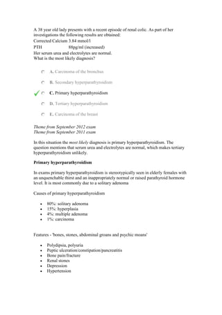
  • 1. A 38 year old lady presents with a recent episode of renal colic. As part of her investigations the following results are obtained: Corrected Calcium 3.84 mmol/l PTH 88pg/ml (increased) Her serum urea and electrolytes are normal. What is the most likely diagnosis? A. Carcinoma of the bronchus B. Secondary hyperparathyroidism C. Primary hyperparathyroidism D. Tertiary hyperparathyroidism E. Carcinoma of the breast Theme from September 2012 exam Theme from September 2011 exam In this situation the most likely diagnosis is primary hyperparathyroidism. The question mentions that serum urea and electrolytes are normal, which makes tertiary hyperparathyroidism unlikely. Primary hyperparathyroidism In exams primary hyperparathyroidism is stereotypically seen in elderly females with an unquenchable thirst and an inappropriately normal or raised parathyroid hormone level. It is most commonly due to a solitary adenoma Causes of primary hyperparathyroidism  80%: solitary adenoma  15%: hyperplasia  4%: multiple adenoma  1%: carcinoma Features - 'bones, stones, abdominal groans and psychic moans'  Polydipsia, polyuria  Peptic ulceration/constipation/pancreatitis  Bone pain/fracture  Renal stones  Depression  Hypertension
  • 2. Associations  Hypertension  Multiple endocrine neoplasia: MEN I and II Investigations  Raised calcium, low phosphate  PTH may be raised or normal  Technetium-MIBI subtraction scan Treatment  Parathyroidectomy, if imaging suggests target gland then a focused approach may be used  Theme: Head and neck lumps A. Branchial cyst B. Cystic hygroma C. Carotid body tumour D. Lymphadenopathy E. Adenolymphoma of the parotid F. Pleomorphic adenoma of the parotid G. Submandibular tumour H. Thyroglossal cyst I. Thoracic outlet syndrome J. Submandibular gland calculus  Please select the most likely lesion to account for the clinical scenario given. Each option may be used once, more than once or not at all. 2. A 60 year old Tibetan immigrant is referred to the surgical clinic with a painless neck swelling. On examination it is located on the left side immediately anterior to the sternocleidomastoid muscle. There are no other abnormalities to find on examination. You answered Branchial cyst The correct answer is Carotid body tumour Carotid body tumours typically present as painless masses. They may compress the vagus or hypoglossal nerves with symptoms attributable to these structures. Over 90% occur spontaneously and are more common in people living at high
  • 3. altitude. In familial cases up to 30% may be bilateral. Treatment is with excision. 3. A 40 year old women presents as an emergency with a painful mass underneath her right mandible. The mass has appeared over the previous week with the pain worsening as the lump has increased in size. On examination there is a 4cm mass underneath her mandible, there is no associated lymphadenopathy. Submandibular gland calculus The sub mandibular gland is the most common site for salivary calculi. Patients will usually complain of pain, which is worse on eating. When the lesion is located distally the duct may be laid open and the stone excised. Otherwise the gland will require removal. 4. A 73 year old male smoker is referred to the clinic by his GP. On examination he has a 3cm soft mass immediately anterior to his ear. It has been present for the past five years and is otherwise associated with no symptoms. You answered Pleomorphic adenoma of the parotid The correct answer is Adenolymphoma of the parotid Warthins tumours (a.k.a. adenolymphoma) are commoner in older men (especially smokers). They are the second commonest benign tumour of the parotid gland, they may be bilateral. They are soft and slow growing and relatively easy to excise. Pleomorphic adenomas typically present in females aged between 40 - 60 years.   Neck lumps  The table below gives characteristic exam question features for conditions causing neck lumps: Reactive lymphadenopathy By far the most common cause of neck swellings. There may be a history of local infection or a generalised viral illness Lymphoma Rubbery, painless lymphadenopathy The phenomenon of pain whilst drinking alcohol is very uncommon There may be associated night sweats and splenomegaly Thyroid swelling May be hypo-, eu- or hyperthyroid symptomatically Moves upwards on swallowing Thyroglossal cyst More common in patients < 20 years old Usually midline, between the isthmus of the thyroid and the hyoid bone
  • 4. Moves upwards with protrusion of the tongue May be painful if infected Pharyngeal pouch More common in older men Represents a posteromedial herniation between thyropharyngeus and cricopharyngeus muscles Usually not seen, but if large then a midline lump in the neck that gurgles on palpation Typical symptoms are dysphagia, regurgitation, aspiration and chronic cough Cystic hygroma A congenital lymphatic lesion (lymphangioma) typically found in the neck, classically on the left side Most are evident at birth, around 90% present before 2 years of age Branchial cyst An oval, mobile cystic mass that develops between the sternocleidomastoid muscle and the pharynx Develop due to failure of obliteration of the second branchial cleft in embryonic development Usually present in early adulthood Cervical rib More common in adult females Around 10% develop thoracic outlet syndrome Carotid aneurysm Pulsatile lateral neck mass which doesn't move on swallowing A 12 year old child is admitted with a 12 hour history of colicky right upper quadrant pain. On examination the child is afebrile and is jaundiced. The abdomen is soft and non tender at the time of examination. What is the most likely cause? A. Infectious hepatitis B. Acute cholecystitis C. Cholangitis D. Hereditary spherocytosis E. Gilberts syndrome Theme from September 2012 Exam The child is most likely to have hereditary spherocytosis. In these individuals there may be disease flares precipitated by acute illness. They form small pigment stones. These may cause biliary colic and some may require cholecystectomy. Hereditary Spherocytosis Most common disorder of the red cell membrane, it has an incidence of 1 in 5000. The abnormally shaped erythrocytes are prone to splenic sequestration and destruction. This can result in hyperbilirubinaemia, jaundice and splenomegaly. In older patients an intercurrent illness may increase the rate of red cell destruction resulting in more acute symptoms. Severe cases may benefit from splenectomy.
  • 5. A 2 day old baby is noted to have voiding difficulties and on closer inspection is noted to have hypospadias. Which of the following abnormalities is most commonly associated with the condition? A. Cryptorchidism B. Diaphragmatic hernia C. Ventricular - septal defect D. Bronchogenic cyst E. Atrial septal defect Theme from January 2012 Exam Hypospadias most commonly occurs as an isolated disorder. Associated urological abnormalities may be seen in up to 40% of infants, of these cryptorchidism is the most frequent (10%). Hypospadias The urethral meatus opens on the ventral surface of the penis. There is also a ventral deficiency of the foreskin. The uretral meatus may open more proximally in the more severe variants. However, 75% of the openings are distally located. The incidence is 1 in 300 male births. Features include:  Absent frenular artery  Ventrally opened glans  Skin tethering to hypoplastic urethra  Splayed columns of spongiosum tissue distal to the meatus  Deficiency of the foreskin ventrally Management:  No routine cultural circumcisions  Urethroplasty  Penile reconstruction The foreskin is often utilised in the reconstructive process. In boys with very distal disease no treatment may be needed. Theme: Liver lesions A. Cystadenoma
  • 6. B. Hyatid cyst C. Amoebic abscess D. Mesenchymal hamartoma E. Liver cell adenoma F. Cavernous haemangioma Please select the most likely lesion for the scenario given. Each option may be used once, more than once or not at all. 7. A 38 year old lady presents with right upper quadrant pain and nausea. She is otherwise well and her only medical therapy is the oral contraceptive pill which she has taken for many years with no ill effects. Her liver function tests are normal. An ultrasound examination demonstrates a hyperechoic well defined lesion in the left lobe of the liver which measures 14 cm in diameter. Cavernous haemangioma Cavernous haemangioma often presents with vague symptoms and signs. They may grow to considerable size. Liver function tests are usually normal. The lesions are typically well defined and hyperechoic on ultrasound. A causative link between OCP use and haemangiomata has yet to be established, but is possible. 8. A 37 year old lady presents with right upper quadrant pain and nausea. She is otherwise well and her only medical therapy is the oral contraceptive pill which she has taken for many years with no ill effects. Her liver function tests and serum alpha feto protein are normal. An ultrasound examination demonstrates a 4cm non encapsulated lesion in the right lobe of the liver which has a mixed echoity and heterogeneous texture. Liver cell adenoma Liver cell adenomas are linked to OCP use and 90% of patients with liver cell adenomas have used the OCP. Liver function tests are often normal. The lesions will typically have a mixed echoity and heterogeneous texture. 9. A 38 year old shepherd presents to the clinic with a 3 month history of malaise and right upper quadrant pain. On examination he is mildly jaundiced. His liver function tests demonstrate a mild elevation in bilirubin and transaminases, his full blood count shows an elevated eosinophil level. An abdominal x-ray is performed by the senior house officer and demonstrates a calcified lesion in the right upper quadrant of the abdomen. Hyatid cyst
  • 7. Similar theme in September 2011 Exam Hyatid disease is more common in those who work with sheep or dogs. Liver function tests may be abnormal and an eosinophilia is often present. Plain radiographs may reveal a calcified cyst wall. Benign liver lesions Benign liver lesions Haemangioma  Most common benign tumours of mesenchymal origin  Incidence in autopsy series is 8%  Cavernous haemangiomas may be enormous  Clinically they are reddish purple hypervascular lesions  Lesions are normally separated from normal liver by ring of fibrous tissue  On ultrasound they are typically hyperechoic Liver cell adenoma  90% develop in women in their third to fifth decade  Linked to use of oral contraceptive pill  Lesions are usually solitary  They are usually sharply demarcated from normal liver although they usually lack a fibrous capsule  On ultrasound the appearances are of mixed echoity and heterogeneous texture. On CT most lesions are hypodense when imaged prior to administration of IV contrast agents  In patients with haemorrhage or symptoms removal of the adenoma may be required Mesenchymal hamartomas Congential and benign, usually present in infants. May compress normal liver Liver abscess  Biliary sepsis is a major predisposing factor  Structures drained by the portal venous system form the second largest source  Common symptoms include fever, right upper quadrant pain. Jaundice may be seen in 50%  Ultrasound will usually show a fluid filled cavity, hyperechoic walls may be seen in chronic abscesses Amoebic abscess  Liver abscess is the most common extra intestinal manifestation of amoebiasis  Between 75 and 90% lesions occur in the right lobe  Presenting complaints typically include fever and right upper quadrant pain  Ultrasonography will usually show a fluid filled structure with poorly defined boundaries  Aspiration yield sterile odourless fluid which has an
  • 8. anchovy paste consistency  Treatment is with metronidazole Hyatid cysts  Seen in cases of Echinococcus infection  Typically an intense fibrotic reaction occurs around sites of infection  The cyst has no epithelial lining  Cysts are commonly unilocular and may grow to 20cm in size. The cyst wall is thick and has an external laminated hilar membrane and an internal enucleated germinal layer  Typically presents with malaise and right upper quadrant pain. Secondary bacterial infection occurs in 10%.  Liver function tests are usually abnormal and eosinophilia is present in 33% cases  Ultrasound may show septa and hyatid sand or daughter cysts.  Percutaneous aspiration is contra indicated  Treatment is by sterilisation of the cyst with mebendazole and may be followed by surgical resection. Hypertonic swabs are packed around the cysts during surgery Polycystic liver disease  Usually occurs in association with polycystic kidney disease  Autosomal dominant disorder  Symptoms may occur as a result of capsular stretch Cystadenoma  Rare lesions with malignant potential  Usually solitary multiloculated lesions  Liver function tests usually normal  Ultrasonography typically shows a large anechoic, fluid filled area with irregular margins. Internal echos may result from septa  Surgical resection is indicated in all cases A 72 year old man presents with symptoms and signs of benign prostatic hyperplasia. Which of the following structures is most likely to be enlarged on digital rectal examination? A. Posterior lobe of the prostate B. Median lobe of the prostate C. Right lateral lobe of the prostate D. Left lateral lobe of the prostate E. Anterior lobe of the prostate Carcinoma of the prostate typically occurs in the posterior lobe. The median lobe is
  • 9. usually enlarged in BPH. The anterior lobe has little in the way of glandular tissue and is seldom enlarged. Benign Prostatic Hyperplasia  Prostatic enlargement occurs in many elderly men  >90% of men aged over 80 will have at least microscopic evidence of benign prostatic hyperplasia Pathology As part of the hyperplastic process increase in both stromal and glandular components are seen. The changes are most notable in the central and periurethral region of the gland. Image showing enlarged prostate removed by transvesical prostatectomy with massive enlargement of the median lobe
  • 10. Image sourced from Wikipedia Presentation The vast majority of men will present with lower urinary tract symptoms. These will typically be:  Poor flow  Nocturia  Hesitancy  Incomplete and double voiding  Terminal dribbling  Urgency  Incontinence Investigation
  • 11.  Digital rectal examination to assess prostatic size and morphology.  Urine dipstick for infections and haematuria.  Uroflowmetry (a flow rate of >15ml/second helps to exclude BOO)  Bladder pressure studies may help identify detrusor failure and whilst may not form part of first line investigations should be included in those with atypical symptoms and prior to redo surgery.  Bladder scanning to demonstrate residual volumes. USS if high pressure chronic retention. Management  Lifestyle changes such as stopping smoking and altering fluid intake may help those with mild symptoms.  Medical therapy includes alpha blockers and 5 alpha reductase inhibitors. The former work quickly on receptor zones located at the bladder neck. Cardiovascular side effects are well documented. The latter work on testosterone metabolising enzymes. Although they have a slower onset of action, the 5 alpha reductase inhibitors may prevent acute urinary retention.  Surgical therapy includes transurethral resection of the prostate and is the treatment of choice in those with severe symptoms and those who fail to respond to medical therapy. More tailored bladder neck incision procedures may be considered in those with small prostates. Retrograde ejaculation may occur following surgery. The change in the type of irrigation solutions used has helped to minimise the TURP syndrome of electrolyte disturbances. A 58 year old man has been suffering from mechanical back pain for several years. One morning he awakes from sleep and feels a sudden onset of pain in his back radiating down his left leg. Which of the following events is most likely to account for his symptoms? A. Prolapse of inner annulus fibrosus B. Prolapse of outer annulus fibrosus C. Prolapse of nucleus pulposus D. Rupture of the ligamentum flavum E. None of the above Theme from 2009 Exam Theme from September 2012 Exam The symptoms would be most likely the result of intervertebral disk prolapse. In disk prolapse the nucleus pulposus is the structure which usually herniates. Intervertebral discs
  • 12.  Consist of an outer annulus fibrosus and an inner nucleus pulposus.  The anulus fibrosus consists of several layers of fibrocartilage.  The nucleus pulposus contains loose fibres suspended in a mucoprotein gel with the consistency of jelly. The nucleus of the disc acts as a shock absorber.  Pressure on the disc causes posterior protrusion of the nucleus pulposus. Most commonly in the lumbrosacral and lower cervical areas.  The discs are separated by hyaline cartilage.  There is one disc between each pair of vertebrae, except for C1/2 and the sacrococcygeal vertebrae. heme: Paediatric neck masses A. Cystic hygroma B. Thyroglossal cyst C. Rhabdomyosarcoma D. Branchial cyst E. Dermoid cyst Please select the most likely underlying diagnosis for the situation that is described. Each option may be used once, more than once, or not at all. 12. A 2 year old boy is brought to the clinic by his mother who has noticed that he has developed a small mass. On examination a small smooth cyst is identified which is located above the hyoid bone. On ultrasound the lesion appears to be a heterogenous and multiloculated mass. You answered Thyroglossal cyst The correct answer is Dermoid cyst Dermoid cysts are usually multiloculated and heterogeneous. Most are located above the hyoid and their appearances on imaging differentiate them from thyroglossal cysts. 13. A 22 month old baby is brought to the clinic by her mother who is concerned that she has developed a swelling in her neck. On examination she has a soft, lesion located in the posterior triangle that transilluminates. Cystic hygroma Cystic hygromas are soft and transilluminate. Most are located in the posterior triangle. 14. A 3 year old boy is brought to the clinic by his mother who has noticed a mass in his neck. On examination he has a smooth mass located on the lateral aspect
  • 13. of his anterior triangle, near to the angle of the mandible. On ultrasound it has a fluid filled, anechoic, appearance. You answered Dermoid cyst The correct answer is Branchial cyst Branchial cysts are usually located laterally and derived from the second branchial cleft. Unless infection has occurred they will usually have an anechoic appearance on ultrasound. Neck Masses in Children Thyroglossal cyst  Located in the anterior triangle, usually in the midline and below the hyoid (65% cases)  Derived from remnants of the thyroglossal duct  Thin walled and anechoic on USS (echogenicity suggests infection of cyst) Branchial cyst  Six branchial arches separated by branchial clefts  Incomplete obliteration of the branchial apparatus may result in cysts, sinuses or fistulae  75% of branchial cysts originate from the second branchial cleft  Usually located anterior to the sternocleidomastoid near the angle of the mandible  Unless infected the fluid of the cyst has a similar consistency to water and is anechoic on USS Dermoids  Derived from pleuripotent stem cells and are located in the midline  Most commonly in a suprahyoid location  They have heterogeneous appearances on imaging and contain variable amounts of calcium and fat Thyroid gland  True thyroid lesions are rare in children and usually represent thyroglossal cysts or tumours like lymphoma Lymphatic malformations  Usually located posterior to the sternocleidomastoid  Cystic hygroma result from occlusion of lymphatic channels  The painless, fluid filled, lesions usually present prior to the age of 2  They are often closely linked to surrounding structures and surgical removal is difficult
  • 14.  They are typically hypoechoic on USS Infantile haemangioma  May present in either triangle of the neck  Grow rapidly initially and then will often spontaneously regress  Plain x-rays will show a mass lesion, usually containing calcified phleboliths  As involution occurs the fat content of the lesions increases Lymphadenopathy  Located in either triangle of the neck  May be reactive or neoplastic  Generalised lymphadenopathy usually secondary to infection in children (very common) An unusually tall 43 year old lady presents to the surgical clinic with bilateral inguinal hernias. She develops chest pain and collapses. As part of her investigations a chest x- ray shows evidence of mediastinal widening. What is the most likely underlying diagnosis? A. Pulmonary embolus B. Aortic dissection C. Tietze syndrome D. Boerhaaves syndrome E. Myocardial infarct Marfans syndrome may present with a variety of connective tissue disorders such as bilateral inguinal hernia. They are at high risk of aortic dissection, as in this case. Aortic dissection  More common than rupture of the abdominal aorta  33% of patients die within the first 24 hours, and 50% die within 48 hours if no treatment received  Associated with hypertension  Features of aortic dissection: tear in the intimal layer, followed by formation and propagation of a subintimal hematoma. Cystic medial necrosis (Marfan's)  Most common site of dissection: 90% occurring within 10 centimetres of the aortic valve Stanford Classification
  • 15. Type Location Treatment A Ascending aorta/ aortic root Surgery- aortic root replacement B Descending aorta Medical therapy with antihypertensives DeBakey classification Type Site affected I Ascending aorta, aortic arch, descending aorta II Ascending aorta only III Descending aorta distal to left subclavian artery Clinical features  Tearing, sudden onset chest pain (painless 10%)  Hypertension or Hypotension  A blood pressure difference greater than 20 mm Hg  Neurologic deficits (20%) Investigations  CXR: widened mediastinum, abnormal aortic knob, ring sign, deviation trachea/oesophagus  CT (spiral)  MRI  Angiography (95% of patients diagnosed) Management  Beta-blockers: aim HR 60-80 bpm and systolic BP 100-120 mm Hg.  Urgent surgical intervention: type A dissections. This will usually involve aortic root replacement. An unusually tall 43 year old lady presents to the surgical clinic with bilateral inguinal hernias. She develops chest pain and collapses. As part of her investigations a chest x- ray shows evidence of mediastinal widening. What is the most likely underlying diagnosis? A. Pulmonary embolus B. Aortic dissection C. Tietze syndrome D. Boerhaaves syndrome E. Myocardial infarct
  • 16. Marfans syndrome may present with a variety of connective tissue disorders such as bilateral inguinal hernia. They are at high risk of aortic dissection, as in this case. Aortic dissection  More common than rupture of the abdominal aorta  33% of patients die within the first 24 hours, and 50% die within 48 hours if no treatment received  Associated with hypertension  Features of aortic dissection: tear in the intimal layer, followed by formation and propagation of a subintimal hematoma. Cystic medial necrosis (Marfan's)  Most common site of dissection: 90% occurring within 10 centimetres of the aortic valve Stanford Classification Type Location Treatment A Ascending aorta/ aortic root Surgery- aortic root replacement B Descending aorta Medical therapy with antihypertensives DeBakey classification Type Site affected I Ascending aorta, aortic arch, descending aorta II Ascending aorta only III Descending aorta distal to left subclavian artery Clinical features  Tearing, sudden onset chest pain (painless 10%)  Hypertension or Hypotension  A blood pressure difference greater than 20 mm Hg  Neurologic deficits (20%) Investigations  CXR: widened mediastinum, abnormal aortic knob, ring sign, deviation trachea/oesophagus  CT (spiral)  MRI  Angiography (95% of patients diagnosed) Management  Beta-blockers: aim HR 60-80 bpm and systolic BP 100-120 mm Hg.
  • 17.  Urgent surgical intervention: type A dissections. This will usually involve aortic root replacement.  A 72 year old man has just undergone an emergency repair for a ruptured abdominal aortic aneurysm. Pre operatively he was taking aspirin, clopidogrel and warfarin. Intra operatively he received 5000 units of unfractionated heparin prior to application of the aortic cross clamp. His blood results on admission to the critical care unit are as follows: Full blood count Hb 8 g/dl Platelets 40 * 109 /l WBC 7.1 * 109 /l  His fibrin degradation products are measured and found to be markedly elevated. Which of the following accounts for these results? A. Anastomotic leak B. Disseminated intravascular coagulation C. Heparin induced thrombocytopenia D. Adverse effect of warfarin E. Adverse effects of antiplatelet agents  Theme from April 2012 Exam The combination of low platelet counts and raised FDP in this setting maked DIC the most likely diagnosis.  Disseminated intravascular coagulation - Diagnosis  Under homeostatic conditions, coagulation and fibrinolysis are coupled. The activation of the coagulation cascade yields thrombin that converts fibrinogen to fibrin; the stable fibrin clot being the final product of hemostasis. The fibrinolytic system breaks down fibrinogen and fibrin. Activation of the fibrinolytic system generates plasmin (in the presence of thrombin), which is responsible for the lysis of fibrin clots. The breakdown of fibrinogen and fibrin results in polypeptides (fibrin degradation products). In a state of homeostasis, the presence of plasmin is critical, as it is the central proteolytic enzyme of coagulation and is also necessary for fibrinolysis. In DIC, the processes of coagulation and fibrinolysis are dysregulated, and the result is widespread clotting with resultant bleeding. Regardless of the triggering event of DIC, once initiated, the pathophysiology of DIC is similar in all conditions. One critical mediator of DIC is the release of a transmembrane glycoprotein (tissue factor =TF). TF is present on the surface of many cell types (including endothelial cells, macrophages, and monocytes) and is not normally in contact with the general circulation, but is exposed to the circulation after vascular damage. For example, TF is released in response
  • 18. to exposure to cytokines (particularly interleukin 1), tumor necrosis factor, and endotoxin. This plays a major role in the development of DIC in septic conditions. TF is also abundant in tissues of the lungs, brain, and placenta. This helps to explain why DIC readily develops in patients with extensive trauma. Upon activation, TF binds with coagulation factors that then triggers the extrinsic pathway (via Factor VII) which subsequently triggers the intrinsic pathway (XII to XI to IX) of coagulation. Diagnosis Fibrin degradation products are often raised. Disorder Prothrombin time APTT Bleeding time Platelet count Warfarin administration Prolonged Normal Normal Normal Aspirin administration Normal Normal Prolonged Normal Heparin Often normal (may be prolonged) Prolonged Normal Normal DIC Prolonged Prolonged Prolonged Low A 53 year old man from Hong Kong presents with symptoms of fatigue, weight loss and recurrent epistaxis. Clinical examination reveals left sided cervical lymphadenopathy and oropharyngeal examination reveals an ulcerated mass in the naso pharynx. Which of the following viral agents is most commonly implicated in the development of this condition? A. Cytomegalovirus B. Epstein Barr virus C. Coxsackie virus D. Herpes simplex virus E. None of the above The clinical scenario is most typical for nasopharyngeal carcinoma. An association with previous Epstein Barr Virus is well established. Infection with the other viruses listed is not a recognised risk factor for the development of the condition. Nasopharyngeal carcinoma  Squamous cell carcinoma of the nasopharynx  Rare in most parts of the world, apart from individuals from Southern China  Associated with Epstein Barr virus infection
  • 19. Presenting features Systemic Local Cervical lymphadenopathy Otalgia Unilateral serous otitis media Nasal obstruction, discharge and/ or epistaxis Cranial nerve palsies e.g. III-VI Imaging Combined CT and MRI. Treatment Radiotherapy is first line therapy. An 18 year old male presents with lethargy, night sweats and on examination is found to have left supraclavicular lymphadenopathy. A surgical registrar performs a left supraclavicular lymph node biopsy. The pathologist identifies Reed- Sternberg cells on the subsequent histology sections, what is the most likely diagnosis? A. Metastatic gastric cancer B. Hodgkins lymphoma C. Non Hodgkins lymphoma D. Tuberculosis E. None of the above Reed-Sternberg cells are characteristic histological cell type found in Hodgkins disease. Lymphadenopathy  Lymphadenopathy in the neck, axillae, groins and abdomen  Need to note: solitary/multiple, defined/indistinct, hard/rubbery/soft, tender/painless Causes of lymphadenopathy Mnemonic: Hodgkins disease H aematological: Hodgkins lymphoma, NHL, Leukaemia O ncological: metastases D ermatopathic lympadenitis G aucher's disease K awasaki disease
  • 20. I nfections: TB, glandular fever, Syphilis N iemann Pick disease S erum sickness D rug reaction (phenytoin) I mmunological (SLE) S arcoidosis E ndocrinological (Hyperthyroidism) A ngioimmunoplastic lymphadenopathy S LE E osinophilic granulomatosis Which of the following lesions is least likely to occur in the presence of severe atrophic gastritis? A. Duodenal ulcer B. Gastric cancer C. Gastric polyp D. Iron deficiency anaemia E. Pernicious anaemia Due the absence of acid a duodenal ulcer is unlikely to occur. Gastric cancer Overview There are 700,000 new cases of gastric cancer worldwide each year. It is most common in Japan and less common in western countries. It is more common in men and incidence rises with increasing age. The exact cause of many sporadic cancer is not known, however, familial cases do occur in HNPCC families. In addition, smoking and smoked or preserved foods increase the risk. Japanese migrants retain their increased risk (decreased in subsequent generations). The distribution of the disease in western countries is changing towards a more proximal location (perhaps due to rising obesity). Pathology There is some evidence of support a stepwise progression of the disease through intestinal metaplasia progressing to atrophic gastritis and subsequent dysplasia, through to cancer. The favoured staging system is TNM. The risk of lymph node involvement is related to size and depth of invasion; early cancers confined to submucosa have a 20% incidence of lymph node metastasis. Tumours of the gastro- oesophageal junction are classified as below: Type 1 True oesophageal cancers and may be associated with Barrett's oesophagus. Type 2 Carcinoma of the cardia, arising from cardiac type epithelium or short segments with intestinal metaplasia at the oesophagogastric junction.
  • 21. Type 3 Sub cardial cancers that spread across the junction. Involve similar nodal stations to gastric cancer. Groups for close endoscopic monitoring  Intestinal metaplasia of columnar type  Atrophic gastritis  Low to medium grade dysplasia  Patients who have previously undergone resections for benign peptic ulcer disease (except highly selective vagotomy). Referral to endoscopy Patients of any age with dyspepsia and any of the following Patients without dyspepsia Worsening dyspepsia Chronic gastrointestinal bleeding Dysphagia Barretts oesophagus Dysphagia Unexplained abdominal pain or weight loss Intestinal metaplasia Weight loss Vomiting Dysplasia Iron deficiency anaemia Upper abdominal mass Atrophic gastritis Upper abdominal mass Jaundice Patient aged over 55 years with unexplained or persistent dyspepsia Upper GI endoscopy performed for dyspepsia. The addition of dye spraying (as shown in the bottom right) may facilitate identification of smaller tumours
  • 22. Image sourced from Wikipedia Staging  CT scanning of the chest abdomen and pelvis is the routine first line staging investigation in most centres.  Laparoscopy to identify occult peritoneal disease  PET CT (particularly for junctional tumours) Treatment  Proximally sited disease greater than 5-10cm from the OG junction may be treated by sub total gastrectomy  Total gastrectomy if tumour is <5cm from OG junction  For type 2 junctional tumours (extending into oesophagus) oesophagogastrectomy is usual  Endoscopic sub mucosal resection may play a role in early gastric cancer confined to the mucosa and perhaps the sub mucosa (this is debated)  Lymphadenectomy should be performed. A D2 lymphadenectomy is widely advocated by the Japanese, the survival advantages of extended lymphadenectomy have been debated. However, the overall recommendation is that a D2 nodal dissection be undertaken.  Most patients will receive chemotherapy either pre or post operatively. Prognosis
  • 23. UK Data Disease extent Percentage 5 year survival All RO resections 54% Early gastric cancer 91% Stage 1 87% Stage 2 65% Stage 3 18% Operative procedure Total Gastrectomy , lymphadenectomy and Roux en Y anastomosis General anaesthesia Prophylactic intravenous antibiotics Incision: Rooftop. Perform a thorough laparotomy to identify any occult disease. Mobilise the left lobe of the liver off the diaphragm and place a large pack over it. Insert a large self retaining retractor e.g. omnitract or Balfour (take time with this, the set up should be perfect). Pack the small bowel away. Begin by mobilising the omentum off the transverse colon. Proceed to detach the short gastric vessels. Mobilise the pylorus and divide it at least 2cm distally using a linear cutter stapling device. Continue the dissection into the lesser sac taking the lesser omentum and left gastric artery flush at its origin. The lymph nodes should be removed en bloc with the specimen where possible. Place 2 stay sutures either side of the distal oesophagus. Ask the anaesthetist to pull back on the nasogastric tube. Divide the distal oesophagus and remove the stomach. The oesphago jejunal anastomosis should be constructed. Identify the DJ flexure and bring a loop of jejunum up to the oesophagus (to check it will reach). Divide the jejunum at this point. Bring the divided jejunum either retrocolic or antecolic to the oesophagus. Anastamose the oesophagus to the jejunum, using either interrupted 3/0 vicryl or a stapling device. Then create the remainder of the Roux en Y reconstruction distally. Place a jejunostomy feeding tube. Wash out the abdomen and insert drains (usually the anastomosis and duodenal stump). Help the anaesthetist insert the nasogastric tube (carefully!) Close the abdomen and skin. Enteral feeding may commence on the first post-operative day. However, most surgeons will leave patients on free NG drainage for several days and keep them nil by mouth. A 28 year old man develops an acute paronychia and subsequent spreading sepsis. The tissue exudate has a higher protein content than normal tissue because?
  • 24. A. Breakdown of tissue cells release protein B. Capillary walls are more permeable C. Increased blood flow transports more protein into the area D. Intracapillary pressure is raised E. Plasma cells release gamma globulin The increased permeability allows the exudation of plasma proteins. Acute inflammation Inflammation is the reaction of the tissue elements to injury. Vascular changes occur, resulting in the generation of a protein rich exudate. So long as the injury does not totally destroy the existing tissue architecture, the episode may resolve with restoration of original tissue architecture. Vascular changes  Vasodilation occurs and persists throughout the inflammatory phase.  Inflammatory cells exit the circulation at the site of injury.  The equilibrium that balances Starlings forces within capillary beds is disrupted and a protein rich exudate will form as the vessel walls also become more permeable to proteins.  The high fibrinogen content of the fluid may form a fibrin clot. This has several important immunomodulatory functions. Sequelae Resolution  Typically occurs with minimal initial injury  Stimulus removed and normal tissue architecture results Organisation  Delayed removed of exudate  Tissues undergo organisation and usually fibrosis Suppuration  Typically formation of an abscess or an empyema  Sequestration of large quantities of dead neutrophils Progression to chronic inflammation  Coupled inflammatory and reparative activities  Usually occurs when initial infection or suppuration has been inadequately managed Causes
  • 25.  Microbacterial infections e.g. Viruses, exotoxins or endotoxins released by bacteria  Chemical agents  Physical agents e.g. Trauma  Hypersensitivity reactions  Tissue necrosis Presence of neutrophil polymorphs is a histological diagnostic feature of acute inflammation As a busy surgical trainee on the colorectal unit you are given the unenviable task of reviewing the unit's histopathology results for colonic polyps. Which of the polyp types described below has the greatest risk of malignancy? A. Hyperplastic polyp B. Tubular adenoma C. Villous adenoma D. Hamartomatous polyp E. Serrated polyp Villous adenomas carry the highest risk of malignant transformation. Hyperplastic polyps carry little in the way of increased risk. Although, patients with hamartomatous polyp syndromes may have a high risk of malignancy, the polyps themselves have little malignant potential. Colonic polyps Colonic Polyps May occur in isolation of greater numbers as part of the polyposis syndromes. In FAP greater than 100 polyps are typically present. The risk of malignancy in association with adenomas is related to size and is the order of 10% in a 1cm adenoma. Isolated adenomas seldom give risk of symptoms (unless large and distal). Distally sited villous lesions may produce mucous and if very large electrolyte disturbances may occur. Follow up of colonic polyps Low risk  1 or 2 adenomas <1cm. No follow up or re-colonoscopy at 5 years. Moderate risk  3 or 4 small adenomas or 1 adenoma >1cm. Re-scope at 3 years.
  • 26. High risk  >5 small adenomas or >3 with 1 of them >1cm. Re scope at 1 year. From Atkins and Saunders Gut 2002 51 (suppl V:V6-V9). It is important to stratify patients appropriately and ensure that a complete colonoscopy with good views was performed. Segmental resection or complete colectomy should be considered when: 1. Incomplete excision of malignant polyp 2. Malignant sessile polyp 3. Malignant pedunculated polyp with submucosal invasion 4. Polyps with poorly differentiated carcinoma 5. Familial polyposis coli -Screening from teenager up to 40 years by 2 yearly sigmoidoscopy/colonoscopy -Panproctocolectomy and Ileostomy or Restorative Panproctocolectomy. Rectal polypoidal lesions may be amenable to trans anal endoscopic microsurgery. A 23 year old man presents to the surgical clinic with an inguinal hernia. On examination he has a small direct hernia. However, you also notice that he has pigmented spots around his mouth, on his palms and soles. In his history he underwent a reduction of an intussusception aged 12 years. Which of the following lesions is most likely to be identified if a colonoscopy were performed? A. Hamartomas B. Tubulovillous adenoma C. Colorectal cancer D. Crohns disease E. Hyperplastic polyps Theme from April 2012 Exam He is most likely to have Peutz-Jeghers syndrome which is associated with Hamartomas. Peutz-Jeghers syndrome Peutz-Jeghers syndrome is an autosomal dominant condition characterised by numerous benign hamartomatous polyps in the gastrointestinal tract. It is also associated with pigmented freckles on the lips, face, palms and soles. Around 50% of patients will have died from a gastrointestinal tract cancer by the age of 60 years. Genetics
  • 27.  Autosomal dominant  Responsible gene encodes serine threonine kinase LKB1 or STK11 Features  Hamartomatous polyps in GI tract (mainly small bowel)  Pigmented lesions on lips, oral mucosa, face, palms and soles  Intestinal obstruction e.g. intussusception (which may lead to diagnosis)  Gastrointestinal bleeding Management  Conservative unless complications develop  A 56 year old surgeon has been successfully operating for many years. Over the past few weeks she has begun to notice that her hands are becoming blistering and weepy. A latex allergy is diagnosed. Which of the following pathological processes accounts for this scenario? A. Type 1 hypersensitivity reaction B. Type 2 hypersensitivity reaction C. Type 4 hypersensitivity reaction D. Type 3 hypersensitivity reaction E. None of the above Hypersensitivity reactions: ACID type 1 --Anaphylactic type 2 --Cytotoxic type 3 --Immune complex type 4 --Delayed hypersensitivity  Theme from 2012 Exam Contact dermatitis of a chronic nature is an example of a type 4 hypersensitivity reaction. Type 4 hypersensitivity reactions are cell mediated rather than antibody mediated.  Hypersensitivity reactions  The Gell and Coombs classification divides hypersensitivity reactions into 4 types Type I Type II Type III Type IV
  • 28. Description Anaphylactic Cytotoxic Immune complex Delayed type Mediator IgE IgG, IgM IgG, IgM T-cells Antigen Exogenous Cell surface Soluble Tissues Response time Minutes Hours Hours 2-3 days Examples Asthma Hay fever Autoimmune haemolytic anaemia Pemphigus Goodpasture's Serum sickness SLE Aspergillosis Graft versus host disease Contact dermatitis  A 56 year old motorcyclist is involved in a road traffic accident and sustains a displaced femoral shaft fracture. Not other injuries are identified on the primary or secondary surveys. The fracture is treated with closed, antegrade intramedullary nailing. The following day the patient becomes increasingly agitated and confused. On examination he is pyrexial, hypoxic SaO2 90% on 6 litres O2, tachycardic and normotensive. Systemic examination demonstrates a non blanching petechial rash present over the torso. What is the most likely explanation for this? A. Pulmonary embolism with paradoxical embolus B. Fat embolism C. Meningococcal sepsis D. Alcohol withdrawl E. Chronic sub dural haematoma This man has a recent injury and physical signs that would be concordant with fat embolism syndrome. Meningococcal sepsis is not usually associated with hypoxia initially. Pulmonary emboli are not typically associated with pyrexia. Fat embolism Diagnosis and clinical features System Feature Cardiothoracic  Early persistent tachycardia  Tachypnoea, dyspnoea, hypoxia usually 72 hours following injury  Pyrexia Dermatological  Red/ brown impalpable petechial rash (usually only in 25- 50%)  Subconjunctival and oral haemorrhage/ petechiae CNS  Confusion and agitation
  • 29.  Retinal haemorrhages and intra-arterial fat globules on fundoscopy Imaging  May be normal  Fat emboli tend to lodge distally and therefore CTPA may not show any vascular occlusion, a ground glass appearance may be seen at the periphery Treatment  Prompt fixation of long bone fractures  Some debate regarding benefit Vs. risk of medullary reaming in femoral shaft/ tibial fractures in terms of increasing risk (probably does not).  DVT prophylaxis  General supportive care Which of these tumour markers is most helpful in identifying an individual with hepatocellular carcinoma? A. Serum AFP B. Serum CA19-9 C. CEA D. Beta HCG E. CA125 Theme from September 2011 Exam Hepatocellular carcinoma is commonly diagnosed with imaging and an elevated alpha fetoprotein. Biopsy may seed the tumour and should be avoided. Up to 80% of hepatocellular carcinoma arise in cirrhotic livers. Liver tumours Primary liver tumours The most common primary tumours are cholangiocarcinoma and hepatocellular carcinoma. Overall metastatic disease accounts for 95% of all liver malignancies making the primary liver tumours comparatively rare. Primary liver tumours include:  Cholangiocarcinoma  Hepatocellular carcinoma  Hepatoblastoma
  • 30.  Sarcomas (Rare)  Lymphomas  Carcinoids (most often secondary although primary may occur) Hepatocellular carcinoma These account for the bulk of primary liver tumours (75% cases). Its worldwide incidence reflects its propensity to occur on a background of chronic inflammatory activity. Most cases arise in cirrhotic livers or those with chronic hepatitis B infection, especially where viral replication is actively occurring. In the UK it accounts for less than 5% of all cancers, although in parts of Asia its incidence is 100 per 100,000. The majority of patients (80%) present with existing liver cirrhosis, with a mass discovered on screening ultrasound. Diagnosis  CT/ MRI (usually both) are the imaging modalities of choice  a-fetoprotein is elevated in almost all cases  Biopsy should be avoided as it seeds tumours cells through a resection plane.  In cases of diagnostic doubt serial CT and aFP measurements are the preferred strategy. Treatment  Patients should be staged with liver MRI and chest, abdomen and pelvic CT scan.  The testis should be examined in males (testicular tumours may cause raised AFP). PET CT may be used to identify occult nodal disease.  Surgical resection is the mainstay of treatment in operable cases. In patients with a small primary tumour in a cirrhotic liver whose primary disease process is controlled, consideration may be given to primary whole liver resection and transplantation.  Liver resections are an option but since most cases occur in an already diseased liver the operative risks and post-operative hepatic dysfunction are far greater than is seen following metastectomy.  These tumours are not particularly chemo or radiosensitive however, both may be used in a palliative setting. Tumour ablation is a more popular strategy. Survival Poor, overall survival is 15% at 5 years. Cholangiocarcinoma This is the second most common type of primary liver malignancy. As its name suggests these tumours arise in the bile ducts. Up to 80% of tumours arise in the extra hepatic biliary tree. Most patients present with jaundice and by this stage the majority will have disease that is not resectable. Primary scelerosing cholangitis is the main risk factor. In deprived countries typhoid and liver flukes are also major risk factors.
  • 31. Diagnosis  Patients will typically have an obstructive picture on liver function tests.  CA 19-9, CEA and CA 125 are often elevated  CT/ MRI and MRCP are the imaging methods of choice. Treatment  Surgical resection offers the best chance of cure. Local invasion of peri hilar tumours is a particular problem and this coupled with lobar atrophy will often contra indicate surgical resection.  Palliation of jaundice is important, although metallic stents should be avoided in those considered for resection. Survival Is poor, approximately 15% 5 year survival. A 39 year old man has suffered from terminal ileal Crohns disease for the past 20 years. Which condition is he least likely to develop? A. Gallstones B. Malabsorption C. Pyoderma gangrenosum D. Amyloidosis E. Feltys syndrome Felteys syndrome:  Rheumatoid disease  Splenomegaly  Neutropenia Feltys syndrome is associated with rheumatoid disease. Individuals with long standing crohns disease are at risk of gallstones because of impairment of the enterohepatic recycling of bile salts. Formation of entero-enteric fistulation may produce malabsorption. Amyloidosis may complicate chronic inflammatory states. Crohns disease Crohns disease is a chronic transmural inflammation of a segment(s) of the gastrointestinal tract and may be associated with extra intestinal manifestations. Frequent disease patterns observed include ileal, ileocolic and colonic disease. Peri-
  • 32. anal disease may occur in association with any of these. The disease is often discontinuous in its distribution. Inflammation may cause ulceration, fissures, fistulas and fibrosis with stricturing. Histology reveals a chronic inflammatory infiltrate that is usually patchy and transmural. Ulcerative colitis Vs Crohns Crohn's disease Ulcerative colitis Distribution Mouth to anus Rectum and colon Macroscopic changes Cobblestone appearance, apthoid ulceration Contact bleeding Depth of disease Transmural inflammation Superficial inflammation Distribution pattern Patchy Continuous Histological features Granulomas (non caseating epithelioid cell aggregates with Langhans' giant cells) Crypt abscesses, Inflammatory cells in the lamina propria Extraintestinal manifestations of Crohns Related to disease extent Unrelated to disease extent Aphthous ulcers (10%) Sacroiliiitis (10-15%) Erythema nodosum (5-10%) Ankylosing spondylitis (1-2%) Pyoderma gangrenosum (0.5%) Primary sclerosing cholangitis (Rare) Acute arthropathy (6-12%) Gallstones (up to 30%) Ocular complications (up to 10%) Renal calculi (up to 10%) Theme: Renal stones A. Calcium oxalate B. Uric acid C. Cystine D. Struvite E. Calcium phosphate Please select the most likely stone type for each of the following urinary tract stone scenarios. Each option may be used once, more than once or not at all. 28. A 73 year old lady is undergoing chemotherapy for treatment of acute leukaemia. She develops symptoms of renal colic. Her urine tests positive for blood. A KUB x-ray shows no evidence of stones. Uric acid Chemotherapy and cell death can increase uric acid levels. In this acute setting the uric acid stones are unlikely to be coated with calcium and will therefore be
  • 33. radiolucent. 29. A 16 year old boy presents with renal colic. His parents both have a similar history of the condition. His urine tests positive for blood. A KUB style x-ray shows a relatively radiodense stone in the region of the mid ureter. Cystine Cystine stones are associated with an inherited metabolic disorder. 30. A 43 year old lady with episodes of recurrent urinary tract sepsis presents with a staghorn calculus of the left kidney. Her urinary pH is 7.3. A KUB x-ray shows a faint outline of the calculus. Struvite Theme from April 2012 Exam Chronic infection with urease producing enzymes can produce an alkaline urine with formation of struvate stone. Renal stones Type of stones Features Percentage of all calculi Calcium oxalate Hypercalciuria is a major risk factor (various causes) Hyperoxaluria may also increase risk Hypocitraturia increases risk because citrate forms complexes with calcium making it more soluble Stones are radio-opaque (though less than calcium phosphate stones) Hyperuricosuria may cause uric acid stones to which calcium oxalate binds 85% Cystine Inherited recessive disorder of transmembrane cystine transport leading to decreased absorption of cystine from intestine and renal tubule Multiple stones may form Relatively radiodense because they contain sulphur 1% Uric acid Uric acid is a product of purine metabolism May precipitate when urinary pH low May be caused by diseases with extensive tissue breakdown e.g. malignancy More common in children with inborn errors of metabolism Radiolucent 5-10%
  • 34. Calcium phosphate May occur in renal tubular acidosis, high urinary pH increases supersaturation of urine with calcium and phosphate Renal tubular acidosis types 1 and 3 increase risk of stone formation (types 2 and 4 do not) Radio-opaque stones (composition similar to bone) 10% Struvite Stones formed from magnesium, ammonium and phosphate Occur as a result of urease producing bacteria (and are thus associated with chronic infections) Under the alkaline conditions produced, the crystals can precipitate Slightly radio-opaque 2-20% Effect of urinary pH on stone formation Urine pH will show individual variation (from pH 5-7). Post prandially the pH falls as purine metabolism will produce uric acid. Then the urine becomes more alkaline (alkaline tide). When the stone is not available for analysis the pH of urine may help to determine which stone was present. Stone type Urine acidity Mean urine pH Calcium phosphate Normal- alkaline >5.5 Calcium oxalate Variable 6 Uric acid Acid 5.5 Struvate Alkaline >7.2 Cystine Normal 6.5 A 64 year old man presents to the clinic with right upper quadrant discomfort. He has never attended the hospital previously and is usually well. He has just retired from full time employment as a machinist in a PVC
  • 35. factory. CT scanning shows a large irregular tumour in the right lobe of his liver. Which of the following lesions is the most likely? A. Liposarcoma B. Angiosarcoma C. Hamartoma D. Hyatid liver disease E. Benign angioma Angiosarcoma of the liver is a rare tumour. However, it is linked to working with vinyl chloride, as in this case. Although modern factories minimise the exposure to this agent, this has not always been the case. Occupational cancers Occupational cancers accounted for 5.3% cancer deaths in 2005. In men the main cancers include:  Mesothelioma  Bladder cancer  Non melanoma skin cancer  Lung cancer  Sino nasal cancer Occupations with high levels of occupational tumours include:  Construction industry  Working with coal tar and pitch  Mining  Metalworkers  Working with asbestos (accounts for 98% of all mesotheliomas)  Working in rubber industry Shift work has been linked to breast cancer in women (Health and safety executive report RR595). The latency between exposure and disease is typically 15 years for solid tumours and 20 for leukaemia. Many occupational cancers are otherwise rare. For example sino nasal cancer is an uncommon tumour, 50% will be SCC. They are linked to conditions such as wood dust exposure and unlike lung cancer is not strongly linked to cigarette smoking. Another typical occupational tumour is angiosarcoma of the liver which is linked to
  • 36. working with vinyl chloride. Again in the non occupational context this is an extremely rare sporadic tumour. A 64 year old man presents to the clinic with right upper quadrant discomfort. He has never attended the hospital previously and is usually well. He has just retired from full time employment as a machinist in a PVC factory. CT scanning shows a large irregular tumour in the right lobe of his liver. Which of the following lesions is the most likely? A. Liposarcoma B. Angiosarcoma C. Hamartoma D. Hyatid liver disease E. Benign angioma Angiosarcoma of the liver is a rare tumour. However, it is linked to working with vinyl chloride, as in this case. Although modern factories minimise the exposure to this agent, this has not always been the case. Occupational cancers Occupational cancers accounted for 5.3% cancer deaths in 2005. In men the main cancers include:  Mesothelioma  Bladder cancer  Non melanoma skin cancer  Lung cancer  Sino nasal cancer Occupations with high levels of occupational tumours include:  Construction industry  Working with coal tar and pitch  Mining  Metalworkers  Working with asbestos (accounts for 98% of all mesotheliomas)  Working in rubber industry Shift work has been linked to breast cancer in women (Health and safety executive report RR595). The latency between exposure and disease is typically 15 years for solid tumours and 20 for leukaemia.
  • 37. Many occupational cancers are otherwise rare. For example sino nasal cancer is an uncommon tumour, 50% will be SCC. They are linked to conditions such as wood dust exposure and unlike lung cancer is not strongly linked to cigarette smoking. Another typical occupational tumour is angiosarcoma of the liver which is linked to working with vinyl chloride. Again in the non occupational context this is an extremely rare sporadic tumour. A 32 year old man is involved in a house fire and sustains extensive partial thickness burns to his torso and thigh. Two weeks post operatively he develops oedema of both lower legs. The most likely cause of this is: A. Iliofemoral deep vein thrombosis B. Venous obstruction due to scarring C. Hypoalbuminaemia D. Excessive administration of intravenous fluids E. None of the above Theme from 2009 Exam Loss of plasma proteins is the most common cause of oedema developing in this time frame. Burns pathology Extensive burns  Haemolysis due to damage of erythrocytes by heat and microangiopathy  Loss of capillary membrane integrity causing plasma leakage into interstitial space  Extravasation of fluids from the burn site causing hypovolaemic shock (up to 48h after injury)- decreased blood volume and increased haematocrit  Protein loss  Secondary infection e.g. Staphylococcus aureus  ARDS  Risk of Curlings ulcer (acute peptic stress ulcers)  Danger of full thickness circumferential burns in an extremity as these may develop compartment syndrome Healing  Superficial burns: keratinocytes migrate to form a new layer over the burn site  Full thickness burns: dermal scarring. Usually need keratinocytes from skin grafts to provide optimal coverage.
  • 38. What is the diagnostic marker for carcinoid syndrome? A. B-HCG B. Histamine C. Chromogranin A D. 5-Hydroxyindoleacetic acid E. 5-Hydroxytryptamine Urinary measurement of 5- HIAA is an important part of clinical follow up. Carcinoid syndrome  Carcinoid tumours secrete serotonin  Originate in neuroendocrine cells mainly in the intestine (midgut-distal ileum/appendix)  Can occur in the rectum, bronchi  Hormonal symptoms mainly occur when disease spreads outside the bowel Clinical features - Onset: years - Flushing face - Palpitations - Tricuspid stenosis causing dyspnoea - Asthma - Severe diarrhoea (secretory, persists despite fasting) Investigation - 5-HIAA in a 24-hour urine collection - Scintigraphy - CT scan Treatment  Octreotide  Surgical removal A 42 year old man from Southern India presents with chronic swelling of both lower legs, they are brawny and indurated with marked skin tophic changes. Which of the following organisms is the most likely origin of this disease process? A. Loa loa
  • 39. B. Wuchereria bancrofti C. Trypanosoma cruzi D. Trypanosoma gambiense E. None of the above W. Bancrofti is the commonest cause of filariasis leading to lymphatic obstruction. Infection with Loa loa typically occurs in the African sub continent and usually results in generalised sub cutaneous infections without lymphatic obstruction. Trypanosomal infections would not produce this clinical picture. Wuchereria bancrofti  Parasitic filarial nematode  Accounts for 90% of cases of filariasis  Usually diagnosed by blood smears  Usually transmitted by mosquitos  Treatment is with diethylcarbamazine A 45 year old lady has recently undergone a thyroidectomy for treatment of medullary thyroid cancer. Which of the following tumour markers is used clinically to screen for recurrence? A. Free T3 B. Thyroglobulin C. Calcitonin D. Free T4 E. Thyroid stimulating hormone Theme from 2011 Exam Calcitonin is clinically utilised to screen for medullary thyroid cancer recurrence. Thyroid function testing does not form part of either diagnosis or follow up from a malignancy perspective. However, routine assessment of TSH may be needed in patients on thyroxine. Thyroid malignancy Papillary carcinoma  Commonest sub-type  Accurately diagnosed on fine needle aspiration cytology
  • 40.  Histologically they may demonstrate psammoma bodies (areas of calcification) and so called 'orphan Annie' nuclei  They typically metastasise via the lymphatics and thus laterally located apparently ectopic thyroid tissue is usually a metastasis from a well differentiated papillary carcinoma. Follicular carcinoma  Are less common than papillary lesions  Like papillary tumours they may present as a discrete nodule. Although they appear to be well encapsulated macroscopically there invasion on microscopic evaluation.  Lymph node metastases are uncommon and these tumours tend to spread haematogenously. This translates into a higher mortality rate.  Follicular lesions cannot be accurately diagnosed on fine needle aspiration cytology and thus all follicular FNA's will require at least a hemi thyroidectomy. Anaplastic carcinoma  Less common and tend to occur in elderly females  Disease is usually advanced at presentation and often only palliative decompression and radiotherapy can be offered. Medullary carcinoma  These are tumours of the parafollicular cells ( C Cells) and are of neural crest origin.  The serum calcitonin may be elevated which is of use when monitoring for recurrence.  They may be familial and occur as part of the MEN -2A disease spectrum.  Spread may be either lymphatic or haematogenous and as these tumours are not derived primarily from thyroid cells they are not responsive to radioiodine. Lymphoma  These respond well to radiotherapy  Radical surgery is unnecessary once the disease has been diagnosed on biopsy material. Such biopsy material is not generated by an FNA and thus a core biopsy has to be obtained (with care!). A 45 year old lady has recently undergone a thyroidectomy for treatment of medullary thyroid cancer. Which of the following tumour markers is used clinically to screen for recurrence?
  • 41. A. Free T3 B. Thyroglobulin C. Calcitonin D. Free T4 E. Thyroid stimulating hormone Theme from 2011 Exam Calcitonin is clinically utilised to screen for medullary thyroid cancer recurrence. Thyroid function testing does not form part of either diagnosis or follow up from a malignancy perspective. However, routine assessment of TSH may be needed in patients on thyroxine. Thyroid malignancy Papillary carcinoma  Commonest sub-type  Accurately diagnosed on fine needle aspiration cytology  Histologically they may demonstrate psammoma bodies (areas of calcification) and so called 'orphan Annie' nuclei  They typically metastasise via the lymphatics and thus laterally located apparently ectopic thyroid tissue is usually a metastasis from a well differentiated papillary carcinoma. Follicular carcinoma  Are less common than papillary lesions  Like papillary tumours they may present as a discrete nodule. Although they appear to be well encapsulated macroscopically there invasion on microscopic evaluation.  Lymph node metastases are uncommon and these tumours tend to spread haematogenously. This translates into a higher mortality rate.  Follicular lesions cannot be accurately diagnosed on fine needle aspiration cytology and thus all follicular FNA's will require at least a hemi thyroidectomy. Anaplastic carcinoma  Less common and tend to occur in elderly females  Disease is usually advanced at presentation and often only palliative decompression and radiotherapy can be offered.
  • 42. Medullary carcinoma  These are tumours of the parafollicular cells ( C Cells) and are of neural crest origin.  The serum calcitonin may be elevated which is of use when monitoring for recurrence.  They may be familial and occur as part of the MEN -2A disease spectrum.  Spread may be either lymphatic or haematogenous and as these tumours are not derived primarily from thyroid cells they are not responsive to radioiodine. Lymphoma  These respond well to radiotherapy  Radical surgery is unnecessary once the disease has been diagnosed on biopsy material. Such biopsy material is not generated by an FNA and thus a core biopsy has to be obtained (with care!). A 22 year old man is kicked in the head during a rugby match. He is temporarily concussed, but then regains consciousness. Half an hour later he develops slurred speech, ataxia and loses consciousnesses. On arrival in hospital he is intubated and ventilated. A CT Scan is performed which shows an extradural haematoma. What is the most likely cause? A. Basilar artery laceration B. Middle meningeal artery laceration C. Laceration of the sigmoid sinus D. Laceration of the anterior cerebral artery E. Laceration of the middle cerebral artery Theme based on September 2011 Exam The most likely vessel from those in the list to cause an acute extra dural haemorrhage is the middle meningeal artery. The anterior and middle cerebral arteries may cause acute sub dural haemorrhage. Acute sub dural haemorrhages usually take slightly longer to evolve than acute extra dural haemorrhages. Middle meningeal artery  Middle meningeal artery is typically the third branch of the first part of the maxillary artery, one of the two terminal branches of the external carotid artery. After branching off the maxillary artery in the infratemporal fossa, it
  • 43. runs through the foramen spinosum to supply the dura mater (the outermost meninges) .  The middle meningeal artery is the largest of the three (paired) arteries which supply the meninges, the others being the anterior meningeal artery and the posterior meningeal artery.  The middle meningeal artery runs beneath the pterion. It is vulnerable to injury at this point, where the skull is thin. Rupture of the artery may give rise to an extra dural hematoma.  In the dry cranium, the middle meningeal, which runs within the dura mater surrounding the brain, makes a deep indention in the calvarium.  The middle meningeal artery is intimately associated with the auriculotemporal nerve which wraps around the artery making the two easily identifiable in the dissection of human cadavers and also easily damaged in surgery. A 22 year old man is kicked in the head during a rugby match. He is temporarily concussed, but then regains consciousness. Half an hour later he develops slurred speech, ataxia and loses consciousnesses. On arrival in hospital he is intubated and ventilated. A CT Scan is performed which shows an extradural haematoma. What is the most likely cause? A. Basilar artery laceration B. Middle meningeal artery laceration C. Laceration of the sigmoid sinus D. Laceration of the anterior cerebral artery E. Laceration of the middle cerebral artery Theme based on September 2011 Exam The most likely vessel from those in the list to cause an acute extra dural haemorrhage is the middle meningeal artery. The anterior and middle cerebral arteries may cause acute sub dural haemorrhage. Acute sub dural haemorrhages usually take slightly longer to evolve than acute extra dural haemorrhages. Middle meningeal artery  Middle meningeal artery is typically the third branch of the first part of the maxillary artery, one of the two terminal branches of the external carotid artery. After branching off the maxillary artery in the infratemporal fossa, it runs through the foramen spinosum to supply the dura mater (the outermost meninges) .  The middle meningeal artery is the largest of the three (paired) arteries which supply the meninges, the others being the anterior meningeal artery and the posterior meningeal artery.
  • 44.  The middle meningeal artery runs beneath the pterion. It is vulnerable to injury at this point, where the skull is thin. Rupture of the artery may give rise to an extra dural hematoma.  In the dry cranium, the middle meningeal, which runs within the dura mater surrounding the brain, makes a deep indention in the calvarium.  The middle meningeal artery is intimately associated with the auriculotemporal nerve which wraps around the artery making the two easily identifiable in the dissection of human cadavers and also easily damaged in surgery. Which of the following is not characteristic of a granuloma? A. Altered macrophages B. Fused macrophages C. Epithelioid cells D. Mixture of chronic inflammatory cells E. Polymorphnuclear leucocytes, cellular debris and fibrin These are typical components of an abscess cavity. Polymorphonuclear leucocytes may be found in a granuloma if there is a focus of suppuration. Chronic inflammation Overview Chronic inflammation may occur secondary to acute inflammation.In most cases chronic inflammation occurs as a primary process. These may be broadly viewed as being one of three main processes:  Persisting infection with certain organisms such as Mycobacterium tuberculosis which results in delayed type hypersensitivity reactions and inflammation.  Prolonged exposure to non-biodegradable substances such as silica or suture materials which may induce an inflammatory response.  Autoimmune conditions involving antibodies formed against host antigens. Acute vs. Chronic inflammation Acute inflammation Chronic inflammation Changes to existing vascular structure and increased permeability of endothelial cells Angiogenesis predominates Infiltration of neutrophils Macrophages, plasma cells and lymphocytes predominate Process may resolve with: Healing by fibrosis is the main result
  • 45.  Suppuration  Complete resolution  Abscess formation  Progression to chronic inflammation  Healing by fibrosis Granulomatous inflammation A granuloma consists of a microscopic aggregation of macrophages (with epithelial type arrangement =epitheliod). Large giant cells may be found at the periphery of granulomas. Mediators Growth factors released by activated macrophages include agents such as interferon and fibroblast growth factor (plus many more). Some of these such as interferons may have systemic features resulting in systemic symptoms and signs, which may be present in individuals with long standing chronic inflammation. The finding of granulomas is pathognomonic of chronic inflammation, as illustrated in this biopsy from a patient with colonic Crohns disease Image sourced from Wikipedia A 42 year old man presents with a painless lump in the left testicle that he noticed on self examination. Clinically there is a firm nodule in the left testicle, ultrasound appearances show an irregular mass lesion. His serum AFP and HCG levels are both within normal limits. What is the most likely diagnosis? A. Yolk sack tumour B. Seminoma C. Testicular teratoma
  • 46. D. Epididymo-orchitis E. Adenomatoid tumour Seminomas typically have normal AFP and HCG. These are usually raised in teratomas and yolk sac tumours This mans age, presenting symptoms and normal tumour markers make a seminoma the most likely diagnosis. Epididymo-orchitis does not produce irregular mass lesions which are painless. Testicular disorders Testicular cancer Testicular cancer is the most common malignancy in men aged 20-30 years. Around 95% of cases of testicular cancer are germ-cell tumours. Germ cell tumours may essentially be divided into: Tumour type Key features Tumour markers Pathology Seminoma  Commonest subtype (50%)  Average age at diagnosis = 40  Even advanced disease associated with 5 year survival of 73%  AFP usually normal  HCG elevated in 10% seminomas  Lactate dehydrogenase; elevated in 10- 20% seminomas (but also in many other conditions) Sheet like lobular patterns of cells with substantial fibrous component. Fibrous septa contain lymphocytic inclusions and granulomas may be seen. Non seminomatous germ cell tumours (42%)  Teratoma  Yolk sac tumour  Choriocarcinoma  Mixed germ cell tumours (10%)  Younger age at presentation =20-30 years  Advanced disease carries worse prognosis (48% at 5 years)  Retroperitoneal lymph node dissection may be needed for residual disease after chemotherapy  AFP elevated in up to 70% of cases  HCG elevated in up to 40% of cases  Other markers rarely helpful Heterogenous texture with occasional ectopic tissue such as hair Image demonstrating a classical seminoma, these tumours are typically more uniform than teratomas
  • 47. Image sourced from Wikipedia Risk factors for testicular cancer  Cryptorchidism  Infertility  Family history  Klinefelter's syndrome  Mumps orchitis Features  A painless lump is the most common presenting symptom  Pain may also be present in a minority of men  Other possible features include hydrocele, gynaecomastia
  • 48. Diagnosis  Ultrasound is first-line  CT scanning of the chest/ abdomen and pelvis is used for staging  Tumour markers (see above) should be measured Management  Orchidectomy (Inguinal approach)  Chemotherapy and radiotherapy may be given depending on staging  Abdominal lesions >1cm following chemotherapy may require retroperitoneal lymph node dissection. Prognosis is generally excellent  5 year survival for seminomas is around 95% if Stage I  5 year survival for teratomas is around 85% if Stage I Benign disease Epididymo-orchitis Acute epididymitis is an acute inflammation of the epididymis, often involving the testis and usually caused by bacterial infection.  Infection spreads from the urethra or bladder. In men <35 years, gonorrhoea or chlamydia are the usual infections.  Amiodarone is a recognised non infective cause of epididymitis, which resolves on stopping the drug.  Tenderness is usually confined to the epididymis, which may facilitate differentiating it from torsion where pain usually affects the entire testis. Testicular torsion  Twist of the spermatic cord resulting in testicular ischaemia and necrosis.  Most common in males aged between 10 and 30 (peak incidence 13-15 years)  Pain is usually severe and of sudden onset.  Cremasteric reflex is lost and elevation of the testis does not ease the pain.  Treatment is with surgical exploration. If a torted testis is identified then both testis should be fixed as the condition of bell clapper testis is often bilateral. A baby is born by normal vaginal delivery at 39 weeks gestation. Initially all appears well and then the clinical staff become concerned because the baby develops recurrent episodes of cyanosis. These are worse during feeding and improve dramatically when the baby cries. The most likely underlying diagnosis is:
  • 49. A. Choanal atresia B. Oesophageal reflux C. Tetralogy of Fallot D. Oesophageal atresia E. Congenital diaphragmatic hernia Question theme from 2011 exam In Choanal atresia the episodes of cyanosis are usually worst during feeding. Improvement may be seen when the baby cries as the oropharyngeal airway is used. Choanal atresia  Congenital disorder with an incidence of 1 in 7000 births.  Posterior nasal airway occluded by soft tissue or bone.  Associated with other congenital malformations e.g. coloboma  Babies with unilateral disease may go unnoticed.  Babies with bilateral disease will present early in life as they are obligate nasal breathers.  Treatment is with fenestration procedures designed to restore patency. A 28 year old lady presents with a pigmented lesion on her calf. Excisional biopsy confirms a diagnosis of melanoma measuring 1cm in diameter with a Breslow thickness of 0.5mm. The lesion is close <1 mm to all resection margins. Which of the following surgical resection margins is acceptable for this lesion? A. 5 cm B. 1 cm C. 0.5 cm D. 2 cm E. 3 cm Malignant melanoma The main diagnostic features (major criteria): Secondary features (minor criteria)
  • 50.  Change in size  Change in shape  Change in colour  Diameter >6mm  Inflammation  Oozing or bleeding  Altered sensation Treatment  Suspicious lesions should undergo excision biopsy. The lesion should be removed in completely as incision biopsy can make subsequent histopathological assessment difficult.  Once the diagnosis is confirmed the pathology report should be reviewed to determine whether further re-exicision of margins is required (see below): Margins of excision-Related to Breslow thickness Lesions 0-1mm thick 1cm Lesions 1-2mm thick 1- 2cm (Depending upon site and pathological features) Lesions 2-4mm thick 2-3 cm (Depending upon site and pathological features) Lesions >4 mm thick 3cm Marsden J et al Revised UK guidelines for management of Melanoma. Br J Dermatol 2010 163:238-256. Further treatments such as sentinel lymph node mapping, isolated limb perfusion and block dissection of regional lymph node groups should be selectively applied. A 20 year old man is involved in a road traffic accident. Following the incident he is unable to extend his wrist. However, this improves over the following weeks. Which type of injury is he most likely to have sustained? A. Radial nerve neurotmesis B. Radial nerve neuropraxia C. Axillary nerve axonotmesis D. Ulnar nerve neuropraxia E. Ulnar nerve axonotmesis Theme from April 2011 Exam Transient loss of function makes neuropraxia the most likely injury. The wrist extensors are innervated by the radial nerve making this the most likely site of injury. Neuropraxia
  • 51.  Nerve intact but electrical conduction is affected  Myelin sheath integrity is preserved  Full recovery  Autonomic function preserved  Wallerian degeneration does not occur  A 53 year old lady has undergone a bilateral breast augmentation procedure many years previously. The implants are tense and uncomfortable and are removed. During their removal the surgeon encounters a dense membrane surrounding the implants, it has a coarse granular appearance. The tissue is sent for histology and it demonstrates fibrosis with the presence of calcification. The underlying process responsible for these changes is: A. Hyperplasia B. Dysplasia C. Metastatic calcification D. Dystrophic calcification E. Necrosis  Breast implants often become surrounded by a pseudocapsule and this may secondarily then be subjected to a process of dystrophic calcification.  Pathological calcification  Dystrophic calcification Deposition of calcium deposits in tissues that have undergone, degeneration, damage or disease in the presence of normal serum calcium levels Metastatic calcification Deposition of calcium deposits in tissues that are otherwise normal in the presence of increased serum calcium levels A 4 year old girl presents with symptoms of right sided loin pain, lethargy and haematuria. On examination she is pyrexial and has a large mass in the right upper quadrant. The most likely underlying diagnosis is: A. Perinephric abscess B. Nephroblastoma C. Renal cortical adenoma D. Grawitz tumour E. Squamous cell carcinoma of the kidney In a child of this age, with the symptoms described a nephroblastoma is the most likely diagnosis. A perinephric abscess is most unlikely. If an abscess were to occur it
  • 52. would be confined to Gertotas fascia in the first instance, and hence anterior extension would be unlikely. Nephroblastoma Nephroblastoma (Wilms tumours)  Usually present in first 4 years of life  May often present as a mass associated with haematuria (pyrexia may occur in 50%)  Often metastasise early (usually to lung)  Treated by nephrectomy  Younger children have better prognosis (<1 year of age =80% overall 5 year survival) Theme: Thyroid neoplasms A. Follicular carcinoma B. Anaplastic carcinoma C. Medullary carcinoma D. Papillary carcinoma E. Lymphoma F. Hashimotos thyroiditis G. Graves disease For the following histological descriptions please select the most likely underlying thyroid neoplasm. Each option may be used once, more than once or not at all. 45. A 22 year old female undergoes a thyroidectomy. The resected specimen shows a non encapsulated tumour with papillary projections and pale empty nuclei. Papillary carcinoma Theme from April 2012 The presence of papillary structures together with the cytoplasmic features described is strongly suggestive of papillary carcinoma. They are seldom encapsulated. 46. A thyroidectomy specimen from a 43 year old lady shows a mass with prominent oxyphil cells and scanty thyroid colloid. You answered Medullary carcinoma
  • 53. The correct answer is Follicular carcinoma Hurthle cell tumours are a variant of follicular neoplasms in which oxyphil cells predominate. They have a poorer prognosis than conventional follicular neoplasms 47. A 32 year old lady undergoes a thyroidectomy for a mild goitre. The resected specimen shows an intense lymphocytic infiltrate with acinar destruction and fibrosis. You answered Lymphoma The correct answer is Hashimotos thyroiditis Lymphocytic infiltrates and fibrosis are typically seen in Hashimotos thyroiditis. In Lymphoma only dense lymphatic type tissue is usually present. Thyroid neoplasms Lesion Common features Follicular adenoma  Usually present as a solitary thyroid nodule  Malignancy can only be excluded on formal histological assessment Papillary carcinoma  Usually contain a mixture of papillary and colloidal filled follicles  Histologically tumour has papillary projections and pale empty nuclei  Seldom encapsulated  Lymph node metastasis predominate  Haematogenous metastasis rare  Account for 60% of thyroid cancers Follicular carcinoma  May appear macroscopically encapsulated, microscopically capsular invasion is seen. Without this finding the lesion is a follicular adenoma.  Vascular invasion predominates  Multifocal disease rare  Account for 20% of all thyroid cancers Anaplastic carcinoma  Most common in elderly females  Local invasion is a common feature
  • 54.  Account for 10% of thyroid cancers  Treatment is by resection where possible, palliation may be achieved through isthmusectomy and radiotherapy. Chemotherapy is ineffective. Medullary carcinoma  Tumours of the parafollicular cells (C Cells)  C cells derived from neural crest and not thyroid tissue  Serum calcitonin levels often raised  Familial genetic disease accounts for up to 20% cases  Both lymphatic and haematogenous metastasis are recognised, nodal disease is associated with a very poor prognosis. Theme: Thyroid neoplasms A. Follicular carcinoma B. Anaplastic carcinoma C. Medullary carcinoma D. Papillary carcinoma E. Lymphoma F. Hashimotos thyroiditis G. Graves disease For the following histological descriptions please select the most likely underlying thyroid neoplasm. Each option may be used once, more than once or not at all. 45. A 22 year old female undergoes a thyroidectomy. The resected specimen shows a non encapsulated tumour with papillary projections and pale empty nuclei. Papillary carcinoma Theme from April 2012 The presence of papillary structures together with the cytoplasmic features described is strongly suggestive of papillary carcinoma. They are seldom encapsulated. 46. A thyroidectomy specimen from a 43 year old lady shows a mass with prominent oxyphil cells and scanty thyroid colloid. You answered Medullary carcinoma The correct answer is Follicular carcinoma
  • 55. Hurthle cell tumours are a variant of follicular neoplasms in which oxyphil cells predominate. They have a poorer prognosis than conventional follicular neoplasms 47. A 32 year old lady undergoes a thyroidectomy for a mild goitre. The resected specimen shows an intense lymphocytic infiltrate with acinar destruction and fibrosis. You answered Lymphoma The correct answer is Hashimotos thyroiditis Lymphocytic infiltrates and fibrosis are typically seen in Hashimotos thyroiditis. In Lymphoma only dense lymphatic type tissue is usually present. Thyroid neoplasms Lesion Common features Follicular adenoma  Usually present as a solitary thyroid nodule  Malignancy can only be excluded on formal histological assessment Papillary carcinoma  Usually contain a mixture of papillary and colloidal filled follicles  Histologically tumour has papillary projections and pale empty nuclei  Seldom encapsulated  Lymph node metastasis predominate  Haematogenous metastasis rare  Account for 60% of thyroid cancers Follicular carcinoma  May appear macroscopically encapsulated, microscopically capsular invasion is seen. Without this finding the lesion is a follicular adenoma.  Vascular invasion predominates  Multifocal disease rare  Account for 20% of all thyroid cancers Anaplastic carcinoma  Most common in elderly females  Local invasion is a common feature  Account for 10% of thyroid cancers  Treatment is by resection where possible, palliation may be
  • 56. achieved through isthmusectomy and radiotherapy. Chemotherapy is ineffective. Medullary carcinoma  Tumours of the parafollicular cells (C Cells)  C cells derived from neural crest and not thyroid tissue  Serum calcitonin levels often raised  Familial genetic disease accounts for up to 20% cases  Both lymphatic and haematogenous metastasis are recognised, nodal disease is associated with a very poor prognosis. A 43 year old lady with hypertension is suspected of having a phaeochromocytoma. Which of the following investigations is most likely to be beneficial in this situation? A. Dexamethasone suppression test B. Urinary 5-Hydroxyindoleacetic Acid (5-HIAA) C. Histamine provocation test D. Tyramine provocation test E. Urinary vanillymandelic acid measurements Theme from September 2011 Exam Theme from September 2012 Exam Urinary VMA measurements are not completely specific but constitute first line assessment. Stimulation tests of any sort are not justified in first line assessments. Phaeochromocytoma and adrenal lesions Phaeochromocytoma Neuroendocrine tumour of the chromaffin cells of the adrenal medulla. Hypertension and hyperglycaemia are often found.  10% of cases are bilateral.  10% occur in children.  11% are malignant (higher when tumour is located outside the adrenal).  10% will not be hypertensive. Familial cases are usually linked to the Multiple endocrine neoplasia syndromes (considered under its own heading). Most tumours are unilateral (often right sided) and smaller than 10cm. Diagnosis
  • 57. Urine analysis of vanillymandelic acid (VMA) is often used (false positives may occur e.g. in patients eating vanilla ice cream!) Blood testing for plasma metanephrine levels. CT and MRI scanning are both used to localise the lesion. Treatment Patients require medical therapy first. An irreversible alpha adrenoreceptor blocker should be given, although minority may prefer reversible bockade(1). Labetolol may be co-administered for cardiac chronotropic control. Isolated beta blockade should not be considered as it will lead to unopposed alpha activity. These patients are often volume depleted and will often require moderate volumes of intra venous normal saline perioperatively. Once medically optimised the phaeochromocytoma should be removed. Most adrenalectomies can now be performed using a laparoscopic approach(2). The adrenals are highly vascular structures and removal can be complicated by catastrophic haemorrhage in the hands of the inexperienced. This is particularly true of right sided resections where the IVC is perilously close. Should the IVC be damaged a laparotomy will be necessary and the defect enclosed within a Satinsky style vascular clamp and the defect closed with prolene sutures. Attempting to interfere with the IVC using any instruments other than vascular clamps will result in vessel trauma and make a bad situation much worse. Incidental adrenal lesions Adrenal lesions may be identified on CT scanning performed for other reasons(3). Factors suggesting benign disease on CT include(4):  Size less than 3cm  Homogeneous texture  Lipid rich tissue  Thin wall to lesion All patients with incidental lesions should be managed jointly with an endocrinologist and full work up as described above. Patients with functioning lesions or those with adverse radiological features (Particularly size >3cm) should proceed to surgery. References 1. Weingarten TN, Cata JP, O'Hara JF, Prybilla DJ, Pike TL, Thompson GB, et al. Comparison of two preoperative medical management strategies for laparoscopic resection of pheochromocytoma. Urology. 2010 Aug;76(2):508 e6-11. 2. Nguyen PH, Keller JE, Novitsky YW, Heniford BT, Kercher KW. Laparoscopic approach to adrenalectomy: review of perioperative outcomes in a single center. Am Surg. 2011 May;77(5):592-6.
  • 58. 3. Ng VW, Ma RC, So WY, Choi KC, Kong AP, Cockram CS, et al. Evaluation of functional and malignant adrenal incidentalomas. Arch Intern Med. 2010 Dec 13;170(22):2017-20. 4. Muth A, Hammarstedt L, Hellstrom M, Sigurjonsdottir HA, Almqvist E, Wangberg B. Cohort study of patients with adrenal lesions discovered incidentally. Br J Surg. 2011 May 27. A 46 year old lady presents with symptoms of diarrhoea, weight loss of 10 Kg and a skin rash of erythematous blisters involving the abdomen and buttocks. The blisters have an irregular border and both intact and ruptured vesicles. What is the most likely diagnosis? A. Colonic adenocarcinoma B. Pancreatic adenocarcinoma C. Tropical sprue D. Glucagonoma E. Insulinoma Theme from September 2011 Exam Theme from September 2012 Exam Glucagonoma is strongly associated with necrolytic migratory erythema. Glucagonoma  Rare pancreatic tumours arising from the alpha cells of the pancreas.  Glucagon levels markedly elevated.  Symptoms include diarrhoea, weight loss and necrolytic migratory erythema.  A serum level of glucagon >1000pg/ml usually suggests the diagnosis, imaging with CT scanning is also required.  Treatment is with surgical resection. A 56 year old man presents with symptoms of neuropathic facial pain and some weakness of the muscles of facial expression on the right side. On examination he has a hard mass approximately 6cm anterior to the right external auditory meatus. What is the most likely diagnosis? A. Pleomorphic adenoma B. Adenocarcinoma
  • 59. C. Mucoepidermoid carcinoma D. Adenoid cystic carcinoma E. Lymphoma Theme from September 2011 Exam The patient is most likely to have a malignant lesion within the parotid. Of the malignancies listed; adenoid cystic carcinoma has the greatest tendency to perineural invasion. Parotid gland malignancy  Most parotid neoplasms (80%) are benign lesions  Most commonly present with painless mass in cheek region  Up to 30% may present with pain, when this is associated with a discrete mass lesion in the parotid it usually indicates perineural invasion.  Perineural invasion is very unlikely to occur in association with benign lesions  80% of patients with facial nerve weakness caused by parotid malignancies will have nodal metastasis and a 5 year survival of 25% Types of malignancy Mucoepidermoid carcinoma 30% of all parotid malignancies Usually low potential for local invasiveness and metastasis (depends mainly on grade) Adenoid cystic carcinoma Unpredictable growth patter Tendency for perineural spread Nerve growth may display skip lesions resulting in incomplete excision Distant metastasis more common (visceral rather than nodal spread) 5 year survival 35% Mixed tumours Often a malignancy occurring in a previously benign parotid lesion Acinic cell carcinoma Intermediate grade malignancy May show perineural invasion Low potential for distant metastasis 5 year survival 80% Adenocarcinoma Develops from secretory portion of gland Risk of regional nodal and distant metastasis 5 year survival depends upon stage at presentation, may be up to 75% with small lesions with no nodal involvement Lymphoma Large rubbery lesion, may occur in association with Warthins tumours
  • 60. Diagnosis should be based on regional nodal biopsy rather than parotid resection Treatment is with chemotherapy (and radiotherapy) A 20 year old African lady undergoes an open appendicectomy. She is reviewed for an unrelated problem 8 months later. On abdominal inspection the wound site is covered by shiny dark protuberant scar tissue that projects beyond the limits of the skin incision. Which of the following is the most likely underlying process? A. Hypertrophic scar B. Keloid scar C. Marjolins ulcer D. Repeated episodes of wound sepsis E. Mycosis fungoides Keloid scars extend beyond the limits of the incision. Mycosis fungoides is a cutaneous T cell lymphoma. Wound healing Surgical wounds are either incisional or excisional and either clean, clean contaminated or dirty. Although the stages of wound healing are broadly similar their contributions will vary according to the wound type. The main stages of wound healing include: Haemostasis  Vasospasm in adjacent vessels, platelet plug formation and generation of fibrin rich clot. Inflammation  Neutrophils migrate into wound (function impaired in diabetes).  Growth factors released, including basic fibroblast growth factor and vascular endothelial growth factor.  Fibroblasts replicate within the adjacent matrix and migrate into wound.  Macrophages and fibroblasts couple matrix regeneration and clot substitution. Regeneration
  • 61.  Platelet derived growth factor and transformation growth factors stimulate fibroblasts and epithelial cells.  Fibroblasts produce a collagen network.  Angiogenesis occurs and wound resembles granulation tissue. Remodeling  Longest phase of the healing process and may last up to one year (or longer).  During this phase fibroblasts become differentiated (myofibroblasts) and these facilitate wound contraction.  Collagen fibres are remodeled.  Microvessels regress leaving a pale scar. The above description represents an idealised scenario. A number of diseases may distort this process. It is obvious that one of the key events is the establishing well vascularised tissue. At a local level angiogenesis occurs, but if arterial inflow and venous return are compromised then healing may be impaired, or simply nor occur at all. The results of vascular compromise are all too evidence in those with peripheral vascular disease or those poorly constructed bowel anastomoses. Conditions such as jaundice will impair fibroblast synthetic function and overall immunity with a detrimental effect in most parts of healing. Problems with scars: Hypertrophic scars Excessive amounts of collagen within a scar. Nodules may be present histologically containing randomly arranged fibrils within and parallel fibres on the surface. The tissue itself is confined to the extent of the wound itself and is usually the result of a full thickness dermal injury. They may go on to develop contractures. Image of hypertrophic scarring. Note that it remains confined to the boundaries of the original wound:
  • 62. Image sourced from Wikipedia Keloid scars Excessive amounts of collagen within a scar. Typically a keloid scar will pass beyond the boundaries of the original injury. They do not contain nodules and may occur following even trivial injury. They do not regress over time and may recur following removal. Image of a keloid scar. Note the extension beyond the boundaries of the original incision: Image sourced from Wikipedia Drugs which impair wound healing:  Non steroidal anti inflammatory drugs
  • 63.  Steroids  Immunosupressive agents  Anti neoplastic drugs Closure Delayed primary closure is the anatomically precise closure that is delayed for a few days but before granulation tissue becomes macroscopically evident. Secondary closure refers to either spontaneous closure or to surgical closure after granulation tissue has formed. The pathogenicity of the tubercle bacillus is due to which of the following? A. Necrosis caused by expanding granulomas B. Ability to multiply within fibroblasts C. Delayed hypersensitivity reaction against bacteria D. Effect of antibody response E. Direct toxic effect on host cells Mycobacteria stimulate a specific T cell response of cell mediated immunity. This is effective in reducing the infection, the delayed hypersensitivity also damages tissues. Necrosis occurs in TB but is usually within the granuloma. Tuberculosis pathology  Is a form of primary chronic inflammation, caused by the inability of macrophages to kill the Mycobacterium tuberculosis.  The macrophages often migrate to regional lymph nodes, the lung lesion plus affected lymph nodes is referred to as a Ghon complex.  This leads to the formation of a granuloma which is a collection of epithelioid histiocytes.  There is the presence of caseous necrosis in the centre.  The inflammatory response is mediated by a type 4 hypersensitivity reaction.  In healthy individuals the disease may be contained, in the immunocompromised disseminated (miliary TB) may occur. Diagnosis  Waxy membrane of mycobacteria prevents binding with normal stains. Ziehl - Neelsen staining is typically used.  Culture based methods take far longer.
  • 64. Image showing acid- alcohol fast mycobacteria stained using the Ziehl- Neelsen method Image sourced from Wikipedia A 45 year old women with a thyroid carcinoma undergoes a total thyroidectomy. The post operative histology report shows a final diagnosis of medullary type thyroid cancer. Which of the tests below is most likely to be of clinical use in screening for disease recurrence? A. Serum CA 19-9 Levels B. Serum thyroglobulin levels C. Serum PTH levels D. Serum calcitonin levels E. Serum TSH levels Theme from September 2012 Exam Medullary thyroid cancers often secrete calcitonin and monitoring the serum levels of this hormone is useful in detecting sub clinical recurrence. Thyroid neoplasms Lesion Common features Follicular adenoma  Usually present as a solitary thyroid nodule  Malignancy can only be excluded on formal histological assessment Papillary carcinoma  Usually contain a mixture of papillary and colloidal filled follicles
  • 65.  Histologically tumour has papillary projections and pale empty nuclei  Seldom encapsulated  Lymph node metastasis predominate  Haematogenous metastasis rare  Account for 60% of thyroid cancers Follicular carcinoma  May appear macroscopically encapsulated, microscopically capsular invasion is seen. Without this finding the lesion is a follicular adenoma.  Vascular invasion predominates  Multifocal disease rare  Account for 20% of all thyroid cancers Anaplastic carcinoma  Most common in elderly females  Local invasion is a common feature  Account for 10% of thyroid cancers  Treatment is by resection where possible, palliation may be achieved through isthmusectomy and radiotherapy. Chemotherapy is ineffective. Medullary carcinoma  Tumours of the parafollicular cells (C Cells)  C cells derived from neural crest and not thyroid tissue  Serum calcitonin levels often raised  Familial genetic disease accounts for up to 20% cases  Both lymphatic and haematogenous metastasis are recognised, nodal disease is associated with a very poor prognosis. A 15 year old boy undergoes an emergency splenectomy for trauma. He makes a full recovery and is discharged home. Eight weeks post operatively the general practitioner performs a full blood count with a blood film. Which of the following is most likely to be present? A. Myofibroblasts B. Howell-Jolly bodies C. Multinucleate giant cells D. Reed Sternberg Cells E. None of the above Post splenectomy blood film features: Howell- Jolly bodies