SlideShare a Scribd company logo
1 of 290
Deramatology MRCGP Qs
A woman who is 31 weeks pregnant presents with a rash on her abdomen and thighs:
The rash is veryitchyand she ishavingdifficultysleepingatnight.Whatis the most likelydiagnosis?
A. Primaryherpessimplex infection
B. Pityriasisrosea
C. Polymorphiceruptionof pregnancy
D. Pemphigoidgestationis
E. Pompholyx
Skin disorders associated with pregnancy
Polymorphic eruption of pregnancy
 pruritic condition associated with last trimester
 lesions often first appear in abdominal striae
 management depends on severity: emollients, mild potency topical steroids and oral
steroids may be used
Pemphigoid gestationis
 pruritic blistering lesions
 often develop in peri-umbilical region, later spreading to the trunk, back, buttocks and
arms
 usually presents 2nd or 3rd trimester and is rarely seen in the first pregnancy
 oral corticosteroids are usually required
A 54-year-oldmanwitha historyof type 2 diabetesmellitusandbenignprostatichyperplasiaisreferred
to dermatologydue toa numberof lesionsoverhisshin.Onexaminationsymmetrical,erythematous,
tender,nodulesare found.The lesionshave startedtoheal withoutscarring.Whatisthe most likely
diagnosis?
A. Necrobiosislipoidicadiabeticorum
B. Erythema nodosum
C. Pyodermagangrenosum
D. Syphilis
E. Pretibial myxoedema
The diagnosis in this question needs to be made on the description of the lesions as the past
medical history is not relevant.
Shin lesions
The differential diagnosis of shin lesions includes the following conditions:
 erythema nodosum
 pretibial myxoedema
 pyoderma gangrenosum
 necrobiosis lipoidica diabeticorum
Below are the characteristic features:
Erythema nodosum
 symmetrical, erythematous, tender, nodules which heal without scarring
 most common causes are streptococcal infections, sarcoidosis, inflammatory bowel
disease and drugs (penicillins, sulphonamides, oral contraceptive pill)
Pretibial myxoedema
 symmetrical, erythematous lesions seen in Graves' disease
 shiny, orange peel skin
Pyoderma gangrenosum
 initially small red papule
 later deep, red, necrotic ulcers with a violaceous border
 idiopathic in 50%, may also be seen in inflammatory bowel disease, connective tissue
disorders and myeloproliferative disorders
Necrobiosis lipoidica diabeticorum
 shiny, painless areas of yellow/red skin typically on the shin of diabetics
 often associated with telangiectasia
Theme:Causesof pruritus
A. Liverdisease
B. Hypothyroidism
C. Diabetesmellitus
D. Pregnancy
E. Chronickidneydisease
F. Polycythaemia
G. Iron deficiencyanaemia
H. Senile pruritus
I. Scabies
J. Lymphoma
For eachof the followingscenariosselectthe mostlikelydiagnosis:
3. A 52-year-oldwomanpresentswithpruritusandlethargy.She hasrecentlyputonweightandis
complainingaboutdryskin
Hypothyroidism
4. A 57-year-oldwomanpresentswithpruritus.She statesshe hasbeengainingweightdespite
eatinglessandcomplainsof constantnausea.Onexaminationshe ispale
You answeredSenile pruritus
The correct answeris Chronickidneydisease
Pregnancyishighlyunlikelygivenherage.
5. A 59-year-oldmancomplainsof pruritusandlethargy.Onexaminationhe hasspoon shaped
nailsanda smoothtongue
Iron deficiencyanaemia
Pruritus
The table below lists the main characteristics of the most important causes of pruritus
Liver disease History of alcohol excess
Stigmata of chronic liver disease:spider naevi, bruising,palmar erythema,gynaecomastia
etc
Evidence of decompensation:ascites,jaundice,encephalopathy
Iron deficiency
anaemia
Pallor
Other signs:koilonychia,atrophic glossitis,post-cricoid webs,angular stomatitis
Polycythaemia Pruritus particularlyafter warm bath
'Ruddy complexion'
Gout
Peptic ulcer disease
Chronic kidney
disease
Lethargy & pallor
Oedema & weightgain
Hypertension
Lymphoma Nightsweats
Lymphadenopathy
Splenomegaly,hepatomegaly
Fatigue
Other causes:
 hyper- and hypothyroidism
 diabetes
 pregnancy
 'senile' pruritus
 urticaria
 skin disorders: eczema, scabies, psoriasis, pityriasis rosea
A 64-year-old man presents with a 'rash' on his legs which has developed over the past few
days:
He complainsof feeling generally'run-down'butreview of systemsisunremarkable.Whatisthe most
likelyunderlyingcause?
A. Vasculitis
B. Erythemamultiforme
C. Necrotisingfasciitis
D. Kaposi sarcoma
E. Venouseczema
Kaposi sarcoma may cause similar skin changes to the larger lesions but would not typically
cause petechiae.
Vasculitis is commonly limited to the skin and may be caused by infections, drugs, autoimmune
disorders and malignancy.
Vasculitides
Large vessel
 temporal arteritis
 Takayasu's arteritis
Medium vessel
 polyarteritis nodosa
 Kawasaki disease
Small vessel
 ANCA-associated vasculitides (Wegener's*, Churg-Strauss*, microscopic polyangiitis)
 Henoch-Schonlein purpura
 cryoglobulinaemic vasculitis
*may also affect medium-sized vessels
A 34-year-oldmanpresentstohisGP withan itchyrash onhis genitalsandpalms.He hasalsonoticed
the rash aroundthe site of a recentscar onhisforearm.Examinationrevealspapuleswithawhite-lace
patternon the surface.What isthe diagnosis?
A. Lichenplanus
B. Scabies
C. Lichensclerosus
D. Morphea
E. Pityriasisrosea
Lichen
 planus: purple, pruritic, papular, polygonal rash on flexor surfaces. Wickham's striae over
surface. Oral involvement common
 sclerosus: itchy white spots typically seen on the vulva of elderly women
This is a typical history of lichen planus
Lichen planus
Lichen planus is a skin disorder of unknown aetiology, most probably being immune mediated
Features
 itchy, papular rash most common on the palms, soles, genitalia and flexor surfaces of
arms
 rash often polygonal in shape, 'white-lace' pattern on the surface (Wickham's striae)
 Koebner phenomenon may be seen (new skin lesions appearing at the site of trauma)
 oral involvement in around 50% of patients
 nails: thinning of nail plate, longitudinal ridging
Lichenoid drug eruptions - causes:
 gold
 quinine
 thiazides
Management
 topical steroids are the mainstay of treatment
 extensive lichen planus may require oral steroids or immunosuppression
A 53-year-old man presents complaining of an itchy scalp and dandruff. On examination he is
noted to have eczema on his scalp, behind his ears and around his nose.
He has tried'Headand Shoulders'and'NeutrogenT-gel'butwithpoorresults.Whichone of the
followingisthe mostappropriate treatmentforhisscalp?
A. Topical hydrocortisone
B. Topical dermovate
C. Topical seleniumsulphide
D. Oral terbinafine
E. Topical ketoconazole
Seborrhoeic dermatitis in adults
Seborrhoeic dermatitis in adults is a chronic dermatitis thought to be caused by an inflammatory
reaction related to a proliferation of a normal skin inhabitant, a fungus called Malassezia furfur
(formerly known as Pityrosporum ovale). It is common, affecting around 2% of the general
population
Features
 eczematous lesions on the sebum-rich areas: scalp (may cause dandruff), periorbital,
auricular and nasolabial folds
 otitis externa and blepharitis may develop
Associated conditions include
 HIV
 Parkinson's disease
Scalp disease management
 over the counter preparations containing zinc pyrithione ('Head & Shoulders') and tar
('Neutrogena T/Gel') are first-line
 the preferred second-line agent is ketoconazole
 selenium sulphide and topical corticosteroid may also be useful
Face and body management
 topical antifungals: e.g. Ketoconazole
 topical steroids: best used for short periods
 difficult to treat - recurrences are common
A 34-year-oldmanpresentsforthe removal of a mole.Where onthe bodyare keloidscarsmostlikelyto
form?
A. Sternum
B. Lowerback
C. Abdomen
D. Flexorsurfacesof limbs
E. Scalp
Keloid scars are most common on the sternum
Keloid scars
Keloid scars are tumour-like lesions that arise from the connective tissue of a scar and extend
beyond the dimensions of the original wound
Predisposing factors
 ethnicity: more common in people with dark skin
 occur more commonly in young adults, rare in the elderly
 common sites (in order of decreasing frequency): sternum, shoulder, neck, face,
extensor surface of limbs, trunk
Keloid scars are less likely if incisions are made along relaxed skin tension lines*
Treatment
 early keloids may be treated with intra-lesional steroids e.g. triamcinolone
 excision is sometimes required
*Langer lines were historically used to determine the optimal incision line. They were based on
procedures done on cadavers but have been shown to produce worse cosmetic results than
when following skin tension lines
A 18-year-old man complains of an itchy sensation around his toes;
What is the most appropriate first line treatment?
A. Topical nystatin
B. Topical miconazole
C. Topical amorolfine
D. Topical steroid
E. Antiperspirant dusting powders
Athlete's foot
Athlete's foot is also known as tinea pedis. It is usually caused by fungi in the genus
Trichophyton.
Features
 typically scaling, flaking, and itching between the toes
Clinical knowledge summaries recommend a topical imidazole, undecenoate, or terbinafine first-
line
A 2-year-old girl develops a rash on her legs. By the time she is brought to surgery the rash has
spread to the rest of her body.
What is the most likely diagnosis?
A. Erythema multiforme
B. Erythema chronica migrans
C. Erythema nodosum
D. Urticaria
E. Dermatitis artefacta
The classic 'target' lesions of erythema multiforme can be seen clearly on this image.
Erythema multiforme
Features
 target lesions
 initially seen on the back of the hands / feet before spreading to the torso
 upper limbs are more commonly affected than the lower limbs
 pruritus is occasionally seen and is usually mild
If symptoms are severe and involve blistering and mucosal involvement the term Stevens-
Johnson syndrome is used.
Causes
 viruses: herpes simplex virus (the most common cause), Orf*
 idiopathic
 bacteria: Mycoplasma, Streptococcus
 drugs: penicillin, sulphonamides, carbamazepine, allopurinol, NSAIDs, oral contraceptive
pill, nevirapine
 connective tissue disease e.g. Systemic lupus erythematosus
 sarcoidosis
 malignancy
*Orf is a skin disease of sheep and goats caused by a parapox virus
A 54-year-oldmanisreferredbyhisGP to the dermatologyoutpatientdepartmentdue toafacial rash
whichhas persistedforthe past12 months.Onexaminationthere isasymmetrical rashconsistingof
extensive pustulesand papuleswhichaffectshisnose,cheeksandforehead.Whatisthe most
appropriate treatment?
A. Ciprofloxacin
B. Isotretinoin
C. Oxytetracycline
D. Hydroxychloroquine
E. Prednisolone
As there is extensive involvement oral oxytetracycline should probably be used rather than
topical metronidazole
Acne rosacea
Acne rosacea is a chronic skin disease of unknown aetiology
Features
 typically affects nose, cheeks and forehead
 flushing is often first symptom
 telangiectasia are common
 later develops into persistent erythema with papules and pustules
 rhinophyma
 ocular involvement: blepharitis
Management
 topical metronidazole may be used for mild symptoms (i.e. Limited number of papules
and pustules, no plaques)
 more severe disease is treated with systemic antibiotics e.g. Oxytetracycline
 recommend daily application of a high-factor sunscreen
 camouflage creams may help conceal redness
 laser therapy may be appropriate for patients with prominent telangiectasia
You review an 82-year-old woman who has developed 'sores' on her legs. For the past two
years she has had dry, itchy skin around her ankles but over the past few weeks the skin has
started to break down.
What isthe mostlikelydiagnosis?
A. Necrobiosislipoidicadiabeticorum
B. Pyodermagangrenosum
C. Arterial ulcers
D. Venousulcers
E. Pretibial myxoedema
The dry, skin represents varicose eczema. Arterial ulcers tend to have a more 'punched-out'
appearance.
Venous ulceration
Venous ulceration is typically seen above the medial malleolus
Investigations
 ankle-brachial pressure index (ABPI) is important in non-healing ulcers to assess for
poor arterial flow which could impair healing
 a 'normal' ABPI may be regarded as between 0.9 - 1.2. Values below 0.9 indicate arterial
disease. Interestingly, values above 1.3 may also indicate arterial disease, in the form of
false-negative results secondary to arterial calcification (e.g. In diabetics)
Management
 compression bandaging, usually four layer (only treatment shown to be of real benefit)
 oral pentoxifylline, a peripheral vasodilator, improves healing rate
 small evidence base supporting use of flavinoids
 little evidence to suggest benefit from hydrocolloid dressings, topical growth factors,
ultrasound therapy and intermittent pneumatic compression
You are examining the chest of a 74-year-old man and notice the following:
What isthe mostlikelydiagnosis?
A. Bowen'sdisease
B. Multiple benignmoles
C. Metastaticmalignantmelanoma
D. Seborrhoeickeratoses
E. Dysplasticnaevussyndrome
This man has multiple seborrhoeic keratoses, also known as basal cell papillomas.
Seborrhoeic keratoses
Seborrhoeic keratoses are benign epidermal skin lesions seen in older people.
Features
 large variation in colour from flesh to light-brown to black
 have a 'stuck-on' appearance
 keratotic plugs may be seen on the surface
Management
 reassurance about the benign nature of the lesion is an option
 options for removal include curettage, cryosurgery and shave biopsy
A 34-year-old man presents with unsightly toes:
What isthe mostlikelycausativeorganism?
A. Microsporumgypseum
B. Trichophytoninterdigitale
C. Candida
D. Non-dermatophyticmoulds
E. Trichophyton rubrum
Fungal nail infections
Onychomycosis is fungal infection of the nails. This may be caused by
 dermatophytes - mainly Trichophyton rubrum, accounts for 90% of cases
 yeasts - such as Candida
 non-dermatophyte moulds
Features
 'unsightly' nails are a common reason for presentation
 thickened, rough, opaque nails are the most common finding
Investigation
 nail clippings
 scrapings of the affected nail
Management
 treatment is successful in around 50-80% of people
 diagnosis should be confirmed by microbiology before starting treatment
 dermatophyte infection: oral terbinafine is currently recommended first-line with oral
itraconazole as an alternative. Six weeks - 3 months therapy is needed for fingernail
infections whilst toenails should be treated for 3 - 6 months
 Candida infection: mild disease should be treated with topical antifungals (e.g.
Amorolfine) whilst more severe infections should be treated with oral itraconazole for a
period of 12 weeks
Please look at this skin lesion below a patient's eye:
Whichone of the following medicationsismostassociatedwiththe developmentof these lesions?
A. Statins
B. Prednisolone
C. Aspirin
D. Amiodarone
E. Combinedoral contraceptive pill
Spider naevi
Spider naevi (also called spider angiomas) describe a central red papule with surrounding
capillaries. The lesions blanch upon pressure. Spider naevi are almost always found on the
upper part of the body.
Around 10-15% of people will have one or more spider naevi and they are more common in
childhood. Other associations
 liver disease
 pregnancy
 combined oral contraceptive pill
A 26-year-oldnewlyqualifiednurse presentsasshe hasdevelopedabilateral erythematousrashon
bothhands.She has recentlyemigratedfromthe Philippinesandhasnopast medical historyof note.A
diagnosisof contactdermatitisissuspected.Whatisthe most suitable totesttoidentifythe underlying
cause?
A. Radioallergosorbenttest(RAST)
B. Latex IgM levels
C. Skinpricktest
D. Urinary porphyrins
E. Skin patch test
The skin patch test is useful in this situation as it may also identify for irritants, not just allergens
Allergy tests
Skin prick test Most commonlyused testas easyto perform and inexpensive.Drops ofdiluted allergen
are placed on the skin after which the skin is pierced using a needle.A large number of
allergens can be tested in one session.Normallyincludes a histamine (positive) and sterile
water (negative) control. A wheal will typically develop if a patienthas an allergy. Can be
interpreted after 15 minutes
Useful for food allergies and also pollen
Radioallergosorbent
test (RAST)
Determines the amountofIgE that reacts specificallywith suspected or known allergens,
for example IgE to egg protein. Results are given in grades from 0 (negative) to 6 (strongly
positive)
Useful for food allergies,inhaled allergens (e.g.Pollen) and wasp/bee venom
Blood tests may be used when skin prick tests are not suitable,for example if there is
extensive eczema or if the patientis taking antihistamines
Skin patch testing Useful for contact dermatitis.Around 30-40 allergens are placed on the back. Irritants may
also be tested for. The patches are removed 48 hours later with the results being read by a
dermatologistafter a further 48 hours
A 27-year-old man with a history of depression and coeliac disease presents with an itchy rash
on his buttocks:
What isthe mostlikelydiagnosis?
A. LinearIgA dermatosis
B. Neuroticexcoriations
C. Scabies
D. Dermatitis herpetiformis
E. SSRI-associated dermatitis
Dermatitis herpetiformis
Dermatitis herpetiformis is an autoimmune blistering skin disorder associated with coeliac
disease. It is caused by deposition of IgA in the dermis.
Features
 itchy, vesicular skin lesions on the extensor surfaces (e.g. elbows, knees buttocks)
Diagnosis
 skin biopsy: direct immunofluorescence shows deposition of IgA in a granular pattern in
the upper dermis
Management
 gluten-free diet
 dapsone
A patientwithahistoryof tineacapitispresentsdue toa raised lesiononhisscalp.The lesionhasbeen
gettinggraduallybiggeroverthe pasttwoweeks.Onexaminationyoufindaraised,pustular,spongy
mass onthe crownof hishead.What isthe most likelydiagnosis
A. Tineacorporis
B. Id reaction(auto-eczematisation)
C. Sebaceouscyst
D. Bacterial skinabscess
E. Kerion
Tinea
Tinea is a term given to dermatophyte fungal infections. Three main types of infection are
described depending on what part of the body is infected
 tinea capitis - scalp
 tinea corporis - trunk, legs or arms
 tinea pedis - feet
Tinea capitis (scalp ringworm)
 a cause of scarring alopecia mainly seen in children
 if untreated a raised, pustular, spongy/boggy mass called a kerion may form
 most common cause is Trichophyton tonsurans in the UK and the USA
 may also be caused by Microsporum canis acquired from cats or dogs
 diagnosis: lesions due to Microsporum canis green fluorescence under Wood's lamp*.
However the most useful investigation is scalp scrapings
 management (based on CKS guidelines): oral antifungals: terbinafine for Trichophyton
tonsurans infections and griseofulvin for Microsporum infections. Topical ketoconazole
shampoo should be given for the first two weeks to reduce transmission
Tinea corporis
 causes include Trichophyton rubrum and Trichophyton verrucosum (e.g. From contact
with cattle)
 well-defined annular, erythematous lesions with pustules and papules
 may be treated with oral fluconazole
Tinea pedis (athlete's foot)
 characterised by itchy, peeling skin between the toes
 common in adolescence
*lesions due to Trichophyton species do not readily fluoresce under Wood's lamp
Please look at the skin lesion shown below:
Whichone of the followingstatementsregardingthistype of skinlesionistrue?
A. Curettage is an acceptable treatmentoption
B. Theyexhibitthe Koebnerphenomenon
C. Theytypicallygrowrapidly
D. Bleedingisunusual
E. Metastasesare presentin10% of patientsatthe time of diagnosi
Basal cell carcinoma
Basal cell carcinoma (BCC) is one of the three main types of skin cancer. Lesions are also
known as rodent ulcers and are characterised by slow-growth and local invasion. Metastases
are extremely rare. BCC is the most common type of cancer in the Western world.
Features
 many types of BCC are described. The most common type is nodular BCC, which is
described here
 sun-exposed sites, especially the head and neck account for the majority of lesions
 initially a pearly, flesh-coloured papule with telangiectasia
 may later ulcerate leaving a central 'crater'
Management options:
 surgical removal
 curettage
 cryotherapy
 topical cream: imiquimod, fluorouracil
 radiotherapy
A 56-year-old woman develops a rash in both axilla:
What isthe mostlikelydiagnosis?
A. Pellagra
B. Erythemagyratumrepens
C. Hidradenitissuppurativa
D. Tineacorporis
E. Acanthosis nigricans
This image shows the typical brown, velvety patches which affect the axilla, neck and groin.
Acanthosis nigricans
Describes symmetrical, brown, velvety plaques that are often found on the neck, axilla and groin
Causes
 gastrointestinal cancer
 insulin-resistant diabetes mellitus
 obesity
 polycystic ovarian syndrome
 acromegaly
 Cushing's disease
 hypothyroidism
 familial
 Prader-Willi syndrome
 drugs: oral contraceptive pill, nicotinic acid
A 23-year-old woman who is 10 weeks pregnant presents with a rapidly growing lesion on her
finger. This has grown from the size of a 'pin-prick' when it first appeared 4 weeks ago.
What isthe mostlikelydiagnosis?
A. Viral wart
B. Orf
C. Pyogenicgranuloma
D. Capillaryhaemangioma
E. Squamouscell carcinoma
Pyogenic granuloma
Pyogenic granuloma is a relatively common benign skin lesion. The name is confusing as they
are neither true granulomas nor pyogenic in nature. There are multiple alternative names but
perhaps 'eruptive haemangioma' is the most useful.
The cause of pyogenic granuloma is not known but a number of factors are linked:
 trauma
 pregnancy
 more common in women and young adults
Features
 most common sites are head/neck, upper trunk and hands. Lesions in the oral mucosa
are common in pregnancy
 initially small red/brown spot
 rapidly progress within days to weeks forming raised, red/brown lesions which are often
spherical in shape
 the lesions may bleed profusely or ulcerate
Management
 lesions associated with pregnancy often resolve spontaneously post-partum
 other lesions usually persist. Removal methods include curettage and cauterisation,
cryotherapy, excision
A 30-year-old man presents with a two-week history of a productive cough. Whilst examining
him you notice a large number of atypical naevi over his torso. On his back you count between
20-25 moles. He reports no change in any of his moles, no bleeding and no itch. One particular
mole is noted due to the irregular border. It is 6 * 4 mm in size.
What isthe mostappropriate action?
A. Refertodermatologyforphotomapping
B. Referunder the two-weekrule to dermatology
C. Advise aboutsunprotection+arrange gene testingforxerodermapigmentosum
D. Advise aboutsunprotection+take a digital photoforhisrecords+ review in1 month
E. Advise aboutsunprotection+take a digital photoforhisrecords
This is very likely to be a melanoma and the patient should be fast-tracked to dermatology. Due
to the location and the number of moles he has it is unlikely that he would have noticed any
change
Malignant melanoma: prognostic factors
The invasion depth of a tumour (Breslow depth) is the single most important factor in
determining prognosis of patients with malignant melanoma
Breslow Thickness Approximate 5 year survival
< 1 mm 95-100%
1 - 2 mm 80-96%
2.1 - 4 mm 60-75%
> 4 mm 50%
Whichone of the followingbestdescribesthe typical distributionof atopic eczemaina10-month-old
child?
A. Nappyarea andflexorsurfacesof armsand legs
B. Face and trunk
C. Nappyarea andtrunk
D. Flexorsurfacesof armsand legs
E. Scalpand arms
Eczema in children
Eczema occurs in around 15-20% of children and is becoming more common. It typically
presents before 6 months but clears in around 50% of children by 5 years of age and in 75% of
children by 10 years of age
Features
 in infants the face and trunk are often affected
 in younger children eczema often occurs on the extensor surfaces
 in older children a more typical distribution is seen, with flexor surfaces affected and the
creases of the face and neck
Management
 avoid irritants
 simple emollients: large quantities should be prescribed (e.g. 250g / week), roughly in a
ratio of with topical steroids of 10:1. If a topical steroid is also being used the emollient
should be applied first followed by waiting at least 30 minutes before applying the topical
steroid. Creams soak into the skin faster than ointments. Emollients can become
contaminated with bacteria - fingers should not be inserted into pots (many brands have
pump dispensers)
 topical steroids
 in severe cases wet wraps and oral ciclosporin may be used
Theme:Acne vulgaris:management
A. Oral trimethoprim
B. Oral flucloxacillin
C. Topical benzoyl peroxide
D. Topical zinc+ erythromycin
E. Oral isotretinoin
F. Oral lymecycline
G. Oral minocycline
H. Oral erythromycin
For eachone of the followingquestionsplease selectthe correctanswerfrom the optionslistedabove:
5. Shouldbe avoideddue toanincreasedriskof drug-inducedlupusandhyperpigmentation
You answeredOral isotretinoin
The correct answeris Oral minocycline
6. Is mostlikelytoaffectthe hepaticmetabolismof othermedications
You answeredOral flucloxacillin
The correct answeris Oral erythromycin
Erythromycinisan inhibitorof the P450 system.
7. Patientsshouldbe warnedaboutphotosensitivity
You answeredOral erythromycin
The correct answeris Oral lymecycline
Care shouldbe takenwithtopical retinoidsaswell.Photosensitivitywithoral isotretinoinislistedasa
'veryrare' side-effectinthe BNF,the lastof a verylonglistof side-effects.
Acne vulgaris: management
Acne vulgaris is a common skin disorder which usually occurs in adolescence. It typically affects
the face, neck and upper trunk and is characterised by the obstruction of the pilosebaceous
follicles with keratin plugs which results in comedones, inflammation and pustules.
Acne may be classified into mild, moderate or severe:
 mild: open and closed comedones with or without sparse inflammatory lesions
 moderate acne: widespread non-inflammatory lesions and numerous papules and
pustules
 severe acne: extensive inflammatory lesions, which may include nodules, pitting, and
scarring
A simple step-up management scheme often used in the treatment of acne is as follows:
 single topical therapy (topical retinoids, benzyl peroxide)
 topical combination therapy (topical antibiotic, benzoyl peroxide, topical retinoid)
 oral antibiotics: e.g. Oxytetracycline, doxycycline. Improvement may not be seen for 3-4
months. Minocycline is now considered less appropriate due to the possibility of
irreversible pigmentation. Gram negative folliculitis may occur as a complication of long-
term antibiotic use - high-dose oral trimethoprim is effective if this occurs
 oral isotretinoin: only under specialist supervision
There is no role for dietary modification in patients with acne
The patient below is being treated for epilepsy:
What isthe mostlikelyunderlyingdiagnosis?
A. HIV
B. Neurofibromatosis
C. Arteriovenousmalformation
D. Tuberous sclerosis
E. Lennox-Gastautsyndrome
These skin lesions represent adenoma sebaceum.
Tuberous sclerosis
Tuberous sclerosis (TS) is a genetic condition of autosomal dominant inheritance. Like
neurofibromatosis, the majority of features seen in TS are neuro-cutaneous
Cutaneous features
 depigmented 'ash-leaf' spots which fluoresce under UV light
 roughened patches of skin over lumbar spine (Shagreen patches)
 adenoma sebaceum: butterfly distribution over nose
 fibromata beneath nails (subungual fibromata)
 café-au-lait spots* may be seen
Neurological features
 developmental delay
 epilepsy (infantile spasms or partial)
 intellectual impairment
Also
 retinal hamartomas: dense white areas on retina (phakomata)
 rhabdomyomas of the heart
 gliomatous changes can occur in the brain lesions
 polycystic kidneys, renal angiomyolipomata
*these of course are more commonly associated with neurofibromatosis. However a 1998 study
of 106 children with TS found café-au-lait spots in 28% of patients
A 17-year-oldfemalepresentswithmultiple comedones,pustulesandpapulesonherface.Whichone
of the followingisleastlikelytoimprove hercondition?
A. Topical retinoids
B. Dietary advice
C. Sunlight
D. Oral trimethoprim
E. Ethinylestradiolwithcyproterone acetate
There is no role for dietary modification in patients with acne vulgaris. Ethinylestradiol with
cyproterone acetate (Dianette) is useful in some female patients with acne unresponsive to
standard treatment. Oral trimethoprim is useful in patients on long-term antibiotics who develop
Gram negative folliculitis
Acne vulgaris: management
Acne vulgaris is a common skin disorder which usually occurs in adolescence. It typically affects
the face, neck and upper trunk and is characterised by the obstruction of the pilosebaceous
follicles with keratin plugs which results in comedones, inflammation and pustules.
Acne may be classified into mild, moderate or severe:
 mild: open and closed comedones with or without sparse inflammatory lesions
 moderate acne: widespread non-inflammatory lesions and numerous papules and
pustules
 severe acne: extensive inflammatory lesions, which may include nodules, pitting, and
scarring
A simple step-up management scheme often used in the treatment of acne is as follows:
 single topical therapy (topical retinoids, benzyl peroxide)
 topical combination therapy (topical antibiotic, benzoyl peroxide, topical retinoid)
 oral antibiotics: e.g. Oxytetracycline, doxycycline. Improvement may not be seen for 3-4
months. Minocycline is now considered less appropriate due to the possibility of
irreversible pigmentation. Gram negative folliculitis may occur as a complication of long-
term antibiotic use - high-dose oral trimethoprim is effective if this occurs
 oral isotretinoin: only under specialist supervision
There is no role for dietary modification in patients with acne
A 23-year-oldmanpresents withathree dayhistoryof general malaise andlow-gradetemperature.
Yesterdayhe developedextensive painfululcerationof hismouthandgums.Onexaminationhis
temperature is37.4ºC, pulse 84 / minand there issubmandibularlymphadenopathy.Whatis the most
likelydiagnosis?
A. EpsteinBarr virus
B. Lichenplanus
C. HIV seroconversionillness
D. Herpessimplexvirusinfection
E. Oral Candida
This man has gingivostomatitis, a characteristic feature of primary herpes simplex virus infection
Herpes simplex virus
There are two strains of the herpes simplex virus (HSV) in humans: HSV-1 and HSV-2. Whilst it
was previously thought HSV-1 accounted for oral lesions (cold sores) and HSV-2 for genital
herpes it is now known there is considerable overlap
Features
 primary infection: may present with a severe gingivostomatitis
 cold sores
 painful genital ulceration
Management
 gingivostomatitis: oral aciclovir, chlorhexidine mouthwash
 cold sores: topical aciclovir although the evidence base for this is modest
 genital herpes: oral aciclovir. Some patients with frequent exacerbations may benefit
from longer term aciclovir
A 59-year-old man complains of dry, sore eyes for the past six months. There has been no
change in his vision and he doesn't wear contact lens. The only past history of note is
hypothyroidism.
What isthe mostlikelydiagnosis?
A. Blepharitis
B. Grave's eye disease
C. Episcleritis
D. Conjunctivitis
E. Hay fever
Blepharitis
Blepharitis is inflammation of the eyelid margins. It may due to either meibomian gland
dysfunction (common, posterior blepharitis) or seborrhoeic dermatitis/staphylococcal infection
(less common, anterior blepharitis). Blepharitis is also more common in patients with rosacea
The meibomian glands secrete oil on to the eye surface to prevent rapid evaporation of the tear
film. Any problem affecting the meibomian glands (as in blepharitis) can hence cause drying of
the eyes which in turns leads to irritation
Features
 symptoms are usually bilateral
 grittiness and discomfort, particularly around the eyelid margins
 eyes may be sticky in the morning
 eyelid margins may be red. Swollen eyelids may be seen in staphylococcal blepharitis
 styes and chalazions are more common in patients with blepharitis
 secondary conjunctivitis may occur
Management
 softening of the lid margin using hot compresses twice a day
 mechanical removal of the debris from lid margins - cotton wool buds dipped in a mixture
of cooled boiled water and baby shampoo is often used*
 artificial tears may be given for symptom relief in people with dry eyes or an abnormal
tear film
*an alternative is sodium bicarbonate, a teaspoonful in a cup of cooled water that has recently
been boiled
You reviewa42-year-oldmanwithchronicplaque psoriasis. He regularlyusesanemollienttocontrol
scaling.Accordingtocurrentclinical guidelines,whichisthe mostappropriate long-termtreatment?
A. Topical calcipotriol
B. Combinedtopical steroid/calcipotriol
C. Topical coal tar
D. Topical dithranol
E. Topical steroid
The 2010 SIGN guidelines recommend topical calcipotriol for the long-term treatment of patients
with chronic plaque psoriasis.
Psoriasis: management
SIGN released guidelines in 2010 on the management of psoriasis and psoriatic arthropathy.
Please see the link for more details.
Chronic plaque psoriasis
 regular emollients may help to reduce scale loss and reduce pruritus
 for acute control SIGN recommend: 'Short term intermittent use of a potent topical
corticosteroid or a combined potent corticosteroid plus calcipotriol
ointment is recommended to gain rapid improvement in plaque psoriasis.'
 'For long term topical treatment of plaque psoriasis a vitamin D analogue (e.g.
Calcipotriol) is recommended.'
 'If a vitamin D analogue is ineffective or not tolerated then consider coal tar (solution,
cream or lotion), tazarotene gel, or short contact dithranol (30 minute exposure in
patients with a small number of relatively large plaques of psoriasis).
Steroids in psoriasis
 topical steroids are commonly used in flexural psoriasis and there is also a role for mild
steroids in facial psoriasis. If steroids are ineffective for these conditions vitamin D
analogues or tacrolimus ointment should be used second line
 SIGN caution against the long term use of potent or very potent topical steroids due to
the risk of side-effects
Scalp psoriasis
 for short term control SIGN recommend either the use of potent topical corticosteroids or
a combination of a potent corticosteroid and a vitamin D
analogue
 'For patients with thick scaling of the scalp, initial treatment with overnight application of
salicylic acid, tar preparations, or oil preparations (eg olive oil, coconut oil) to remove
thick scale is recommended.
Secondary care management
Phototherapy
 narrow band ultraviolet B light (311-313nm) is now the treatment of choice
 photochemotherapy is also used - psoralen + ultraviolet A light (PUVA)
 adverse effects: skin ageing, squamous cell cancer (not melanoma)
Systemic therapy
 methotrexate: useful if associated joint disease
 ciclosporin
 systemic retinoids
 biological agents: infliximab, etanercept and adalimumab
 ustekinumab (IL-12 and IL-23 blocker) is showing promise in early trials
Mechanism of action of commonly used drugs:
 coal tar: probably inhibit DNA synthesis
 calcipotriol: vitamin D analogue which reduces epidermal proliferation and restores a
normal horny layer
 dithranol: inhibits DNA synthesis, wash off after 30 mins, SE: burning, staining
Theme:Skindisordersaffectingthe solesof the feet
A. Pittedkeratolysis
B. Mosaic wart
C. Acquiredkeratoderma
D. Juvenileplantardermatosis
E. Palmoplantarpustulosis
F. Tineapedis
G. Callus
H. Idiopathicplantarhidradenitis
I. Exfoliative keratolysis
J. Contact dermatitis
For eachone of the followingscenariosplease selectthe mostlikelydiagnosis:
13. A 23-year-oldfemalepresentswithred,thickenedskinonthe soles.Oncloserinspectionacrop
of raisedlesionsare seen.
You answeredExfoliative keratolysis
The correct answeris Palmoplantarpustulosis
14. A 22-year-oldmanpresentswitha3 cm areaof hyperkeratoticskinonthe heel of hisrightfoot.
A numberof pinpointpetechiae are seeninthe lesion.
You answeredPalmoplantarpustulosis
The correct answeris Mosaicwart
15. A 15-year-oldcomplainsof excessivelysmellyfeet.Onexaminationhe haswhite skinoverthe
sole of the forefootbilaterally.Small holescanbe seenonthe surface of the affectedskin.
You answeredTineapedis
The correct answeris Pitted keratolysis
Skin disorders affecting the soles of the feet
The table below gives characteristic exam question features for conditions affecting the soles of
the feet
Verrucas Secondary to the human papilloma virus
Firm, hyperkeratotic lesions
Pinpointpetechiae centrallywithin the lesions
May coalesce with surrounding warts to form mosaic warts
Tinea pedis More commonlycalled Athlete's foot
Affected skin is moist,flaky and itchy
Corn and calluses A corn is small areas ofvery thick skin secondaryto a reactive hyperkeratosis
A callus is larger,broader and has a less well defined edge than a corn
Keratoderma May be acquired or congenital
Describes a thickening ofthe skin of the palms and soles
Acquired causes include reactive arthritis (keratoderma blennorrhagica)
Pitted keratolysis Affects people who sweatexcessively
Patients may complain ofdamp and excessivelysmellyfeet
Usuallycaused by Corynebacterium
Heel and forefoot may become white with clusters ofpunched-outpits
Palmoplantar pustulosis Crops of sterile pustules affecting the palms and soles
The skin is thickened,red. Scaly and may crack
More common in smokers
Juvenile plantar dermatosis Affects children.More common in atopic patients with a history of eczema
Soles become shinyand hard. Cracks may develop causing pain
Worse during the summer
You review a 27-year-old man who is under the care of the dermatology department.
Whichone of the followingconditionsismostassociatedwiththisskindisorder?
A. Addison'sdisease
B. Asthma
C. Iron-deficiencyanaemia
D. Toxicmultinodulargoitre
E. Type 2 diabetesmellitus
Vitiligo is associated with other autoimmune conditions such as Addison's disease, type 1
diabetes mellitus and autoimmune thyroid disorders.
Vitiligo
Vitiligo is an autoimmune condition which results in the loss of melanocytes and consequent
depigmentation of the skin. It is thought to affect around 1% of the population and symptoms
typically develop by the age of 20-30 years.
Features
 well demarcated patches of depigmented skin
 the peripheries tend to be most affected
 trauma may precipitate new lesions (Koebner phenomenon)
Associated conditions
 type 1 diabetes mellitus
 Addison's disease
 autoimmune thyroid disorders
 pernicious anaemia
 alopecia areata
Management
 sun block for affected areas of skin
 camouflage make-up
 topical corticosteroids may reverse the changes if applied early
 there may also be a role for topical tacrolimus and phototherapy, although caution needs
to be exercised with light-skinned patients
A 54-year-oldmanpresentswithatwomonthhistoryof a rapidlygrowinglesiononhisrightforearm.
The lesioninitiallyappearedasared papule butinthe lasttwo weekshasbecome acrater filled
centrallywithyellow/brownmaterial.Onexaminationthe manhasskintype II,the lesionis4mm in
diameterandismorphologicallyasdescribedabove.Whatisthe mostlikelydiagnosis?
A. Seborrhoeickeratosis
B. Keratoacanthoma
C. Pyodermagangrenosum
D. Basal cell carcinoma
E. Malignantmelanoma
Keratoacanthoma
Keratoacanthoma is a benign epithelial tumour. They are more frequent in middle age and do
not become more common in old age (unlike basal cell and squamous cell carcinoma)
Features - said to look like a volcano or crater
 initially a smooth dome-shaped papule
 rapidly grows to become a crater centrally-filled with keratin
Spontaneous regression of keratoacanthoma within 3 months is common, often resulting in a
scar. Such lesions should however be urgently excised as it is difficult clinically to exclude
squamous cell carcinoma. Removal also may prevent scarring
A womanburnsher arm on the ovendoorand phonesthe surgeryforadvice.She reportsa'2 inch by
half an inchred line'onherright forearm.The burnis painful butshe isotherwisewellandhasno
breathingproblems.Youbookheran appointmentforlateroninthe surgery.What isthe most
appropriate firstaidadvice?
A. Run under cool (noticed) water for 20 mins+ cover in layersof cling film
B. Run undercool (noticed) waterfor10 mins+ applyliberal amountsof E45
C. Do nothinguntil she isseen
D. Applya frozenbagof food(e.g.peas) for10 mins+ coverinlayersof clingfilm
E. Applya bandage thathas beensoakedincoldwater
Burns
The following is based on guidance issued by Clinical Knowledge Summaries (please see the
link for more details).
Immediate first aid
 airway, breathing, circulation
 burns caused by heat: remove the person from the source. Within 20 minutes of the
injury irrigate the burn with cool (not iced) water for between 10 and 30 minutes. Cover
the burn using cling film, layered, rather than wrapped around a limb
 electrical burns: switch off power supply, remove the person from the source
 chemical burns: brush any powder off then irrigate with water. Attempts to neutralise the
chemical are not recommended
Assessing the extent of the burn
 Wallace's Rule of Nines: head + neck = 9%, each arm = 9%, each anterior part of leg =
9%, each posterior part of leg = 9%, anterior chest = 9%, posterior chest = 9%, anterior
abdomen = 9%, posterior abdomen = 9%
 Lund and Browder chart: the most accurate method
 the palmar surface is roughly equivalent to 1% of total body surface area (TBSA). Not
accurate for burns > 15% TBSA
Assessing the depth of the burn
Modern terminology Former
terminology
Appearance
Superficial epidermal First degree Red and painful
Partial thickness (superficial
dermal)
Second degree Pale pink, painful,blistered
Partial thickness (deep
dermal)
Second degree Typically white but may have patches of non-blanching erythema.
Reduced sensation
Full thickness Third degree White/brown/black in colour,no blisters,no pain
Referral to secondary care
 all deep dermal and full-thickness burns.
 superficial dermal burns of more than 10% TBSA in adults, or more than 5% TBSA in
children
 superficial dermal burns involving the face, hands, feet, perineum, genitalia, or any
flexure, or circumferential burns of the limbs, torso, or neck
 any inhalation injury
 any electrical or chemical burn injury
 suspicion of non-accidental injury
Management of burns
 initial first aid as above
 review referral criteria to ensure can be managed in primary care
 superficial epidermal: symptomatic relief - analgesia, emollients etc
 superficial dermal: cleanse wound, leave blister intact, non-adherent dressing, avoid
topical creams, review in 24 hours
A 85-year-oldladypresentstoherGPcomplainingof itchywhite plaquesaffectinghervulva.There isno
historyof vaginal discharge orbleeding.A similarplaqueisalsoseenonherinnerthigh.Whatisthe
likelydiagnosis?
A. Candida
B. Lichenplanus
C. Lichensclerosus
D. Herpessimplex
E. Seborrhoeicdermatitis
Lichen
 planus: purple, pruritic, papular, polygonal rash on flexor surfaces. Wickham's striae over
surface. Oral involvement common
 sclerosus: itchy white spots typically seen on the vulva of elderly women
The correct answer is lichen sclerosus. Candida may cause pruritus and white plaques but
lesions would not also be seen on her inner thigh
Lichen sclerosus
Lichen sclerosus was previously termed lichen sclerosus et atrophicus. It is an inflammatory
condition which usually affects the genitalia and is more common in elderly females. Lichen
sclerosus leads to atrophy of the epidermis with white plaques forming
Features
 itch is prominent
A biopsy is often performed to exclude other diagnoses
Management
 topical steroids and emollients
 increased risk of vulval cancer
A 30-year-oldfemaleinherthirdtrimesterof pregnancymentionsduringanantenatal appointmentthat
she has noticedanitchyrash around herumbilicus.Thisishersecondpregnancyandshe hadno similar
problemsinherfirstpregnancy.Examinationrevealsblistering lesionsinthe peri-umbilical regionand
on herarms. What isthe likelydiagnosis?
A. Seborrhoeicdermatitis
B. Pompholyx
C. Polymorphiceruptionof pregnancy
D. Lichenplanus
E. Pemphigoidgestationis
Polymorphic eruption of pregnancy is not associated with blistering
Pemphigoid gestationis is the correct answer. Polymorphic eruption of pregnancy is not
associated with blistering
Skin disorders associated with pregnancy
Polymorphic eruption of pregnancy
 pruritic condition associated with last trimester
 lesions often first appear in abdominal striae
 management depends on severity: emollients, mild potency topical steroids and oral
steroids may be used
Pemphigoid gestationis
 pruritic blistering lesions
 often develop in peri-umbilical region, later spreading to the trunk, back, buttocks and
arms
 usually presents 2nd or 3rd trimester and is rarely seen in the first pregnancy
 oral corticosteroids are usually required
A 31-year-oldwomandevelopspainful,purplelesionsonhershins.Whichone of the following
medicationsismostlikelytobe responsible?
A. Montelukast
B. Lansoprazole
C. Combinedoral contraceptive pill
D. Sodiumvalproate
E. Carbimazole
Erythema nodosum
Overview
 inflammation of subcutaneous fat
 typically causes tender, erythematous, nodular lesions
 usually occurs over shins, may also occur elsewhere (e.g. forearms, thighs)
 usually resolves within 6 weeks
 lesions heal without scarring
Causes
 infection: streptococci, TB, brucellosis
 systemic disease: sarcoidosis, inflammatory bowel disease, Behcet's
 malignancy/lymphoma
 drugs: penicillins, sulphonamides, combined oral contraceptive pill
 pregnancy
A 60-year-old man presents with a painful lesion on his right ear:
What isthe mostlikelydiagnosis?
A. Actinickeratosis
B. Pseudocystof the auricle
C. Chondrodermatitisnodularishelicis
D. Basal cell carcinoma
E. Keratoacanthoma
Chondrodermatitis nodularis helicis
Chondrodermatitis nodularis helicis (CNH) is a common and benign condition characterised by
the development of a painful nodule on the ear. It is thought to be caused by factors such as
persistent pressure on the ear (e.g. secondary to sleep, headsets), trauma or cold. CNH is more
common in men and with increasing age.
Management
 reducing pressure on the ear: foam 'ear protectors' may be used during sleep
 other treatment options include cryotherapy, steroid injection, collagen injection
 surgical treatment may be used but there is a high recurrence rate
A 34-year-oldmanwitha history of polyarthralgia,backpainanddiarrhoeaisfoundtohave a 3 cm red
lesiononhisshinwhichisstartingtoulcerate.Whatis the most likelydiagnosis?
A. SystemicShigellainfection
B. Syphilis
C. Metastaticcoloncancer
D. Erythemanodosum
E. Pyoderma gangrenosum
This patient is likely to have ulcerative colitis, which has a known association with large-joint
arthritis, sacroilitis and pyoderma gangrenosum
Pyoderma gangrenosum
Features
 typically on the lower limbs
 initially small red papule
 later deep, red, necrotic ulcers with a violaceous border
 may be accompanied systemic symptoms e.g. Fever, myalgia
Causes*
 idiopathic in 50%
 inflammatory bowel disease: ulcerative colitis, Crohn's
 rheumatoid arthritis, SLE
 myeloproliferative disorders
 lymphoma, myeloid leukaemias
 monoclonal gammopathy (IgA)
 primary biliary cirrhosis
Management
 the potential for rapid progression is high in most patients and most doctors advocate
oral steroids as first-line treatment
 other immunosuppressive therapy, for example ciclosporin and infliximab, have a role in
difficult cases
*note whilst pyoderma gangrenosum can occur in diabetes mellitus it is rare and is generally not
included in a differential of potential causes
You are teachingamotherabout the use of topical steroidsforherchildwithatopiceczema.She has
heardabout the use of FingerTipUnits (FTU) whendetermininghow muchsteroidtouse.Whatdoes1
FTU equate to?
A. Sufficienttotreata skinareaabout thatof a forearm
B. Sufficienttotreat a skin area about twice that of the flat of an adult hand
C. Sufficienttotreata skinareaabout 5 * 5 cm (2 * 2 inches)
D. Sufficienttotreata skinareaabout twice thatof the forearm
E. Sufficienttotreata skinareaabout thatof the flat of an adulthand
Finger tip unit (FTU) for steroids = twice area of the flat of an adult hand
Eczema: topical steroids
Use weakest steroid cream which controls patients symptoms
The table below shows topical steroids by potency
Mild Moderate Potent Very potent
Hydrocortisone 0.5-
2.5%
Betamethasone valerate 0.025%
(Betnovate RD)
Fluticasone propionate 0.05%
(Cutivate)
Clobetasol propionate
0.05% (Dermovate)
Clobetasone butyrate 0.05%
(Eumovate)
Betamethasone valerate
0.1% (Betnovate)
Finger tip rule
 1 finger tip unit (FTU) = 0.5 g, sufficient to treat a skin area about twice that of the flat of
an adult hand
Topical steroid doses for eczema in adults
Area of skin Fingertip units per dose
Hand and fingers (front and back) 1.0
A foot (all over) 2.0
Front of chestand abdomen 7.0
Back and buttocks 7.0
Face and neck 2.5
An entire arm and hand 4.0
An entire leg and foot 8.0
A 67-year-old man who is a retired builder presents following the development of a number of
red, scaly lesions on his forehead. These were initially small and flat but are now erythematous
and rough to touch.
What isthe mostlikelydiagnosis?
A. Pityriasisversicolor
B. Seborrhoeickeratosis
C. Polymorphouslighteruption
D. Actinic keratoses
E. Malignantmelanoma
Actinic keratoses
Actinic, or solar, keratoses (AK) is a common premalignant skin lesion that develops as a
consequence of chronic sun exposure
Features
 small, crusty or scaly, lesions
 may be pink, red, brown or the same colour as the skin
 typically on sun-exposed areas e.g. temples of head
 multiple lesions may be present
Management options include
 prevention of further risk: e.g. sun avoidance, sun cream
 fluorouracil cream: typically a 2 to 3 week course. The skin will become red and inflamed
- sometimes topical hydrocortisone is given following fluorouracil to help settle the
inflammation
 topical diclofenac: may be used for mild AKs. Moderate efficacy but much fewer side-
effects
 topical imiquimod: trials have shown good efficacy
 cryotherapy
 curettage and cautery
A 72-year-oldmancomplainsof askinlesiononhistrunk.On examinationithasthe typical appearance
of a seborrhoeickeratosis.Whichone of the followingmanagementoptionsisleastsuitable?
A. Cryosurgery
B. Reassurance aboutbenignnature
C. Shave biopsy
D. Curettage
E. Excision
Scalpel excision is not usually performed on seborrhoeic keratoses due to the success of other
simpler methods.
Seborrhoeic keratoses
Seborrhoeic keratoses are benign epidermal skin lesions seen in older people.
Features
 large variation in colour from flesh to light-brown to black
 have a 'stuck-on' appearance
 keratotic plugs may be seen on the surface
Management
 reassurance about the benign nature of the lesion is an option
 options for removal include curettage, cryosurgery and shave biopsy
A 62-year-oldwithahistoryof acne rosaceapresentsforadvice regardingtreatment.Whichone of the
followinginterventionshasthe leastrole inmanagement?
A. Camouflage creams
B. Topical metronidazole
C. Low-dose topical corticosteroids
D. Laser therapy
E. Use of high-factorsunblock
Acne rosacea
Acne rosacea is a chronic skin disease of unknown aetiology
Features
 typically affects nose, cheeks and forehead
 flushing is often first symptom
 telangiectasia are common
 later develops into persistent erythema with papules and pustules
 rhinophyma
 ocular involvement: blepharitis
Management
 topical metronidazole may be used for mild symptoms (i.e. Limited number of papules
and pustules, no plaques)
 more severe disease is treated with systemic antibiotics e.g. Oxytetracycline
 recommend daily application of a high-factor sunscreen
 camouflage creams may help conceal redness
 laser therapy may be appropriate for patients with prominent telangiectasia
You reviewa24-year-oldwoman.Six daysagoshe developedapink,itchyrashoverhertorso and arms.
The followingdayshe startedtotake loratidine 10mgodbut thishas onlyledtoa slightimprovementin
hersymptoms.Onreview todayshe hasa widespreadurticarial rashwhichisextremelyitchy.There is
no lipor tongue swelling,herchestisclearandvital signsare unremarkable.Whatisthe most
appropriate nextstepinmanagement?
A. Referforallergytesting
B. Prescribe topical hydrocortisonetorelievethe itch
C. Switchto cetirizine10mgod
D. Start a five day course of oral prednisolone
E. Increase loratidineto10mg bd
Urticaria
Urticaria describes a local or generalised superficial swelling of the skin. The most common
cause of urticaria is allergy although non-allergic causes are seen.
Features
 pale, pink raised skin. Variously described as 'hives', 'wheals', 'nettle rash'
 pruritic
Management
 non-sedating antihistamines are first-line
 prednisolone is used for severe or resistent episodes
A 19-year-old man presents as he has developed a number of skin lesions similar to the one
below:
You advise himtouse regularemollientstocontrol the itchandscale.What is the mostappropriate
first-linemanagement?
A. Topical steroidorcombinedtopical steroid/dithranol
B. Topical dithranol
C. Topical coal tar or topical calcipotriol
D. Topical calcipotriol orcombinedtopical steroid/dithranol
E. Topical steroidor combinedtopical steroid/calcipotriol
Psoriasis: management
SIGN released guidelines in 2010 on the management of psoriasis and psoriatic arthropathy.
Please see the link for more details.
Chronic plaque psoriasis
 regular emollients may help to reduce scale loss and reduce pruritus
 for acute control SIGN recommend: 'Short term intermittent use of a potent topical
corticosteroid or a combined potent corticosteroid plus calcipotriol
ointment is recommended to gain rapid improvement in plaque psoriasis.'
 'For long term topical treatment of plaque psoriasis a vitamin D analogue (e.g.
Calcipotriol) is recommended.'
 'If a vitamin D analogue is ineffective or not tolerated then consider coal tar (solution,
cream or lotion), tazarotene gel, or short contact dithranol (30 minute exposure in
patients with a small number of relatively large plaques of psoriasis).
Steroids in psoriasis
 topical steroids are commonly used in flexural psoriasis and there is also a role for mild
steroids in facial psoriasis. If steroids are ineffective for these conditions vitamin D
analogues or tacrolimus ointment should be used second line
 SIGN caution against the long term use of potent or very potent topical steroids due to
the risk of side-effects
Scalp psoriasis
 for short term control SIGN recommend either the use of potent topical corticosteroids or
a combination of a potent corticosteroid and a vitamin D
analogue
 'For patients with thick scaling of the scalp, initial treatment with overnight application of
salicylic acid, tar preparations, or oil preparations (eg olive oil, coconut oil) to remove
thick scale is recommended.
Secondary care management
Phototherapy
 narrow band ultraviolet B light (311-313nm) is now the treatment of choice
 photochemotherapy is also used - psoralen + ultraviolet A light (PUVA)
 adverse effects: skin ageing, squamous cell cancer (not melanoma)
Systemic therapy
 methotrexate: useful if associated joint disease
 ciclosporin
 systemic retinoids
 biological agents: infliximab, etanercept and adalimumab
 ustekinumab (IL-12 and IL-23 blocker) is showing promise in early trials
Mechanism of action of commonly used drugs:
 coal tar: probably inhibit DNA synthesis
 calcipotriol: vitamin D analogue which reduces epidermal proliferation and restores a
normal horny layer
 dithranol: inhibits DNA synthesis, wash off after 30 mins, SE: burning, staining
A 27-year-old woman who is 34 weeks pregnant presents with an itchy, blistering rash over her
abdomen. Initially she had a red rash around her umbilicus but it later spread.
What isthe mostlikelydiagnosis?
A. Pemphigoidgestationis
B. Seborrhoeicdermatitis
C. Polymorphiceruptionof pregnancy
D. HELLP syndrome
E. Pompholyx
Skin disorders associated with pregnancy
Polymorphic eruption of pregnancy
 pruritic condition associated with last trimester
 lesions often first appear in abdominal striae
 management depends on severity: emollients, mild potency topical steroids and oral
steroids may be used
Pemphigoid gestationis
 pruritic blistering lesions
 often develop in peri-umbilical region, later spreading to the trunk, back, buttocks and
arms
 usually presents 2nd or 3rd trimester and is rarely seen in the first pregnancy
 oral corticosteroids are usually required
A 26-year-oldmanwhoisHIV positive isnotedtohave developedseborrhoeicdermatitis.Whichof the
followingtwocomplicationsare mostassociatedwiththiscondition?
A. Alopeciaandotitisexterna
B. Blepharitisand otitis externa
C. Photosensitivityandalopecia
D. Photosensitivityandblepharitis
E. Blepharitisandalopecia
Alopecia is not commonly seen in seborrhoeic dermatitis, but may develop if a severe
secondary infection develops
Seborrhoeic dermatitis in adults
Seborrhoeic dermatitis in adults is a chronic dermatitis thought to be caused by an inflammatory
reaction related to a proliferation of a normal skin inhabitant, a fungus called Malassezia furfur
(formerly known as Pityrosporum ovale). It is common, affecting around 2% of the general
population
Features
 eczematous lesions on the sebum-rich areas: scalp (may cause dandruff), periorbital,
auricular and nasolabial folds
 otitis externa and blepharitis may develop
Associated conditions include
 HIV
 Parkinson's disease
Scalp disease management
 over the counter preparations containing zinc pyrithione ('Head & Shoulders') and tar
('Neutrogena T/Gel') are first-line
 the preferred second-line agent is ketoconazole
 selenium sulphide and topical corticosteroid may also be useful
Face and body management
 topical antifungals: e.g. Ketoconazole
 topical steroids: best used for short periods
 difficult to treat - recurrences are common
A 62-year-oldfemaleisreferreddue toalong-standingulcerabove the rightmedial malleolus.Ankle-
brachial pressure index readingsare asfollows:
Right 0.95
Left 0.95
To date it has beenmanagedbythe DistrictNurse withstandarddressings.Whatisthe most
appropriate managementtomaximize the likelihoodof the ulcerhealing?
A. Compressionbandaging
B. Intermittentpneumaticcompression
C. Hydrocolloiddressings
D. Refertovascular surgeon
E. Topical flucloxacillin
Management of venous ulceration - compression bandaging
The ankle-brachial pressure index readings indicate a reasonable arterial supply and suggest
the ulcers are venous in nature.
Venous ulceration
Venous ulceration is typically seen above the medial malleolus
Investigations
 ankle-brachial pressure index (ABPI) is important in non-healing ulcers to assess for
poor arterial flow which could impair healing
 a 'normal' ABPI may be regarded as between 0.9 - 1.2. Values below 0.9 indicate arterial
disease. Interestingly, values above 1.3 may also indicate arterial disease, in the form of
false-negative results secondary to arterial calcification (e.g. In diabetics)
Management
 compression bandaging, usually four layer (only treatment shown to be of real benefit)
 oral pentoxifylline, a peripheral vasodilator, improves healing rate
 small evidence base supporting use of flavinoids
 little evidence to suggest benefit from hydrocolloid dressings, topical growth factors,
ultrasound therapy and intermittent pneumatic compression
A 41-year-old man presents with an itchy rash over his arms and abdomen. It has got gradually
worse over the past three days.
What isthe mostlikely diagnosis?
A. Scabies
B. Pityriasisrosea
C. Erythemamultiforme
D. Urticaria
E. Guttate psoriasis
The linear burrows of the scabies mite are clearly seen on this image.
Scabies
Scabies is caused by the mite Sarcoptes scabiei and is spread by prolonged skin contact. It
typically affects children and young adults.
The scabies mite burrows into the skin, laying its eggs in the stratum corneum. The intense
pruritus associated with scabies is due to a delayed type IV hypersensitivity reaction to
mites/eggs which occurs about 30 days after the initial infection.
Features
 widespread pruritus
 linear burrows on the side of fingers, interdigital webs and flexor aspects of the wrist
 in infants the face and scalp may also be affected
 secondary features are seen due to scratching: excoriation, infection
Management
 permethrin 5% is first-line
 malathion 0.5% is second-line
 give appropriate guidance on use (see below)
 pruritus persists for up to 4-6 weeks post eradication
Patient guidance on treatment (from Clinical Knowledge Summaries)
 avoid close physical contact with others until treatment is complete
 all household and close physical contacts should be treated at the same time, even if
asymptomatic
 launder, iron or tumble dry clothing, bedding, towels, etc., on the first day of treatment to
kill off mites.
The BNF advises to apply the insecticide to all areas, including the face and scalp, contrary to
the manufacturer's recommendation. Patients should be given the following instructions:
 apply the insecticide cream or liquid to cool, dry skin
 pay close attention to areas between fingers and toes, under nails, armpit area, creases
of the skin such as at the wrist and elbow
 allow to dry and leave on the skin for 8-12 hours for permethrin, or for 24 hours for
malathion, before washing off
 reapply if insecticide is removed during the treatment period, e.g. If wash hands, change
nappy, etc
 repeat treatment 7 days later
A 41-year-old woman shows you a rash on her legs:
What isthe mostlikelycause of sucha rash?
A. Domesticabuse
B. Excessive ultravioletlight
C. Drug reaction
D. Infrared radiation
E. Syphilis
This patient has erythema ab igne, a skin reaction caused by excessive infrared radiation.
Erythema ab igne
Erythema ab igne is a skin disorder caused by over exposure to infrared radiation.
Characteristic features include erythematous patches with hyperpigmentation and
telangiectasia. A typical history would be an elderly women who always sits next to an open fire
If the cause is not treated then patients may go on to develop squamous cell skin cancer
Whichone of the followingfactorswouldpredisposeapatienttoformingkeloidscars?
A. Havingwhite skin
B. Incisionsalongrelaxedskintensionlines
C. Beingaged 20-40 years
D. Beingfemale
E. Havinga woundon the lowerback
Keloid scars - more common in young, black, male adults
Keloid scars
Keloid scars are tumour-like lesions that arise from the connective tissue of a scar and extend
beyond the dimensions of the original wound
Predisposing factors
 ethnicity: more common in people with dark skin
 occur more commonly in young adults, rare in the elderly
 common sites (in order of decreasing frequency): sternum, shoulder, neck, face,
extensor surface of limbs, trunk
Keloid scars are less likely if incisions are made along relaxed skin tension lines*
Treatment
 early keloids may be treated with intra-lesional steroids e.g. triamcinolone
 excision is sometimes required
*Langer lines were historically used to determine the optimal incision line. They were based on
procedures done on cadavers but have been shown to produce worse cosmetic results than
when following skin tension lines
A 62-year-old man asks you to look at a lesion on his face:
What isthe mostlikelydiagnosis?
A. Keratoacanthoma
B. Seborrhoeickeratoses
C. Actinickeratosis
D. Basal cell carcinoma
E. Pyodermagangrenosum
This patient should be fast-tracked to exclude a squamous cell carcinoma.
Keratoacanthoma
Keratoacanthoma is a benign epithelial tumour. They are more frequent in middle age and do
not become more common in old age (unlike basal cell and squamous cell carcinoma)
Features - said to look like a volcano or crater
 initially a smooth dome-shaped papule
 rapidly grows to become a crater centrally-filled with keratin
Spontaneous regression of keratoacanthoma within 3 months is common, often resulting in a
scar. Such lesions should however be urgently excised as it is difficult clinically to exclude
squamous cell carcinoma. Removal also may prevent scarring
A 24-year-oldwomanpresentsdue toarash on herneckand forehead.She returnedfromaholidayin
Cyprus1 weekagoand hadher hairdyed2 daysago. On examinationthere isaweepy,vesicularrash
aroundher hairline althoughthe scalpitself isnotbadlyaffected.Whatisthe mostlikelydiagnosis?
A. Cutaneousleishmaniasis
B. Irritantcontact dermatitis
C. Allergiccontact dermatitis
D. Syphilis
E. Photocontactdermatitis
Contact dermatitis
There are two main types of contact dermatitis
 irritant contact dermatitis: common - non-allergic reaction due to weak acids or alkalis
(e.g. detergents). Often seen on the hands. Erythema is typical, crusting and vesicles
are rare
 allergic contact dermatitis: type IV hypersensitivity reaction. Uncommon - often seen on
the head following hair dyes. Presents as an acute weeping eczema which
predominately affects the margins of the hairline rather than the hairy scalp itself. Topical
treatment with a potent steroid is indicated
Cement is a frequent cause of contact dermatitis. The alkaline nature of cement may cause an
irritant contact dermatitis whilst the dichromates in cement also can cause an allergic contact
dermatitis
Whichone of the followingstatementsregarding the DermatologyLifeQualityIndex (DLQI) iscorrect?
A. Takesaround 10-15 minutestocomplete
B. Shouldnotbe usedfor patientswithpsoriasis
C. A lowerscore indicatesamore significantimpactonqualityof life
D. Maximumscore is 60
E. Comprisesof 10 questions
Dermatology Life Quality Index (DLQI)
The Dermatology Life Quality Index (DLQI) is a widely used scoring system which assesses the
impact of chronic skin disorders on patients lives.
Basics
 10 questions each scored out of 3. Maximum score is therefore 30, with a higher score
indicating a more significant impact on quality of life
 usually takes around 1-2 minutes to complete
 covers 6 areas: Symptoms and feelings, Daily activities, Leisure Questions , Work and
School , Personal relationships and Treatment
DLQI questions:
 Over the last week, how itchy, sore, painful or stinging has your skin been?
 Over the last week, how embarrassed or self conscious have you been because of your
skin?
 Over the last week, how much has your skin interfered with you going shopping or
looking after your home or garden?
 Over the last week, how much has your skin influenced the clothes you wear?
 Over the last week, how much has your skin affected any social or leisure activities?
 Over the last week, how much has your skin made it difficult for you to do any sport?
 Over the last week, has your skin prevented you from working or studying?
 Over the last week, how much has your skin created problems with your partner or any
of your close friends or relatives?
 Over the last week, how much has your skin caused any sexual difficulties?
 Over the last week, how much of a problem has the treatment for your skin been, for
example by making your home messy, or by taking up time?
Interpreting DLQI scores:
0-1 no effect at all on patient's life
2-5 small effecton patient's life
6-10 moderate effect on patient's life
11-20 very large effect on patient's life
21-30 extremely large effect on patient's life
An 18-year-oldmanpresentsdue anumberof itchyskinlesionsonhisarmsand trunk.Onexamination
the lesionsare copperybrownincolourandscaly.A diagnosisof pityriasisversicolorissuspected.Which
one of the followingisthe mostappropriate treatment?
A. Topical dapsone
B. Topical fusidicacid
C. Topical seleniumsulphide
D. Topical hydrocortisone
E. PhototherapywithUVB
Pityriasis versicolor
Pityriasis versicolor, also called tinea versicolor, is a superficial cutaneous fungal infection
caused by Malassezia furfur (formerly termed Pityrosporum ovale)
Features
 most commonly affects trunk
 patches may be hypopigmented, pink or brown (hence versicolor)
 scale is common
 mild pruritus
Predisposing factors
 occurs in healthy individuals
 immunosuppression
 malnutrition
 Cushing's
Management
 topical antifungal e.g. terbinafine or selenium sulphide
 if extensive disease or failure to respond to topical treatment then consider oral
itraconazole
A 7-year-old boy with a history of atopic eczema is brought to the surgery. Overnight he has
developed a painful blistering rash affecting his face and neck. His temperature is 38.1deg.
Whichone of the followingismostlikelytobe responsible forthispresentation?
A. Varicellazostervirus
B. Streptococcuspneumoniae
C. Pox virus
D. Staphylococcusaureus
E. Herpessimplexvirus
The widespread nature of the rash and systemic upset points away from a diagnosis of
impetigo.
Eczema herpeticum
Eczema herpeticum describes a severe primary infection of the skin by herpes simplex virus 1
or 2. It is more commonly seen in children with atopic eczema. As it is potentially life threatening
children should be admitted for IV acyclovir
A patientpresentstohisGPfollowingthe developmentof anurticarial skinrashfollowingthe
introductionof anewdrug. Whichone of the followingismostlikelytobe responsible?
A. Omeprazole
B. Sodiumvalproate
C. Aspirin
D. Paracetamol
E. Simvastatin
Aspirin is a common cause of urticaria
Although all medications can potentially cause urticaria it is commonly seen secondary to
aspirin
Drug causes of urticaria
The following drugs commonly cause urticaria:
 aspirin
 penicillins
 NSAIDs
 opiates
Please look at the image below:
Whichone of the followingisthispatientmostlikelytogoon anddevelop?
A. Parkinson'sdisease
B. Hypothyroidism
C. Rheumatoidarthritis
D. Sarcoidosis
E. Metabolicsyndrome
Along with psoriatic arthritis, metabolic syndrome is one of the most common and significant
complications of psoriasis.
Psoriasis
Psoriasis is a common and chronic skin disorder. It generally presents with red, scaly patches
on the skin although it is now recognised that patients with psoriasis are at increased risk of
arthritis and cardiovascular disease.
Pathophysiology
 multifactorial and not yet fully understood
 genetic: associated HLA-B13, -B17, and -Cw6. Strong concordance (70%) in identical
twins
 immunological: abnormal T cell activity stimulates keratinocyte proliferation. There is
increasing evidence this may be mediated by a novel group of T helper cells producing
IL-17, designated Th17. These cells seem to be a third T-effector cell subset in addition
to Th1 and Th2
 environmental: it is recognised that psoriasis may be worsened (e.g. Skin trauma,
stress), triggered (e.g. Streptococcal infection) or improved (e.g. Sunlight) by
environmental factors
Recognised subtypes of psoriasis
 plaque psoriasis: the most common sub-type resulting in the typical well demarcated
red, scaly patches affecting the extensor surfaces, sacrum and scalp
 flexural psoriasis: in contrast to plaque psoriasis the skin is smooth
 guttate psoriasis: transient psoriatic rash frequently triggered by a streptococcal
infection. Multiple red, teardrop lesions appear on the body
 pustular psoriasis: commonly occurs on the palms and soles
Other features
 nail signs: pitting, onycholysis
 arthritis
Complications
 psoriatic arthropathy (around 10%)
 increased incidence of metabolic syndrome
 increased incidence of cardiovascular disease
 psychological distress
A 54-year-oldwomanpresentswithanunsightlytoenail.Nail scrapingsdemonstratedermatophyte
infection.Whatisthe treatmentof choice?
A. Oral terbinafine for12 weeks
B. Oral itraconazole for4 weeks
C. Topical itraconazole for2 weeks
D. Topical amorolfine for6weeks
E. Oral itraconazole for1 weeks
Fungal nail infections
Onychomycosis is fungal infection of the nails. This may be caused by
 dermatophytes - mainly Trichophyton rubrum, accounts for 90% of cases
 yeasts - such as Candida
 non-dermatophyte moulds
Features
 'unsightly' nails are a common reason for presentation
 thickened, rough, opaque nails are the most common finding
Investigation
 nail clippings
 scrapings of the affected nail
Management
 treatment is successful in around 50-80% of people
 diagnosis should be confirmed by microbiology before starting treatment
 dermatophyte infection: oral terbinafine is currently recommended first-line with oral
itraconazole as an alternative. Six weeks - 3 months therapy is needed for fingernail
infections whilst toenails should be treated for 3 - 6 months
 Candida infection: mild disease should be treated with topical antifungals (e.g.
Amorolfine) whilst more severe infections should be treated with oral itraconazole for a
period of 12 weeks
A 19-year-oldfemalewhohasjuststartedworkas a cleanerpresentswitharash onher hands.On
examinationthere isageneralised erythematousrashonthe dorsumof bothhands.There isno
evidence of scalingorvesicles.Whatisthe mostlikelydiagnosis?
A. Tineamanuum
B. Irritant contact dermatitis
C. Allergiccontactdermatitis
D. Ichthyosisvulgaris
E. Pustularpsoriasis
The strong alkalis and acids found in cleaning solutions are common triggers of irritant contact
dermatitis
Contact dermatitis
There are two main types of contact dermatitis
 irritant contact dermatitis: common - non-allergic reaction due to weak acids or alkalis
(e.g. detergents). Often seen on the hands. Erythema is typical, crusting and vesicles
are rare
 allergic contact dermatitis: type IV hypersensitivity reaction. Uncommon - often seen on
the head following hair dyes. Presents as an acute weeping eczema which
predominately affects the margins of the hairline rather than the hairy scalp itself. Topical
treatment with a potent steroid is indicated
Cement is a frequent cause of contact dermatitis. The alkaline nature of cement may cause an
irritant contact dermatitis whilst the dichromates in cement also can cause an allergic contact
dermatitis
A 34-year-old man comes to surgery. He has been generally unwell since an episode of
diarrhoea four weeks ago, with joint pains, pain on passing water and a rash on the soles of his
feet:
What doesthisrash likelyrepresent?
A. Pompholyx
B. HIV-associateddermopathy
C. Plantarpustularpsoriasis
D. Mosaic warts
E. Keratoderma blennorrhagica
Reactive arthritis:features
Reactive arthritis is one of the HLA-B27 associated seronegative spondyloarthropathies. It
encompasses Reiter's syndrome, a term which described a classic triad of urethritis,
conjunctivitis and arthritis following a dysenteric illness during the Second World War. Later
studies identified patients who developed symptoms following a sexually transmitted infection
(post-STI, now sometimes referred to as sexually acquired reactive arthritis, SARA).
Reactive arthritis is defined as an arthritis that develops following an infection where the
organism cannot be recovered from the joint.
Features
 typically develops within 4 weeks of initial infection - symptoms generally last around 4-6
months
 arthritis is typically an asymmetrical oligoarthritis of lower limbs
 dactylitis
 symptoms of urethritis
 eye: conjunctivitis (seen in 50%), anterior uveitis
 skin: circinate balanitis (painless vesicles on the coronal margin of the prepuce),
keratoderma blenorrhagica (waxy yellow/brown papules on palms and soles)
Around 25% of patients have recurrent episodes whilst 10% of patients develop chronic disease
You reviewa50-year-oldmanwhohas psoriasis.Whichone of the followingmedicationsismostlikely
exacerbate hiscondition?
A. Nicorandil
B. Simvastatin
C. Verapamil
D. Atenolol
E. Isosorbide mononitrate
Psoriasis: common triggers are beta-blockers and lithium
Psoriasis: exacerbating factors
The following factors may exacerbate psoriasis:
 trauma
 alcohol
 drugs: beta blockers, lithium, antimalarials (chloroquine and hydroxychloroquine),
NSAIDs and ACE inhibitors
 withdrawal of systemic steroids
A 4-year-oldboydevelopsmultipletear-droppapulesonhistrunkandlimbs.He isotherwise well.A
diagnosisof guttate psoriasisissuspected.Whatisthe most appropriate management?
A. Oral penicillinfor14 days
B. Reassurance + topical treatment iflesionsare symptomatic
C. Oral penicillinfor14 days + topical treatmentif lesionsare symptomatic
D. Referral tosecondarycare
E. Oral corticosteroids
The British Association of Dermatologists state in their psoriasis guidelines that 'evidence does
not support a therapeutic benefit from antibiotic therapy'.
Psoriasis: guttate
Guttate psoriasis is more common in children and adolescents. It may be precipitated by a
streptococcal infection 2-4 weeks prior to the lesions appearing
Features
 tear drop papules on the trunk and limbs
Management
 most cases resolve spontaneously within 2-3 months
 there is no firm evidence to support the use of antibiotics to eradicate streptococcal
infection
 topical agents as per psoriasis
 UVB phototherapy
 tonsillectomy may be necessary with recurrent episodes
Whichone of the followingstatementsregardinghirsuitismiscorrect?
A. Cushing'ssyndrome isthe mostcommon cause
B. Topical eflornithinemaybe safelyusedduringpregnancy
C. Weightlossmaymake hirsuitismworse inobesepatients
D. The Ferriman-Gallweyscoringsystemisusedtoassessthe psychological impactof
hirsuitism
E. Co-cyprindiol (Dianette) maya useful treatment for patientsmoderate-severe
hirsuitism
Polycystic ovarian syndrome is by far the most common cause in women.
Hirsutism and hypertrichosis
/hirsutism is often used to describe androgen-dependent hair growth in women, with
hypertrichosis being used for androgen-independent hair growth
Polycystic ovarian syndrome is the most common causes of hirsutism. Other causes include:
 Cushing's syndrome
 congenital adrenal hyperplasia
 androgen therapy
 obesity: due to peripheral conversion oestrogens to androgens
 adrenal tumour
 androgen secreting ovarian tumour
 drugs: phenytoin
Assessment of hirsutism
 Ferriman-Gallwey scoring system: 9 body areas are assigned a score of 0 - 4, a score >
15 is considered to indicate moderate or severe hirsutism
Management of hirsutism
 advise weight loss if overweight
 cosmetic techniques such as waxing/bleaching - not available on the NHS
 consider using combined oral contraceptive pills such as co-cyprindiol (Dianette) or
ethinylestradiol and drospirenone (Yasmin). Co-cyprindiol should not be used long-term
due to the increased risk of venous thromboembolism
 facial hirsutism: topical eflornithine - contraindicated in pregnancy and breast-feeding
Causes of hypertrichosis
 drugs: minoxidil, ciclosporin, diazoxide
 congenital hypertrichosis lanuginosa, congenital hypertrichosis terminalis
 porphyria cutanea tarda
 anorexia nervosa
A 50-year-oldmanwitha historyof ulcerative colitiscomesforreview.Six yearsagohe hadan
ileostomyformedwhichhasbeen functioningwell until now.Unfortunatelyhe iscurrentlysuffering
significantpainaroundthe stomasite.Onexaminationadeeperythematousulcerisnotedwitha
raggededge.The surroundingskiniserythematousandswollen.Whatisthe mostlikelydiagnosis?
A. Munchausen'ssyndrome
B. Irritantcontact dermatitis
C. Pyoderma gangrenosum
D. Dermatitisartefacta
E. Stomal granuloma
Pyoderma gangrenosum is associated with inflammatory bowel disease and may be seen
around the stoma site. Treatment is usually with immunosuppressants as surgery may worsen
the problem
A differential diagnosis would be malignancy and hence lesions should be referred for specialist
opinion to evaluate the need for a biopsy. Irritant contact dermatitis is common but would not be
expected to cause such a deep ulcer.
Pyoderma gangrenosum
Features
 typically on the lower limbs
 initially small red papule
 later deep, red, necrotic ulcers with a violaceous border
 may be accompanied systemic symptoms e.g. Fever, myalgia
Causes*
 idiopathic in 50%
 inflammatory bowel disease: ulcerative colitis, Crohn's
 rheumatoid arthritis, SLE
 myeloproliferative disorders
 lymphoma, myeloid leukaemias
 monoclonal gammopathy (IgA)
 primary biliary cirrhosis
Management
 the potential for rapid progression is high in most patients and most doctors advocate
oral steroids as first-line treatment
 other immunosuppressive therapy, for example ciclosporin and infliximab, have a role in
difficult cases
*note whilst pyoderma gangrenosum can occur in diabetes mellitus it is rare and is generally not
included in a differential of potential causes
A 15-year-oldmale returnstothe surgeryforreview.He hasa past historyof acne andis currently
treatedwithoral lymecycline.There hasbeennoresponsetotreatmentandexaminationreveals
evidence of scarringonhisface.What isthe mostsuitable treatment?
A. Oral doxycycline
B. Oral cyproterone acetate
C. Referral for oral retinoin
D. Referral forUV lighttherapy
E. Topical retinoids
Patients with scarring should be referred for oral retinoin
Acne vulgaris: management
Acne vulgaris is a common skin disorder which usually occurs in adolescence. It typically affects
the face, neck and upper trunk and is characterised by the obstruction of the pilosebaceous
follicles with keratin plugs which results in comedones, inflammation and pustules.
Acne may be classified into mild, moderate or severe:
 mild: open and closed comedones with or without sparse inflammatory lesions
 moderate acne: widespread non-inflammatory lesions and numerous papules and
pustules
 severe acne: extensive inflammatory lesions, which may include nodules, pitting, and
scarring
A simple step-up management scheme often used in the treatment of acne is as follows:
 single topical therapy (topical retinoids, benzyl peroxide)
 topical combination therapy (topical antibiotic, benzoyl peroxide, topical retinoid)
 oral antibiotics: e.g. Oxytetracycline, doxycycline. Improvement may not be seen for 3-4
months. Minocycline is now considered less appropriate due to the possibility of
irreversible pigmentation. Gram negative folliculitis may occur as a complication of long-
term antibiotic use - high-dose oral trimethoprim is effective if this occurs
 oral isotretinoin: only under specialist supervision
There is no role for dietary modification in patients with acne
A 50-year-oldmanpresentswithshiny,flat-toppedpapulesonthe palmaraspectof the wrists.He is
mainlybotheredbythe troublesome andpersistentitching.A diagnosisof lichenplanusissuspected.
What isthe mostappropriate treatment?
A. Referforpunchbiopsy
B. Emollients+oral antihistamine
C. Topical dapsone
D. Topical clotrimazole
E. Topical clobetasone butyrate
Lichen planus
Lichen planus is a skin disorder of unknown aetiology, most probably being immune mediated
Features
 itchy, papular rash most common on the palms, soles, genitalia and flexor surfaces of
arms
 rash often polygonal in shape, 'white-lace' pattern on the surface (Wickham's striae)
 Koebner phenomenon may be seen (new skin lesions appearing at the site of trauma)
 oral involvement in around 50% of patients
 nails: thinning of nail plate, longitudinal ridging
Lichenoid drug eruptions - causes:
 gold
 quinine
 thiazides
Management
 topical steroids are the mainstay of treatment
 extensive lichen planus may require oral steroids or immunosuppression
A 50-year-old man who has had a cough for the past week develops a rash. It initially appeared
on his arms but has now spread to the torso:
What isthe mostlikelyunderlyingdiagnosis?
A. Goodpasture'ssyndrome
B. Adenovirus
C. Mycoplasma pneumoniae
D. Legionellapneumophilia
E. Rhinovirus
This patient has developed erythema multiforme (EM), a known complication of Mycoplasma
infection. Mycoplasma pneumoniae is the second most common trigger of EM after the herpes
simplex virus.
Erythema multiforme
Features
 target lesions
 initially seen on the back of the hands / feet before spreading to the torso
 upper limbs are more commonly affected than the lower limbs
 pruritus is occasionally seen and is usually mild
If symptoms are severe and involve blistering and mucosal involvement the term Stevens-
Johnson syndrome is used.
Causes
 viruses: herpes simplex virus (the most common cause), Orf*
 idiopathic
 bacteria: Mycoplasma, Streptococcus
 drugs: penicillin, sulphonamides, carbamazepine, allopurinol, NSAIDs, oral contraceptive
pill, nevirapine
 connective tissue disease e.g. Systemic lupus erythematosus
 sarcoidosis
 malignancy
*Orf is a skin disease of sheep and goats caused by a parapox virus
A 5-week-old girl is brought to the surgery due to a rash on her scalp:
What isthe mostappropriate management?
A. Referral topaediatricdermatologist
B. Swabrash and prescribe topical fusidicacid
C. Baby shampooand baby oil
D. Topical hydrocortisone
E. Topical ketoconazole
Seborrhoeic dermatitis in children
Seborrhoeic dermatitis is a relatively common skin disorder seen in children. It typically affects
the scalp ('Cradle cap'), nappy area, face and limb flexures.
Cradle cap is an early sign which may develop in the first few weeks of life. It is characterised by
an erythematous rash with coarse yellow scales.
Management depends on severity
 mild-moderate: baby shampoo and baby oils
 severe: mild topical steroids e.g. 1% hydrocortisone
Seborrhoeic dermatitis in children tends to resolve spontaneously by around 8 months of age
A 34-year-old who has recently returned from a business trip to New York presents with a one-
day history of a painful rash on his neck:
What isthe mostappropriate management?
A. Topical fusidicacid
B. Topical clotrimazole +hydrocortisone
C. Oral aciclovir+ prednisolone
D. Oral aciclovir
E. Sendbloodforantibodiesto Borreliaburgdorferi
One of the main clues in the question is the combination of a rash with pain. Other than
shingles, there are not many conditions which cause both.
Whilst there is some evidence that systemic steroids speed up the healing of shingles,
consensus guidelines do not advocate their use as adverse effects probably outweigh potential
benefits.
Herpes zoster
Shingles is an acute, unilateral, painful blistering rash caused by reactivation of the Varicella
Zoster Virus (VZV)
Management
 oral aciclovir
The districtnursesinformyouthat one of your patientshasdevelopedapressure ulceroverthe sacrum.
The patientiselderlyandfrail butsystemicallywell.Whichone of the followingshouldnotbe partof
your managementplan?
A. Referral forsurgical debridement
B. Hydrocolloiddressings
C. Referral tothe tissue viabilitynurse
D. Detaileddocumentationof the ulcerincludingaphotograph
E. Routine swabbing of the wound
Pressure ulcers
The following is based on a 2009 NHS Best Practice Statement. Please see the link for further
details. Some selected points are listed below. NICE also published guidelines in 2005.
Pressure ulcers develop in patients who are unable to move parts of their body due to illness,
paralysis or advancing age. They typically develop over bony prominences such as the sacrum
or heel. The following factors predispose to the development of pressure ulcers:
 malnourishment
 incontinence
 lack of mobility
 pain (leads to a reduction in mobility)
Grading of pressure ulcers - the following is taken from the European Pressure Ulcer Advisory
Panel classification system.
Grade
1
Non-blanchable erythema ofintact skin.Discolouration ofthe skin,warmth,oedema,induration or hardness
may also be used as indicators,particularlyon individuals with darker skin
Grade
2
Partial thickness skin loss involving epidermis or dermis,or both.The
ulcer is superficial and presents clinicallyas an abrasion or blister
Grade
3
Full thickness skin loss involving damage to or necrosis ofsubcutaneous tissue thatmayextend down to,
but not through,underlying fascia.
Grade
4
Extensive destruction,tissue necrosis,or damage to muscle,bone or
supporting structures with or without full thickness skin loss
Management
 a moist wound environment encourages ulcer healing. Hydrocolloid dressings and
hydrogels may help facilitate this. The use of soap should be discouraged to avoid
drying the wound
 wound swabs should not be done routinely as the vast majority of pressure ulcers are
colonised with bacteria. The decision to use systemic antibiotics should be taken on a
clinical basis (e.g. Evidence of surrounding cellulitis)
 consider referral to the tissue viability nurse
 surgical debridement may be beneficial for selected wounds
An elderly man complains about the appearance of his nose:
What isthe mostlikelydiagnosis?
A. Sarcoidosis
B. Basal cell carcinoma
C. Systemiclupuserythematosus
D. Alcohol excess
E. Acne rosacea
This man has a rhinophyma, a complication of acne rosacea.
Acne rosacea
Acne rosacea is a chronic skin disease of unknown aetiology
Features
 typically affects nose, cheeks and forehead
 flushing is often first symptom
 telangiectasia are common
 later develops into persistent erythema with papules and pustules
 rhinophyma
 ocular involvement: blepharitis
Management
 topical metronidazole may be used for mild symptoms (i.e. Limited number of papules
and pustules, no plaques)
 more severe disease is treated with systemic antibiotics e.g. Oxytetracycline
 recommend daily application of a high-factor sunscreen
 camouflage creams may help conceal redness
 laser therapy may be appropriate for patients with prominent telangiectasia
A 59-year-old man presents with a new skin lesion which has developed over the past few
months:
You planto referthe patienttodermatology.Whatisthe mostlikelydiagnosis?
A. Malignantmelanoma
B. Actinickeratosis
C. Bowen'sdisease
D. Basal cell carcinoma
E. Seborrhoeickeratosis
Bowen's disease
Bowen's disease is a type of intraepidermal squamous cell carcinoma. More common in elderly
females. There is around a 3% chance of developing invasive skin cancer
Features
- red, scaly patches
 often occur on the lower limbs
A 30-year-old man is investigated for recurrent nose bleeds and iron deficiency anaemia. You
notice a number of erythematous lesions on his skin:
What isthe mostlikelyunderlyingdiagnosis?
A. Peutz-Jegherssyndrome
B. Alcohol excess
C. Hereditary haemorrhagictelangiectasia
D. HaemophiliaA
E. Idiopathicthrombocytopenicpurpura
Hereditaryhaemorrhagic telangiectasia
Also known as Osler-Weber-Rendu syndrome, hereditary haemorrhagic telangiectasia (HHT) is
an autosomal dominant condition characterised by (as the name suggests) multiple
telangiectasia over the skin and mucous membranes. Twenty percent of cases occur
spontaneously without prior family history.
There are 4 main diagnostic criteria. If the patient has 2 then they are said to have a possible
diagnosis of HHT. If they meet 3 or more of the criteria they are said to have a definite diagnosis
of HHT:
 epistaxis : spontaneous, recurrent nosebleeds
 telangiectases: multiple at characteristic sites (lips, oral cavity, fingers, nose)
 visceral lesions: for example gastrointestinal telangiectasia (with or without bleeding),
pulmonary arteriovenous malformations (AVM), hepatic AVM, cerebral AVM, spinal AVM
 family history: a first-degree relative with HHT
Whichone of the following complicationsismostassociatedwithpsoralen+ultravioletA light(PUVA)
therapy?
A. Squamouscell cancer
B. Osteoporosis
C. Basal cell cancer
D. Dermoidcysts
E. Malignantmelanoma
The most significant complication of PUVA therapy for psoriasis is squamous cell skin cancer.
Psoriasis: management
SIGN released guidelines in 2010 on the management of psoriasis and psoriatic arthropathy.
Please see the link for more details.
Chronic plaque psoriasis
 regular emollients may help to reduce scale loss and reduce pruritus
 for acute control SIGN recommend: 'Short term intermittent use of a potent topical
corticosteroid or a combined potent corticosteroid plus calcipotriol
ointment is recommended to gain rapid improvement in plaque psoriasis.'
 'For long term topical treatment of plaque psoriasis a vitamin D analogue (e.g.
Calcipotriol) is recommended.'
 'If a vitamin D analogue is ineffective or not tolerated then consider coal tar (solution,
cream or lotion), tazarotene gel, or short contact dithranol (30 minute exposure in
patients with a small number of relatively large plaques of psoriasis).
Steroids in psoriasis
 topical steroids are commonly used in flexural psoriasis and there is also a role for mild
steroids in facial psoriasis. If steroids are ineffective for these conditions vitamin D
analogues or tacrolimus ointment should be used second line
 SIGN caution against the long term use of potent or very potent topical steroids due to
the risk of side-effects
Scalp psoriasis
 for short term control SIGN recommend either the use of potent topical corticosteroids or
a combination of a potent corticosteroid and a vitamin D
analogue
 'For patients with thick scaling of the scalp, initial treatment with overnight application of
salicylic acid, tar preparations, or oil preparations (eg olive oil, coconut oil) to remove
thick scale is recommended.
Secondary care management
Phototherapy
 narrow band ultraviolet B light (311-313nm) is now the treatment of choice
 photochemotherapy is also used - psoralen + ultraviolet A light (PUVA)
 adverse effects: skin ageing, squamous cell cancer (not melanoma)
Systemic therapy
 methotrexate: useful if associated joint disease
 ciclosporin
 systemic retinoids
 biological agents: infliximab, etanercept and adalimumab
 ustekinumab (IL-12 and IL-23 blocker) is showing promise in early trials
Mechanism of action of commonly used drugs:
 coal tar: probably inhibit DNA synthesis
 calcipotriol: vitamin D analogue which reduces epidermal proliferation and restores a
normal horny layer
 dithranol: inhibits DNA synthesis, wash off after 30 mins, SE: burning, staining
Whichone of the followingstatementsregardingacne vulgarisisincorrect?
A. Follicularepidermalhyperproliferationresultsinobstructionof the pilosebaceous
follicle
B. Acne vulgarisaffectsatleast80% of teenagers
C. Propionibacteriumacnesisananaerobicbacterium
D. Typical lesionsinclude comedonesandpustules
E. Beyond the age of 25 years acne vulgaris ismore common in males
Acne is actually more common in females after the age of 25 years
Acne vulgaris
Acne vulgaris is a common skin disorder which usually occurs in adolescence. It typically affects
the face, neck and upper trunk and is characterised by the obstruction of the pilosebaceous
follicle with keratin plugs which results in comedones, inflammation and pustules.
Epidemiology
 affects around 80-90% of teenagers, 60% of whom seek medical advice
 acne may also persist beyond adolescence, with 10-15% of females and 5% of males
over 25 years old being affected
Pathophysiology is multifactorial
 follicular epidermal hyperproliferation resulting in the formation of a keratin plug. This in
turn causes obstruction of the pilosebaceous follicle. Activity of sebaceous glands may
be controlled by androgen, although levels are often normal in patients with acne
 colonisation by the anaerobic bacterium Propionibacterium acnes
 inflammation
Whichof the followingisleastassociatedwithacanthosisnigricans?
A. Gastric adenocarcinoma
B. Cushing'sdisease
C. Polycysticovariansyndrome
D. Anorexianervosa
E. Acromegaly
Acanthosis nigricans
Describes symmetrical, brown, velvety plaques that are often found on the neck, axilla and groin
Causes
 gastrointestinal cancer
 insulin-resistant diabetes mellitus
 obesity
 polycystic ovarian syndrome
 acromegaly
 Cushing's disease
 hypothyroidism
 familial
 Prader-Willi syndrome
 drugs: oral contraceptive pill, nicotinic acid
A 26-year-oldmale presentswitharash.Examinationrevealserythematousoval lesionsonhisbackand
upperarms whichhave a slightscale justinside the edge.Theyvaryinsize from1 to 5 cm in diameter.
What isthe mostlikelydiagnosis?
A. Lichenplanus
B. Guttate psoriasis
C. Lichensclerosus
D. Pityriasisrosea
E. Pityriasisversicolor
The skin lesions seen in pityriasis rosea are generally larger than those found in guttate
psoriasis and scaling is typically confined to the edges
Pityriasis rosea
Overview
 cause unknown, herpes hominis virus 7 (HHV-7) a possibility
 tends to affect young adults
Features
 herald patch (usually on trunk)
 followed by erythematous, oval, scaly patches which follow a characteristic distribution
with the longitudinal diameters of the oval lesions running parallel to the line of Langer.
This may produce a 'fir-tree' appearance
Management
 self-limiting, usually disappears after 4-6 weeks
A 35-year-oldfemalepresentswithtender,erythematousnodulesoverherforearms.Bloodtestsreveal:
Calcium2.78 mmol/l
What isthe mostlikely diagnosis?
A. Granulomaannulare
B. Erythema nodosum
C. Lupuspernio
D. Erythemamultiforme
E. Necrobiosislipoidica
The likely underlying diagnosis is sarcoidosis
Erythema nodosum
Overview
 inflammation of subcutaneous fat
 typically causes tender, erythematous, nodular lesions
 usually occurs over shins, may also occur elsewhere (e.g. forearms, thighs)
 usually resolves within 6 weeks
 lesions heal without scarring
Causes
 infection: streptococci, TB, brucellosis
 systemic disease: sarcoidosis, inflammatory bowel disease, Behcet's
 malignancy/lymphoma
 drugs: penicillins, sulphonamides, combined oral contraceptive pill
 pregnancy
Pregnancy Skin Rash & Seborrheic Dermatitis

More Related Content

What's hot

Odontogenic infections (4)
Odontogenic infections (4)Odontogenic infections (4)
Odontogenic infections (4)Chelsea Mareé
 
Ulceration slides-090331
Ulceration slides-090331Ulceration slides-090331
Ulceration slides-090331Shaymaa Rafat
 
Sur mucosal lesions
Sur mucosal lesionsSur mucosal lesions
Sur mucosal lesionsNINAN THOMAS
 
Otitis media intracranial complications
Otitis media intracranial complicationsOtitis media intracranial complications
Otitis media intracranial complicationsSurendra Poudel
 
Complications of csom dr.sithanandha kumar 29.02.2016
Complications of csom   dr.sithanandha kumar 29.02.2016Complications of csom   dr.sithanandha kumar 29.02.2016
Complications of csom dr.sithanandha kumar 29.02.2016ophthalmgmcri
 
Vesicular and bullous lesions power point
Vesicular and bullous lesions power pointVesicular and bullous lesions power point
Vesicular and bullous lesions power pointMoh Adnan
 
Complications of chronic otitis media
Complications of chronic otitis mediaComplications of chronic otitis media
Complications of chronic otitis mediaHimanshu Mishra
 
Odontogenic infection
Odontogenic infectionOdontogenic infection
Odontogenic infectionislam kassem
 
Ulcerative and infective vesiculobullous lesions
Ulcerative and infective vesiculobullous lesionsUlcerative and infective vesiculobullous lesions
Ulcerative and infective vesiculobullous lesionsSsemagandaAddinan
 
Oral ulceration Dr mohamed assadawy
Oral ulceration  Dr mohamed assadawyOral ulceration  Dr mohamed assadawy
Oral ulceration Dr mohamed assadawyDrMohamed Assadawy
 
Priya seminar on ulcerative,vesicular and bullous lesions
Priya seminar on ulcerative,vesicular and bullous lesionsPriya seminar on ulcerative,vesicular and bullous lesions
Priya seminar on ulcerative,vesicular and bullous lesionspriyadershini rangari
 
ACUTE GINGIVAL INFECTIONS
ACUTE GINGIVAL INFECTIONSACUTE GINGIVAL INFECTIONS
ACUTE GINGIVAL INFECTIONSVishnu C
 
spread of oral infections
spread of oral infectionsspread of oral infections
spread of oral infectionsipshadhali
 
Complications of suppurative otitis media
Complications of suppurative otitis mediaComplications of suppurative otitis media
Complications of suppurative otitis mediaDr Asmatullah Achakzai
 
Desquamative gingivitis ppt
Desquamative gingivitis pptDesquamative gingivitis ppt
Desquamative gingivitis pptShrutiPatil123
 
Malignant otitis externa
Malignant otitis externaMalignant otitis externa
Malignant otitis externagoogle
 
Different ear conditions
Different ear conditions Different ear conditions
Different ear conditions Deepesh Maurya
 

What's hot (20)

Odontogenic infections (4)
Odontogenic infections (4)Odontogenic infections (4)
Odontogenic infections (4)
 
Ulceration slides-090331
Ulceration slides-090331Ulceration slides-090331
Ulceration slides-090331
 
Sur mucosal lesions
Sur mucosal lesionsSur mucosal lesions
Sur mucosal lesions
 
Otitis media intracranial complications
Otitis media intracranial complicationsOtitis media intracranial complications
Otitis media intracranial complications
 
Malignant otitis externa
Malignant otitis externaMalignant otitis externa
Malignant otitis externa
 
Malignant otitis externa
Malignant otitis externaMalignant otitis externa
Malignant otitis externa
 
Complications of csom dr.sithanandha kumar 29.02.2016
Complications of csom   dr.sithanandha kumar 29.02.2016Complications of csom   dr.sithanandha kumar 29.02.2016
Complications of csom dr.sithanandha kumar 29.02.2016
 
Otitis externa - ENT
Otitis externa - ENTOtitis externa - ENT
Otitis externa - ENT
 
Vesicular and bullous lesions power point
Vesicular and bullous lesions power pointVesicular and bullous lesions power point
Vesicular and bullous lesions power point
 
Complications of chronic otitis media
Complications of chronic otitis mediaComplications of chronic otitis media
Complications of chronic otitis media
 
Odontogenic infection
Odontogenic infectionOdontogenic infection
Odontogenic infection
 
Ulcerative and infective vesiculobullous lesions
Ulcerative and infective vesiculobullous lesionsUlcerative and infective vesiculobullous lesions
Ulcerative and infective vesiculobullous lesions
 
Oral ulceration Dr mohamed assadawy
Oral ulceration  Dr mohamed assadawyOral ulceration  Dr mohamed assadawy
Oral ulceration Dr mohamed assadawy
 
Priya seminar on ulcerative,vesicular and bullous lesions
Priya seminar on ulcerative,vesicular and bullous lesionsPriya seminar on ulcerative,vesicular and bullous lesions
Priya seminar on ulcerative,vesicular and bullous lesions
 
ACUTE GINGIVAL INFECTIONS
ACUTE GINGIVAL INFECTIONSACUTE GINGIVAL INFECTIONS
ACUTE GINGIVAL INFECTIONS
 
spread of oral infections
spread of oral infectionsspread of oral infections
spread of oral infections
 
Complications of suppurative otitis media
Complications of suppurative otitis mediaComplications of suppurative otitis media
Complications of suppurative otitis media
 
Desquamative gingivitis ppt
Desquamative gingivitis pptDesquamative gingivitis ppt
Desquamative gingivitis ppt
 
Malignant otitis externa
Malignant otitis externaMalignant otitis externa
Malignant otitis externa
 
Different ear conditions
Different ear conditions Different ear conditions
Different ear conditions
 

Similar to Pregnancy Skin Rash & Seborrheic Dermatitis

Common Skin Diseases
Common Skin DiseasesCommon Skin Diseases
Common Skin Diseasesdoctorshazly
 
Dermatolody quizzes
Dermatolody quizzesDermatolody quizzes
Dermatolody quizzesFayzaRayes
 
Previous year question on lichen planus based on neet pg, usmle, plab and fmg...
Previous year question on lichen planus based on neet pg, usmle, plab and fmg...Previous year question on lichen planus based on neet pg, usmle, plab and fmg...
Previous year question on lichen planus based on neet pg, usmle, plab and fmg...Abhishek Gupta
 
Common Skin Disorders Of The Penis
Common Skin Disorders Of The PenisCommon Skin Disorders Of The Penis
Common Skin Disorders Of The PenisAhmad Kharrouby
 
Erysipelas jainish patel
Erysipelas  jainish patelErysipelas  jainish patel
Erysipelas jainish patelVasyl Sorokhan
 
Bacterial , viral, parasitic infections
Bacterial , viral, parasitic infectionsBacterial , viral, parasitic infections
Bacterial , viral, parasitic infectionsMustafa Al Mously
 
SURGICAL INFECTIONS.pptx
SURGICAL INFECTIONS.pptxSURGICAL INFECTIONS.pptx
SURGICAL INFECTIONS.pptxKhansamohamed2
 
Papulosequamous disorder
Papulosequamous disorder Papulosequamous disorder
Papulosequamous disorder ssuser9127b3
 
Section b dermatology
Section b dermatologySection b dermatology
Section b dermatologyMUBOSScz
 
leishmania 4.pptx
leishmania 4.pptxleishmania 4.pptx
leishmania 4.pptxagho john
 
dermatology.Bact .inf 5th.(dr.ali)
dermatology.Bact .inf 5th.(dr.ali)dermatology.Bact .inf 5th.(dr.ali)
dermatology.Bact .inf 5th.(dr.ali)student
 
Cervical mases DDx and Radio-imaging by B.H.A.A Malik
Cervical mases DDx and Radio-imaging by  B.H.A.A MalikCervical mases DDx and Radio-imaging by  B.H.A.A Malik
Cervical mases DDx and Radio-imaging by B.H.A.A Malikbushra a malik
 
Skin And Soft Tissue Infections
Skin And Soft Tissue InfectionsSkin And Soft Tissue Infections
Skin And Soft Tissue InfectionsMiami Dade
 

Similar to Pregnancy Skin Rash & Seborrheic Dermatitis (20)

Common Skin Diseases
Common Skin DiseasesCommon Skin Diseases
Common Skin Diseases
 
Dermatolody quizzes
Dermatolody quizzesDermatolody quizzes
Dermatolody quizzes
 
Previous year question on lichen planus based on neet pg, usmle, plab and fmg...
Previous year question on lichen planus based on neet pg, usmle, plab and fmg...Previous year question on lichen planus based on neet pg, usmle, plab and fmg...
Previous year question on lichen planus based on neet pg, usmle, plab and fmg...
 
Previous year question on lichen planus based on neet pg, usmle, plab and fmg...
Previous year question on lichen planus based on neet pg, usmle, plab and fmg...Previous year question on lichen planus based on neet pg, usmle, plab and fmg...
Previous year question on lichen planus based on neet pg, usmle, plab and fmg...
 
Common Skin Disorders Of The Penis
Common Skin Disorders Of The PenisCommon Skin Disorders Of The Penis
Common Skin Disorders Of The Penis
 
Erysipelas jainish patel
Erysipelas  jainish patelErysipelas  jainish patel
Erysipelas jainish patel
 
Bacterial , viral, parasitic infections
Bacterial , viral, parasitic infectionsBacterial , viral, parasitic infections
Bacterial , viral, parasitic infections
 
Neck lumps
Neck lumpsNeck lumps
Neck lumps
 
SURGICAL INFECTIONS.pptx
SURGICAL INFECTIONS.pptxSURGICAL INFECTIONS.pptx
SURGICAL INFECTIONS.pptx
 
Dermatology 5th year, 3rd lecture (Dr. Kazhan)
Dermatology 5th year, 3rd lecture (Dr. Kazhan)Dermatology 5th year, 3rd lecture (Dr. Kazhan)
Dermatology 5th year, 3rd lecture (Dr. Kazhan)
 
Papulosequamous disorder
Papulosequamous disorder Papulosequamous disorder
Papulosequamous disorder
 
7. Fungal infections
7. Fungal infections7. Fungal infections
7. Fungal infections
 
Erysipelas
ErysipelasErysipelas
Erysipelas
 
Section b dermatology
Section b dermatologySection b dermatology
Section b dermatology
 
leishmania 4.pptx
leishmania 4.pptxleishmania 4.pptx
leishmania 4.pptx
 
Dermatology 5th year, 5ht lecture (Dr. Ali El-Ethawi)
Dermatology 5th year, 5ht lecture (Dr. Ali El-Ethawi)Dermatology 5th year, 5ht lecture (Dr. Ali El-Ethawi)
Dermatology 5th year, 5ht lecture (Dr. Ali El-Ethawi)
 
dermatology.Bact .inf 5th.(dr.ali)
dermatology.Bact .inf 5th.(dr.ali)dermatology.Bact .inf 5th.(dr.ali)
dermatology.Bact .inf 5th.(dr.ali)
 
Cervical mases DDx and Radio-imaging by B.H.A.A Malik
Cervical mases DDx and Radio-imaging by  B.H.A.A MalikCervical mases DDx and Radio-imaging by  B.H.A.A Malik
Cervical mases DDx and Radio-imaging by B.H.A.A Malik
 
Leprosy & syphilis
Leprosy & syphilisLeprosy & syphilis
Leprosy & syphilis
 
Skin And Soft Tissue Infections
Skin And Soft Tissue InfectionsSkin And Soft Tissue Infections
Skin And Soft Tissue Infections
 

More from ssnsharifa

Evidence Update in UTI
Evidence Update in UTIEvidence Update in UTI
Evidence Update in UTIssnsharifa
 
Drug Interaction
Drug InteractionDrug Interaction
Drug Interactionssnsharifa
 
High Alert Medication
 High Alert Medication  High Alert Medication
High Alert Medication ssnsharifa
 
Case Presentation
Case PresentationCase Presentation
Case Presentationssnsharifa
 
Vaginal Discharge
Vaginal DischargeVaginal Discharge
Vaginal Dischargessnsharifa
 
Mediations during month of ramadhan
Mediations during month of ramadhanMediations during month of ramadhan
Mediations during month of ramadhanssnsharifa
 
Trauma Management in Primary Care Setting
Trauma Management in Primary Care SettingTrauma Management in Primary Care Setting
Trauma Management in Primary Care Settingssnsharifa
 
Occupational asthma
Occupational asthmaOccupational asthma
Occupational asthmassnsharifa
 
Low carbs diet
Low carbs dietLow carbs diet
Low carbs dietssnsharifa
 
Medicine MRCGP Endocrinology.
Medicine MRCGP  Endocrinology.Medicine MRCGP  Endocrinology.
Medicine MRCGP Endocrinology.ssnsharifa
 

More from ssnsharifa (19)

Evidence Update in UTI
Evidence Update in UTIEvidence Update in UTI
Evidence Update in UTI
 
Drug Interaction
Drug InteractionDrug Interaction
Drug Interaction
 
High Alert Medication
 High Alert Medication  High Alert Medication
High Alert Medication
 
Case Presentation
Case PresentationCase Presentation
Case Presentation
 
Vaginal Discharge
Vaginal DischargeVaginal Discharge
Vaginal Discharge
 
Mediations during month of ramadhan
Mediations during month of ramadhanMediations during month of ramadhan
Mediations during month of ramadhan
 
Womens Health
Womens HealthWomens Health
Womens Health
 
Trauma Management in Primary Care Setting
Trauma Management in Primary Care SettingTrauma Management in Primary Care Setting
Trauma Management in Primary Care Setting
 
Short Stature
Short StatureShort Stature
Short Stature
 
PCOS
PCOSPCOS
PCOS
 
Osteoprosis
OsteoprosisOsteoprosis
Osteoprosis
 
Occupational asthma
Occupational asthmaOccupational asthma
Occupational asthma
 
Monoarthritis
MonoarthritisMonoarthritis
Monoarthritis
 
Menopause
MenopauseMenopause
Menopause
 
Low carbs diet
Low carbs dietLow carbs diet
Low carbs diet
 
Insomnia
InsomniaInsomnia
Insomnia
 
Fever & Rash
Fever & RashFever & Rash
Fever & Rash
 
Medicine MRCGP Endocrinology.
Medicine MRCGP  Endocrinology.Medicine MRCGP  Endocrinology.
Medicine MRCGP Endocrinology.
 
HPV
HPVHPV
HPV
 

Recently uploaded

Russian Call Girls in Chennai Pallavi 9907093804 Independent Call Girls Servi...
Russian Call Girls in Chennai Pallavi 9907093804 Independent Call Girls Servi...Russian Call Girls in Chennai Pallavi 9907093804 Independent Call Girls Servi...
Russian Call Girls in Chennai Pallavi 9907093804 Independent Call Girls Servi...Nehru place Escorts
 
Kesar Bagh Call Girl Price 9548273370 , Lucknow Call Girls Service
Kesar Bagh Call Girl Price 9548273370 , Lucknow Call Girls ServiceKesar Bagh Call Girl Price 9548273370 , Lucknow Call Girls Service
Kesar Bagh Call Girl Price 9548273370 , Lucknow Call Girls Servicemakika9823
 
Call Girls Horamavu WhatsApp Number 7001035870 Meeting With Bangalore Escorts
Call Girls Horamavu WhatsApp Number 7001035870 Meeting With Bangalore EscortsCall Girls Horamavu WhatsApp Number 7001035870 Meeting With Bangalore Escorts
Call Girls Horamavu WhatsApp Number 7001035870 Meeting With Bangalore Escortsvidya singh
 
Artifacts in Nuclear Medicine with Identifying and resolving artifacts.
Artifacts in Nuclear Medicine with Identifying and resolving artifacts.Artifacts in Nuclear Medicine with Identifying and resolving artifacts.
Artifacts in Nuclear Medicine with Identifying and resolving artifacts.MiadAlsulami
 
Russian Call Girls Chickpet - 7001305949 Booking and charges genuine rate for...
Russian Call Girls Chickpet - 7001305949 Booking and charges genuine rate for...Russian Call Girls Chickpet - 7001305949 Booking and charges genuine rate for...
Russian Call Girls Chickpet - 7001305949 Booking and charges genuine rate for...narwatsonia7
 
Call Girls In Andheri East Call 9920874524 Book Hot And Sexy Girls
Call Girls In Andheri East Call 9920874524 Book Hot And Sexy GirlsCall Girls In Andheri East Call 9920874524 Book Hot And Sexy Girls
Call Girls In Andheri East Call 9920874524 Book Hot And Sexy Girlsnehamumbai
 
Call Girls Service Surat Samaira ❤️🍑 8250192130 👄 Independent Escort Service ...
Call Girls Service Surat Samaira ❤️🍑 8250192130 👄 Independent Escort Service ...Call Girls Service Surat Samaira ❤️🍑 8250192130 👄 Independent Escort Service ...
Call Girls Service Surat Samaira ❤️🍑 8250192130 👄 Independent Escort Service ...CALL GIRLS
 
Call Girl Bangalore Nandini 7001305949 Independent Escort Service Bangalore
Call Girl Bangalore Nandini 7001305949 Independent Escort Service BangaloreCall Girl Bangalore Nandini 7001305949 Independent Escort Service Bangalore
Call Girl Bangalore Nandini 7001305949 Independent Escort Service Bangalorenarwatsonia7
 
CALL ON ➥9907093804 🔝 Call Girls Baramati ( Pune) Girls Service
CALL ON ➥9907093804 🔝 Call Girls Baramati ( Pune)  Girls ServiceCALL ON ➥9907093804 🔝 Call Girls Baramati ( Pune)  Girls Service
CALL ON ➥9907093804 🔝 Call Girls Baramati ( Pune) Girls ServiceMiss joya
 
Russian Call Girl Brookfield - 7001305949 Escorts Service 50% Off with Cash O...
Russian Call Girl Brookfield - 7001305949 Escorts Service 50% Off with Cash O...Russian Call Girl Brookfield - 7001305949 Escorts Service 50% Off with Cash O...
Russian Call Girl Brookfield - 7001305949 Escorts Service 50% Off with Cash O...narwatsonia7
 
Housewife Call Girls Hoskote | 7001305949 At Low Cost Cash Payment Booking
Housewife Call Girls Hoskote | 7001305949 At Low Cost Cash Payment BookingHousewife Call Girls Hoskote | 7001305949 At Low Cost Cash Payment Booking
Housewife Call Girls Hoskote | 7001305949 At Low Cost Cash Payment Bookingnarwatsonia7
 
Call Girls Service In Shyam Nagar Whatsapp 8445551418 Independent Escort Service
Call Girls Service In Shyam Nagar Whatsapp 8445551418 Independent Escort ServiceCall Girls Service In Shyam Nagar Whatsapp 8445551418 Independent Escort Service
Call Girls Service In Shyam Nagar Whatsapp 8445551418 Independent Escort Serviceparulsinha
 
VIP Call Girls Pune Vani 9907093804 Short 1500 Night 6000 Best call girls Ser...
VIP Call Girls Pune Vani 9907093804 Short 1500 Night 6000 Best call girls Ser...VIP Call Girls Pune Vani 9907093804 Short 1500 Night 6000 Best call girls Ser...
VIP Call Girls Pune Vani 9907093804 Short 1500 Night 6000 Best call girls Ser...Miss joya
 
Bangalore Call Girls Majestic 📞 9907093804 High Profile Service 100% Safe
Bangalore Call Girls Majestic 📞 9907093804 High Profile Service 100% SafeBangalore Call Girls Majestic 📞 9907093804 High Profile Service 100% Safe
Bangalore Call Girls Majestic 📞 9907093804 High Profile Service 100% Safenarwatsonia7
 
Low Rate Call Girls Pune Esha 9907093804 Short 1500 Night 6000 Best call girl...
Low Rate Call Girls Pune Esha 9907093804 Short 1500 Night 6000 Best call girl...Low Rate Call Girls Pune Esha 9907093804 Short 1500 Night 6000 Best call girl...
Low Rate Call Girls Pune Esha 9907093804 Short 1500 Night 6000 Best call girl...Miss joya
 
VIP Call Girls Tirunelveli Aaradhya 8250192130 Independent Escort Service Tir...
VIP Call Girls Tirunelveli Aaradhya 8250192130 Independent Escort Service Tir...VIP Call Girls Tirunelveli Aaradhya 8250192130 Independent Escort Service Tir...
VIP Call Girls Tirunelveli Aaradhya 8250192130 Independent Escort Service Tir...narwatsonia7
 
Russian Call Girls in Bangalore Manisha 7001305949 Independent Escort Service...
Russian Call Girls in Bangalore Manisha 7001305949 Independent Escort Service...Russian Call Girls in Bangalore Manisha 7001305949 Independent Escort Service...
Russian Call Girls in Bangalore Manisha 7001305949 Independent Escort Service...narwatsonia7
 
Call Girl Chennai Indira 9907093804 Independent Call Girls Service Chennai
Call Girl Chennai Indira 9907093804 Independent Call Girls Service ChennaiCall Girl Chennai Indira 9907093804 Independent Call Girls Service Chennai
Call Girl Chennai Indira 9907093804 Independent Call Girls Service ChennaiNehru place Escorts
 
Aspirin presentation slides by Dr. Rewas Ali
Aspirin presentation slides by Dr. Rewas AliAspirin presentation slides by Dr. Rewas Ali
Aspirin presentation slides by Dr. Rewas AliRewAs ALI
 

Recently uploaded (20)

Russian Call Girls in Chennai Pallavi 9907093804 Independent Call Girls Servi...
Russian Call Girls in Chennai Pallavi 9907093804 Independent Call Girls Servi...Russian Call Girls in Chennai Pallavi 9907093804 Independent Call Girls Servi...
Russian Call Girls in Chennai Pallavi 9907093804 Independent Call Girls Servi...
 
Kesar Bagh Call Girl Price 9548273370 , Lucknow Call Girls Service
Kesar Bagh Call Girl Price 9548273370 , Lucknow Call Girls ServiceKesar Bagh Call Girl Price 9548273370 , Lucknow Call Girls Service
Kesar Bagh Call Girl Price 9548273370 , Lucknow Call Girls Service
 
Call Girls Horamavu WhatsApp Number 7001035870 Meeting With Bangalore Escorts
Call Girls Horamavu WhatsApp Number 7001035870 Meeting With Bangalore EscortsCall Girls Horamavu WhatsApp Number 7001035870 Meeting With Bangalore Escorts
Call Girls Horamavu WhatsApp Number 7001035870 Meeting With Bangalore Escorts
 
Artifacts in Nuclear Medicine with Identifying and resolving artifacts.
Artifacts in Nuclear Medicine with Identifying and resolving artifacts.Artifacts in Nuclear Medicine with Identifying and resolving artifacts.
Artifacts in Nuclear Medicine with Identifying and resolving artifacts.
 
Russian Call Girls Chickpet - 7001305949 Booking and charges genuine rate for...
Russian Call Girls Chickpet - 7001305949 Booking and charges genuine rate for...Russian Call Girls Chickpet - 7001305949 Booking and charges genuine rate for...
Russian Call Girls Chickpet - 7001305949 Booking and charges genuine rate for...
 
Call Girls In Andheri East Call 9920874524 Book Hot And Sexy Girls
Call Girls In Andheri East Call 9920874524 Book Hot And Sexy GirlsCall Girls In Andheri East Call 9920874524 Book Hot And Sexy Girls
Call Girls In Andheri East Call 9920874524 Book Hot And Sexy Girls
 
Call Girls Service Surat Samaira ❤️🍑 8250192130 👄 Independent Escort Service ...
Call Girls Service Surat Samaira ❤️🍑 8250192130 👄 Independent Escort Service ...Call Girls Service Surat Samaira ❤️🍑 8250192130 👄 Independent Escort Service ...
Call Girls Service Surat Samaira ❤️🍑 8250192130 👄 Independent Escort Service ...
 
Call Girl Bangalore Nandini 7001305949 Independent Escort Service Bangalore
Call Girl Bangalore Nandini 7001305949 Independent Escort Service BangaloreCall Girl Bangalore Nandini 7001305949 Independent Escort Service Bangalore
Call Girl Bangalore Nandini 7001305949 Independent Escort Service Bangalore
 
CALL ON ➥9907093804 🔝 Call Girls Baramati ( Pune) Girls Service
CALL ON ➥9907093804 🔝 Call Girls Baramati ( Pune)  Girls ServiceCALL ON ➥9907093804 🔝 Call Girls Baramati ( Pune)  Girls Service
CALL ON ➥9907093804 🔝 Call Girls Baramati ( Pune) Girls Service
 
Russian Call Girl Brookfield - 7001305949 Escorts Service 50% Off with Cash O...
Russian Call Girl Brookfield - 7001305949 Escorts Service 50% Off with Cash O...Russian Call Girl Brookfield - 7001305949 Escorts Service 50% Off with Cash O...
Russian Call Girl Brookfield - 7001305949 Escorts Service 50% Off with Cash O...
 
Housewife Call Girls Hoskote | 7001305949 At Low Cost Cash Payment Booking
Housewife Call Girls Hoskote | 7001305949 At Low Cost Cash Payment BookingHousewife Call Girls Hoskote | 7001305949 At Low Cost Cash Payment Booking
Housewife Call Girls Hoskote | 7001305949 At Low Cost Cash Payment Booking
 
Call Girls Service In Shyam Nagar Whatsapp 8445551418 Independent Escort Service
Call Girls Service In Shyam Nagar Whatsapp 8445551418 Independent Escort ServiceCall Girls Service In Shyam Nagar Whatsapp 8445551418 Independent Escort Service
Call Girls Service In Shyam Nagar Whatsapp 8445551418 Independent Escort Service
 
VIP Call Girls Pune Vani 9907093804 Short 1500 Night 6000 Best call girls Ser...
VIP Call Girls Pune Vani 9907093804 Short 1500 Night 6000 Best call girls Ser...VIP Call Girls Pune Vani 9907093804 Short 1500 Night 6000 Best call girls Ser...
VIP Call Girls Pune Vani 9907093804 Short 1500 Night 6000 Best call girls Ser...
 
Bangalore Call Girls Majestic 📞 9907093804 High Profile Service 100% Safe
Bangalore Call Girls Majestic 📞 9907093804 High Profile Service 100% SafeBangalore Call Girls Majestic 📞 9907093804 High Profile Service 100% Safe
Bangalore Call Girls Majestic 📞 9907093804 High Profile Service 100% Safe
 
Low Rate Call Girls Pune Esha 9907093804 Short 1500 Night 6000 Best call girl...
Low Rate Call Girls Pune Esha 9907093804 Short 1500 Night 6000 Best call girl...Low Rate Call Girls Pune Esha 9907093804 Short 1500 Night 6000 Best call girl...
Low Rate Call Girls Pune Esha 9907093804 Short 1500 Night 6000 Best call girl...
 
sauth delhi call girls in Bhajanpura 🔝 9953056974 🔝 escort Service
sauth delhi call girls in Bhajanpura 🔝 9953056974 🔝 escort Servicesauth delhi call girls in Bhajanpura 🔝 9953056974 🔝 escort Service
sauth delhi call girls in Bhajanpura 🔝 9953056974 🔝 escort Service
 
VIP Call Girls Tirunelveli Aaradhya 8250192130 Independent Escort Service Tir...
VIP Call Girls Tirunelveli Aaradhya 8250192130 Independent Escort Service Tir...VIP Call Girls Tirunelveli Aaradhya 8250192130 Independent Escort Service Tir...
VIP Call Girls Tirunelveli Aaradhya 8250192130 Independent Escort Service Tir...
 
Russian Call Girls in Bangalore Manisha 7001305949 Independent Escort Service...
Russian Call Girls in Bangalore Manisha 7001305949 Independent Escort Service...Russian Call Girls in Bangalore Manisha 7001305949 Independent Escort Service...
Russian Call Girls in Bangalore Manisha 7001305949 Independent Escort Service...
 
Call Girl Chennai Indira 9907093804 Independent Call Girls Service Chennai
Call Girl Chennai Indira 9907093804 Independent Call Girls Service ChennaiCall Girl Chennai Indira 9907093804 Independent Call Girls Service Chennai
Call Girl Chennai Indira 9907093804 Independent Call Girls Service Chennai
 
Aspirin presentation slides by Dr. Rewas Ali
Aspirin presentation slides by Dr. Rewas AliAspirin presentation slides by Dr. Rewas Ali
Aspirin presentation slides by Dr. Rewas Ali
 

Pregnancy Skin Rash & Seborrheic Dermatitis

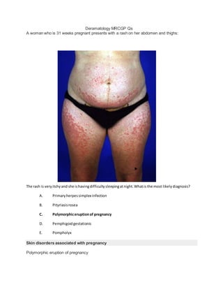
  • 1. Deramatology MRCGP Qs A woman who is 31 weeks pregnant presents with a rash on her abdomen and thighs: The rash is veryitchyand she ishavingdifficultysleepingatnight.Whatis the most likelydiagnosis? A. Primaryherpessimplex infection B. Pityriasisrosea C. Polymorphiceruptionof pregnancy D. Pemphigoidgestationis E. Pompholyx Skin disorders associated with pregnancy Polymorphic eruption of pregnancy
  • 2.  pruritic condition associated with last trimester  lesions often first appear in abdominal striae  management depends on severity: emollients, mild potency topical steroids and oral steroids may be used Pemphigoid gestationis  pruritic blistering lesions  often develop in peri-umbilical region, later spreading to the trunk, back, buttocks and arms  usually presents 2nd or 3rd trimester and is rarely seen in the first pregnancy  oral corticosteroids are usually required A 54-year-oldmanwitha historyof type 2 diabetesmellitusandbenignprostatichyperplasiaisreferred to dermatologydue toa numberof lesionsoverhisshin.Onexaminationsymmetrical,erythematous, tender,nodulesare found.The lesionshave startedtoheal withoutscarring.Whatisthe most likely diagnosis? A. Necrobiosislipoidicadiabeticorum B. Erythema nodosum C. Pyodermagangrenosum D. Syphilis E. Pretibial myxoedema The diagnosis in this question needs to be made on the description of the lesions as the past medical history is not relevant. Shin lesions
  • 3. The differential diagnosis of shin lesions includes the following conditions:  erythema nodosum  pretibial myxoedema  pyoderma gangrenosum  necrobiosis lipoidica diabeticorum Below are the characteristic features: Erythema nodosum  symmetrical, erythematous, tender, nodules which heal without scarring  most common causes are streptococcal infections, sarcoidosis, inflammatory bowel disease and drugs (penicillins, sulphonamides, oral contraceptive pill) Pretibial myxoedema  symmetrical, erythematous lesions seen in Graves' disease  shiny, orange peel skin Pyoderma gangrenosum  initially small red papule  later deep, red, necrotic ulcers with a violaceous border  idiopathic in 50%, may also be seen in inflammatory bowel disease, connective tissue disorders and myeloproliferative disorders Necrobiosis lipoidica diabeticorum  shiny, painless areas of yellow/red skin typically on the shin of diabetics  often associated with telangiectasia
  • 4. Theme:Causesof pruritus A. Liverdisease B. Hypothyroidism C. Diabetesmellitus D. Pregnancy E. Chronickidneydisease F. Polycythaemia G. Iron deficiencyanaemia H. Senile pruritus I. Scabies J. Lymphoma For eachof the followingscenariosselectthe mostlikelydiagnosis: 3. A 52-year-oldwomanpresentswithpruritusandlethargy.She hasrecentlyputonweightandis complainingaboutdryskin Hypothyroidism 4. A 57-year-oldwomanpresentswithpruritus.She statesshe hasbeengainingweightdespite eatinglessandcomplainsof constantnausea.Onexaminationshe ispale
  • 5. You answeredSenile pruritus The correct answeris Chronickidneydisease Pregnancyishighlyunlikelygivenherage. 5. A 59-year-oldmancomplainsof pruritusandlethargy.Onexaminationhe hasspoon shaped nailsanda smoothtongue Iron deficiencyanaemia Pruritus The table below lists the main characteristics of the most important causes of pruritus Liver disease History of alcohol excess Stigmata of chronic liver disease:spider naevi, bruising,palmar erythema,gynaecomastia etc Evidence of decompensation:ascites,jaundice,encephalopathy Iron deficiency anaemia Pallor Other signs:koilonychia,atrophic glossitis,post-cricoid webs,angular stomatitis Polycythaemia Pruritus particularlyafter warm bath 'Ruddy complexion' Gout Peptic ulcer disease Chronic kidney disease Lethargy & pallor Oedema & weightgain Hypertension Lymphoma Nightsweats Lymphadenopathy Splenomegaly,hepatomegaly Fatigue Other causes:  hyper- and hypothyroidism
  • 6.  diabetes  pregnancy  'senile' pruritus  urticaria  skin disorders: eczema, scabies, psoriasis, pityriasis rosea A 64-year-old man presents with a 'rash' on his legs which has developed over the past few days: He complainsof feeling generally'run-down'butreview of systemsisunremarkable.Whatisthe most likelyunderlyingcause? A. Vasculitis B. Erythemamultiforme
  • 7. C. Necrotisingfasciitis D. Kaposi sarcoma E. Venouseczema Kaposi sarcoma may cause similar skin changes to the larger lesions but would not typically cause petechiae. Vasculitis is commonly limited to the skin and may be caused by infections, drugs, autoimmune disorders and malignancy. Vasculitides Large vessel  temporal arteritis  Takayasu's arteritis Medium vessel  polyarteritis nodosa  Kawasaki disease Small vessel  ANCA-associated vasculitides (Wegener's*, Churg-Strauss*, microscopic polyangiitis)  Henoch-Schonlein purpura  cryoglobulinaemic vasculitis *may also affect medium-sized vessels
  • 8. A 34-year-oldmanpresentstohisGP withan itchyrash onhis genitalsandpalms.He hasalsonoticed the rash aroundthe site of a recentscar onhisforearm.Examinationrevealspapuleswithawhite-lace patternon the surface.What isthe diagnosis? A. Lichenplanus B. Scabies C. Lichensclerosus D. Morphea E. Pityriasisrosea Lichen  planus: purple, pruritic, papular, polygonal rash on flexor surfaces. Wickham's striae over surface. Oral involvement common  sclerosus: itchy white spots typically seen on the vulva of elderly women This is a typical history of lichen planus Lichen planus Lichen planus is a skin disorder of unknown aetiology, most probably being immune mediated Features  itchy, papular rash most common on the palms, soles, genitalia and flexor surfaces of arms  rash often polygonal in shape, 'white-lace' pattern on the surface (Wickham's striae)  Koebner phenomenon may be seen (new skin lesions appearing at the site of trauma)  oral involvement in around 50% of patients  nails: thinning of nail plate, longitudinal ridging Lichenoid drug eruptions - causes:
  • 9.  gold  quinine  thiazides Management  topical steroids are the mainstay of treatment  extensive lichen planus may require oral steroids or immunosuppression A 53-year-old man presents complaining of an itchy scalp and dandruff. On examination he is noted to have eczema on his scalp, behind his ears and around his nose. He has tried'Headand Shoulders'and'NeutrogenT-gel'butwithpoorresults.Whichone of the followingisthe mostappropriate treatmentforhisscalp?
  • 10. A. Topical hydrocortisone B. Topical dermovate C. Topical seleniumsulphide D. Oral terbinafine E. Topical ketoconazole Seborrhoeic dermatitis in adults Seborrhoeic dermatitis in adults is a chronic dermatitis thought to be caused by an inflammatory reaction related to a proliferation of a normal skin inhabitant, a fungus called Malassezia furfur (formerly known as Pityrosporum ovale). It is common, affecting around 2% of the general population Features  eczematous lesions on the sebum-rich areas: scalp (may cause dandruff), periorbital, auricular and nasolabial folds  otitis externa and blepharitis may develop Associated conditions include  HIV  Parkinson's disease Scalp disease management  over the counter preparations containing zinc pyrithione ('Head & Shoulders') and tar ('Neutrogena T/Gel') are first-line  the preferred second-line agent is ketoconazole  selenium sulphide and topical corticosteroid may also be useful Face and body management
  • 11.  topical antifungals: e.g. Ketoconazole  topical steroids: best used for short periods  difficult to treat - recurrences are common A 34-year-oldmanpresentsforthe removal of a mole.Where onthe bodyare keloidscarsmostlikelyto form? A. Sternum B. Lowerback C. Abdomen D. Flexorsurfacesof limbs E. Scalp Keloid scars are most common on the sternum Keloid scars Keloid scars are tumour-like lesions that arise from the connective tissue of a scar and extend beyond the dimensions of the original wound Predisposing factors  ethnicity: more common in people with dark skin  occur more commonly in young adults, rare in the elderly  common sites (in order of decreasing frequency): sternum, shoulder, neck, face, extensor surface of limbs, trunk
  • 12. Keloid scars are less likely if incisions are made along relaxed skin tension lines* Treatment  early keloids may be treated with intra-lesional steroids e.g. triamcinolone  excision is sometimes required *Langer lines were historically used to determine the optimal incision line. They were based on procedures done on cadavers but have been shown to produce worse cosmetic results than when following skin tension lines A 18-year-old man complains of an itchy sensation around his toes; What is the most appropriate first line treatment? A. Topical nystatin B. Topical miconazole
  • 13. C. Topical amorolfine D. Topical steroid E. Antiperspirant dusting powders Athlete's foot Athlete's foot is also known as tinea pedis. It is usually caused by fungi in the genus Trichophyton. Features  typically scaling, flaking, and itching between the toes Clinical knowledge summaries recommend a topical imidazole, undecenoate, or terbinafine first- line A 2-year-old girl develops a rash on her legs. By the time she is brought to surgery the rash has spread to the rest of her body.
  • 14. What is the most likely diagnosis? A. Erythema multiforme B. Erythema chronica migrans C. Erythema nodosum D. Urticaria E. Dermatitis artefacta The classic 'target' lesions of erythema multiforme can be seen clearly on this image. Erythema multiforme Features  target lesions  initially seen on the back of the hands / feet before spreading to the torso  upper limbs are more commonly affected than the lower limbs  pruritus is occasionally seen and is usually mild If symptoms are severe and involve blistering and mucosal involvement the term Stevens- Johnson syndrome is used. Causes
  • 15.  viruses: herpes simplex virus (the most common cause), Orf*  idiopathic  bacteria: Mycoplasma, Streptococcus  drugs: penicillin, sulphonamides, carbamazepine, allopurinol, NSAIDs, oral contraceptive pill, nevirapine  connective tissue disease e.g. Systemic lupus erythematosus  sarcoidosis  malignancy *Orf is a skin disease of sheep and goats caused by a parapox virus A 54-year-oldmanisreferredbyhisGP to the dermatologyoutpatientdepartmentdue toafacial rash whichhas persistedforthe past12 months.Onexaminationthere isasymmetrical rashconsistingof extensive pustulesand papuleswhichaffectshisnose,cheeksandforehead.Whatisthe most appropriate treatment? A. Ciprofloxacin B. Isotretinoin C. Oxytetracycline D. Hydroxychloroquine E. Prednisolone As there is extensive involvement oral oxytetracycline should probably be used rather than topical metronidazole Acne rosacea Acne rosacea is a chronic skin disease of unknown aetiology Features
  • 16.  typically affects nose, cheeks and forehead  flushing is often first symptom  telangiectasia are common  later develops into persistent erythema with papules and pustules  rhinophyma  ocular involvement: blepharitis Management  topical metronidazole may be used for mild symptoms (i.e. Limited number of papules and pustules, no plaques)  more severe disease is treated with systemic antibiotics e.g. Oxytetracycline  recommend daily application of a high-factor sunscreen  camouflage creams may help conceal redness  laser therapy may be appropriate for patients with prominent telangiectasia You review an 82-year-old woman who has developed 'sores' on her legs. For the past two years she has had dry, itchy skin around her ankles but over the past few weeks the skin has started to break down.
  • 17. What isthe mostlikelydiagnosis? A. Necrobiosislipoidicadiabeticorum B. Pyodermagangrenosum C. Arterial ulcers D. Venousulcers E. Pretibial myxoedema The dry, skin represents varicose eczema. Arterial ulcers tend to have a more 'punched-out' appearance. Venous ulceration Venous ulceration is typically seen above the medial malleolus Investigations
  • 18.  ankle-brachial pressure index (ABPI) is important in non-healing ulcers to assess for poor arterial flow which could impair healing  a 'normal' ABPI may be regarded as between 0.9 - 1.2. Values below 0.9 indicate arterial disease. Interestingly, values above 1.3 may also indicate arterial disease, in the form of false-negative results secondary to arterial calcification (e.g. In diabetics) Management  compression bandaging, usually four layer (only treatment shown to be of real benefit)  oral pentoxifylline, a peripheral vasodilator, improves healing rate  small evidence base supporting use of flavinoids  little evidence to suggest benefit from hydrocolloid dressings, topical growth factors, ultrasound therapy and intermittent pneumatic compression You are examining the chest of a 74-year-old man and notice the following: What isthe mostlikelydiagnosis?
  • 19. A. Bowen'sdisease B. Multiple benignmoles C. Metastaticmalignantmelanoma D. Seborrhoeickeratoses E. Dysplasticnaevussyndrome This man has multiple seborrhoeic keratoses, also known as basal cell papillomas. Seborrhoeic keratoses Seborrhoeic keratoses are benign epidermal skin lesions seen in older people. Features  large variation in colour from flesh to light-brown to black  have a 'stuck-on' appearance  keratotic plugs may be seen on the surface Management  reassurance about the benign nature of the lesion is an option  options for removal include curettage, cryosurgery and shave biopsy A 34-year-old man presents with unsightly toes:
  • 20. What isthe mostlikelycausativeorganism? A. Microsporumgypseum B. Trichophytoninterdigitale C. Candida D. Non-dermatophyticmoulds E. Trichophyton rubrum Fungal nail infections Onychomycosis is fungal infection of the nails. This may be caused by  dermatophytes - mainly Trichophyton rubrum, accounts for 90% of cases  yeasts - such as Candida  non-dermatophyte moulds Features  'unsightly' nails are a common reason for presentation  thickened, rough, opaque nails are the most common finding Investigation
  • 21.  nail clippings  scrapings of the affected nail Management  treatment is successful in around 50-80% of people  diagnosis should be confirmed by microbiology before starting treatment  dermatophyte infection: oral terbinafine is currently recommended first-line with oral itraconazole as an alternative. Six weeks - 3 months therapy is needed for fingernail infections whilst toenails should be treated for 3 - 6 months  Candida infection: mild disease should be treated with topical antifungals (e.g. Amorolfine) whilst more severe infections should be treated with oral itraconazole for a period of 12 weeks Please look at this skin lesion below a patient's eye:
  • 22. Whichone of the following medicationsismostassociatedwiththe developmentof these lesions? A. Statins B. Prednisolone C. Aspirin D. Amiodarone E. Combinedoral contraceptive pill Spider naevi Spider naevi (also called spider angiomas) describe a central red papule with surrounding capillaries. The lesions blanch upon pressure. Spider naevi are almost always found on the upper part of the body. Around 10-15% of people will have one or more spider naevi and they are more common in childhood. Other associations  liver disease  pregnancy  combined oral contraceptive pill A 26-year-oldnewlyqualifiednurse presentsasshe hasdevelopedabilateral erythematousrashon bothhands.She has recentlyemigratedfromthe Philippinesandhasnopast medical historyof note.A diagnosisof contactdermatitisissuspected.Whatisthe most suitable totesttoidentifythe underlying cause? A. Radioallergosorbenttest(RAST) B. Latex IgM levels
  • 23. C. Skinpricktest D. Urinary porphyrins E. Skin patch test The skin patch test is useful in this situation as it may also identify for irritants, not just allergens Allergy tests Skin prick test Most commonlyused testas easyto perform and inexpensive.Drops ofdiluted allergen are placed on the skin after which the skin is pierced using a needle.A large number of allergens can be tested in one session.Normallyincludes a histamine (positive) and sterile water (negative) control. A wheal will typically develop if a patienthas an allergy. Can be interpreted after 15 minutes Useful for food allergies and also pollen Radioallergosorbent test (RAST) Determines the amountofIgE that reacts specificallywith suspected or known allergens, for example IgE to egg protein. Results are given in grades from 0 (negative) to 6 (strongly positive) Useful for food allergies,inhaled allergens (e.g.Pollen) and wasp/bee venom Blood tests may be used when skin prick tests are not suitable,for example if there is extensive eczema or if the patientis taking antihistamines Skin patch testing Useful for contact dermatitis.Around 30-40 allergens are placed on the back. Irritants may also be tested for. The patches are removed 48 hours later with the results being read by a dermatologistafter a further 48 hours A 27-year-old man with a history of depression and coeliac disease presents with an itchy rash on his buttocks:
  • 24. What isthe mostlikelydiagnosis? A. LinearIgA dermatosis B. Neuroticexcoriations C. Scabies D. Dermatitis herpetiformis E. SSRI-associated dermatitis Dermatitis herpetiformis Dermatitis herpetiformis is an autoimmune blistering skin disorder associated with coeliac disease. It is caused by deposition of IgA in the dermis. Features  itchy, vesicular skin lesions on the extensor surfaces (e.g. elbows, knees buttocks) Diagnosis
  • 25.  skin biopsy: direct immunofluorescence shows deposition of IgA in a granular pattern in the upper dermis Management  gluten-free diet  dapsone A patientwithahistoryof tineacapitispresentsdue toa raised lesiononhisscalp.The lesionhasbeen gettinggraduallybiggeroverthe pasttwoweeks.Onexaminationyoufindaraised,pustular,spongy mass onthe crownof hishead.What isthe most likelydiagnosis A. Tineacorporis B. Id reaction(auto-eczematisation) C. Sebaceouscyst D. Bacterial skinabscess E. Kerion Tinea Tinea is a term given to dermatophyte fungal infections. Three main types of infection are described depending on what part of the body is infected  tinea capitis - scalp  tinea corporis - trunk, legs or arms  tinea pedis - feet Tinea capitis (scalp ringworm)
  • 26.  a cause of scarring alopecia mainly seen in children  if untreated a raised, pustular, spongy/boggy mass called a kerion may form  most common cause is Trichophyton tonsurans in the UK and the USA  may also be caused by Microsporum canis acquired from cats or dogs  diagnosis: lesions due to Microsporum canis green fluorescence under Wood's lamp*. However the most useful investigation is scalp scrapings  management (based on CKS guidelines): oral antifungals: terbinafine for Trichophyton tonsurans infections and griseofulvin for Microsporum infections. Topical ketoconazole shampoo should be given for the first two weeks to reduce transmission Tinea corporis  causes include Trichophyton rubrum and Trichophyton verrucosum (e.g. From contact with cattle)  well-defined annular, erythematous lesions with pustules and papules  may be treated with oral fluconazole Tinea pedis (athlete's foot)  characterised by itchy, peeling skin between the toes  common in adolescence *lesions due to Trichophyton species do not readily fluoresce under Wood's lamp Please look at the skin lesion shown below:
  • 27. Whichone of the followingstatementsregardingthistype of skinlesionistrue? A. Curettage is an acceptable treatmentoption B. Theyexhibitthe Koebnerphenomenon C. Theytypicallygrowrapidly D. Bleedingisunusual E. Metastasesare presentin10% of patientsatthe time of diagnosi Basal cell carcinoma Basal cell carcinoma (BCC) is one of the three main types of skin cancer. Lesions are also known as rodent ulcers and are characterised by slow-growth and local invasion. Metastases are extremely rare. BCC is the most common type of cancer in the Western world. Features  many types of BCC are described. The most common type is nodular BCC, which is described here
  • 28.  sun-exposed sites, especially the head and neck account for the majority of lesions  initially a pearly, flesh-coloured papule with telangiectasia  may later ulcerate leaving a central 'crater' Management options:  surgical removal  curettage  cryotherapy  topical cream: imiquimod, fluorouracil  radiotherapy A 56-year-old woman develops a rash in both axilla: What isthe mostlikelydiagnosis?
  • 29. A. Pellagra B. Erythemagyratumrepens C. Hidradenitissuppurativa D. Tineacorporis E. Acanthosis nigricans This image shows the typical brown, velvety patches which affect the axilla, neck and groin. Acanthosis nigricans Describes symmetrical, brown, velvety plaques that are often found on the neck, axilla and groin Causes  gastrointestinal cancer  insulin-resistant diabetes mellitus  obesity  polycystic ovarian syndrome  acromegaly  Cushing's disease  hypothyroidism  familial  Prader-Willi syndrome  drugs: oral contraceptive pill, nicotinic acid A 23-year-old woman who is 10 weeks pregnant presents with a rapidly growing lesion on her finger. This has grown from the size of a 'pin-prick' when it first appeared 4 weeks ago.
  • 30. What isthe mostlikelydiagnosis? A. Viral wart B. Orf C. Pyogenicgranuloma D. Capillaryhaemangioma E. Squamouscell carcinoma Pyogenic granuloma Pyogenic granuloma is a relatively common benign skin lesion. The name is confusing as they are neither true granulomas nor pyogenic in nature. There are multiple alternative names but perhaps 'eruptive haemangioma' is the most useful. The cause of pyogenic granuloma is not known but a number of factors are linked:  trauma  pregnancy  more common in women and young adults Features
  • 31.  most common sites are head/neck, upper trunk and hands. Lesions in the oral mucosa are common in pregnancy  initially small red/brown spot  rapidly progress within days to weeks forming raised, red/brown lesions which are often spherical in shape  the lesions may bleed profusely or ulcerate Management  lesions associated with pregnancy often resolve spontaneously post-partum  other lesions usually persist. Removal methods include curettage and cauterisation, cryotherapy, excision A 30-year-old man presents with a two-week history of a productive cough. Whilst examining him you notice a large number of atypical naevi over his torso. On his back you count between 20-25 moles. He reports no change in any of his moles, no bleeding and no itch. One particular mole is noted due to the irregular border. It is 6 * 4 mm in size.
  • 32. What isthe mostappropriate action? A. Refertodermatologyforphotomapping B. Referunder the two-weekrule to dermatology C. Advise aboutsunprotection+arrange gene testingforxerodermapigmentosum D. Advise aboutsunprotection+take a digital photoforhisrecords+ review in1 month E. Advise aboutsunprotection+take a digital photoforhisrecords This is very likely to be a melanoma and the patient should be fast-tracked to dermatology. Due to the location and the number of moles he has it is unlikely that he would have noticed any change Malignant melanoma: prognostic factors The invasion depth of a tumour (Breslow depth) is the single most important factor in determining prognosis of patients with malignant melanoma Breslow Thickness Approximate 5 year survival < 1 mm 95-100% 1 - 2 mm 80-96% 2.1 - 4 mm 60-75% > 4 mm 50% Whichone of the followingbestdescribesthe typical distributionof atopic eczemaina10-month-old child? A. Nappyarea andflexorsurfacesof armsand legs B. Face and trunk C. Nappyarea andtrunk D. Flexorsurfacesof armsand legs
  • 33. E. Scalpand arms Eczema in children Eczema occurs in around 15-20% of children and is becoming more common. It typically presents before 6 months but clears in around 50% of children by 5 years of age and in 75% of children by 10 years of age Features  in infants the face and trunk are often affected  in younger children eczema often occurs on the extensor surfaces  in older children a more typical distribution is seen, with flexor surfaces affected and the creases of the face and neck Management  avoid irritants  simple emollients: large quantities should be prescribed (e.g. 250g / week), roughly in a ratio of with topical steroids of 10:1. If a topical steroid is also being used the emollient should be applied first followed by waiting at least 30 minutes before applying the topical steroid. Creams soak into the skin faster than ointments. Emollients can become contaminated with bacteria - fingers should not be inserted into pots (many brands have pump dispensers)  topical steroids  in severe cases wet wraps and oral ciclosporin may be used Theme:Acne vulgaris:management A. Oral trimethoprim B. Oral flucloxacillin
  • 34. C. Topical benzoyl peroxide D. Topical zinc+ erythromycin E. Oral isotretinoin F. Oral lymecycline G. Oral minocycline H. Oral erythromycin For eachone of the followingquestionsplease selectthe correctanswerfrom the optionslistedabove: 5. Shouldbe avoideddue toanincreasedriskof drug-inducedlupusandhyperpigmentation You answeredOral isotretinoin The correct answeris Oral minocycline 6. Is mostlikelytoaffectthe hepaticmetabolismof othermedications You answeredOral flucloxacillin The correct answeris Oral erythromycin Erythromycinisan inhibitorof the P450 system. 7. Patientsshouldbe warnedaboutphotosensitivity You answeredOral erythromycin
  • 35. The correct answeris Oral lymecycline Care shouldbe takenwithtopical retinoidsaswell.Photosensitivitywithoral isotretinoinislistedasa 'veryrare' side-effectinthe BNF,the lastof a verylonglistof side-effects. Acne vulgaris: management Acne vulgaris is a common skin disorder which usually occurs in adolescence. It typically affects the face, neck and upper trunk and is characterised by the obstruction of the pilosebaceous follicles with keratin plugs which results in comedones, inflammation and pustules. Acne may be classified into mild, moderate or severe:  mild: open and closed comedones with or without sparse inflammatory lesions  moderate acne: widespread non-inflammatory lesions and numerous papules and pustules  severe acne: extensive inflammatory lesions, which may include nodules, pitting, and scarring A simple step-up management scheme often used in the treatment of acne is as follows:  single topical therapy (topical retinoids, benzyl peroxide)  topical combination therapy (topical antibiotic, benzoyl peroxide, topical retinoid)  oral antibiotics: e.g. Oxytetracycline, doxycycline. Improvement may not be seen for 3-4 months. Minocycline is now considered less appropriate due to the possibility of irreversible pigmentation. Gram negative folliculitis may occur as a complication of long- term antibiotic use - high-dose oral trimethoprim is effective if this occurs  oral isotretinoin: only under specialist supervision There is no role for dietary modification in patients with acne
  • 36. The patient below is being treated for epilepsy: What isthe mostlikelyunderlyingdiagnosis? A. HIV B. Neurofibromatosis C. Arteriovenousmalformation D. Tuberous sclerosis E. Lennox-Gastautsyndrome These skin lesions represent adenoma sebaceum. Tuberous sclerosis Tuberous sclerosis (TS) is a genetic condition of autosomal dominant inheritance. Like neurofibromatosis, the majority of features seen in TS are neuro-cutaneous Cutaneous features  depigmented 'ash-leaf' spots which fluoresce under UV light  roughened patches of skin over lumbar spine (Shagreen patches)  adenoma sebaceum: butterfly distribution over nose  fibromata beneath nails (subungual fibromata)
  • 37.  café-au-lait spots* may be seen Neurological features  developmental delay  epilepsy (infantile spasms or partial)  intellectual impairment Also  retinal hamartomas: dense white areas on retina (phakomata)  rhabdomyomas of the heart  gliomatous changes can occur in the brain lesions  polycystic kidneys, renal angiomyolipomata *these of course are more commonly associated with neurofibromatosis. However a 1998 study of 106 children with TS found café-au-lait spots in 28% of patients A 17-year-oldfemalepresentswithmultiple comedones,pustulesandpapulesonherface.Whichone of the followingisleastlikelytoimprove hercondition? A. Topical retinoids B. Dietary advice C. Sunlight D. Oral trimethoprim E. Ethinylestradiolwithcyproterone acetate
  • 38. There is no role for dietary modification in patients with acne vulgaris. Ethinylestradiol with cyproterone acetate (Dianette) is useful in some female patients with acne unresponsive to standard treatment. Oral trimethoprim is useful in patients on long-term antibiotics who develop Gram negative folliculitis Acne vulgaris: management Acne vulgaris is a common skin disorder which usually occurs in adolescence. It typically affects the face, neck and upper trunk and is characterised by the obstruction of the pilosebaceous follicles with keratin plugs which results in comedones, inflammation and pustules. Acne may be classified into mild, moderate or severe:  mild: open and closed comedones with or without sparse inflammatory lesions  moderate acne: widespread non-inflammatory lesions and numerous papules and pustules  severe acne: extensive inflammatory lesions, which may include nodules, pitting, and scarring A simple step-up management scheme often used in the treatment of acne is as follows:  single topical therapy (topical retinoids, benzyl peroxide)  topical combination therapy (topical antibiotic, benzoyl peroxide, topical retinoid)  oral antibiotics: e.g. Oxytetracycline, doxycycline. Improvement may not be seen for 3-4 months. Minocycline is now considered less appropriate due to the possibility of irreversible pigmentation. Gram negative folliculitis may occur as a complication of long- term antibiotic use - high-dose oral trimethoprim is effective if this occurs  oral isotretinoin: only under specialist supervision There is no role for dietary modification in patients with acne
  • 39. A 23-year-oldmanpresents withathree dayhistoryof general malaise andlow-gradetemperature. Yesterdayhe developedextensive painfululcerationof hismouthandgums.Onexaminationhis temperature is37.4ºC, pulse 84 / minand there issubmandibularlymphadenopathy.Whatis the most likelydiagnosis? A. EpsteinBarr virus B. Lichenplanus C. HIV seroconversionillness D. Herpessimplexvirusinfection E. Oral Candida This man has gingivostomatitis, a characteristic feature of primary herpes simplex virus infection Herpes simplex virus There are two strains of the herpes simplex virus (HSV) in humans: HSV-1 and HSV-2. Whilst it was previously thought HSV-1 accounted for oral lesions (cold sores) and HSV-2 for genital herpes it is now known there is considerable overlap Features  primary infection: may present with a severe gingivostomatitis  cold sores  painful genital ulceration Management  gingivostomatitis: oral aciclovir, chlorhexidine mouthwash  cold sores: topical aciclovir although the evidence base for this is modest  genital herpes: oral aciclovir. Some patients with frequent exacerbations may benefit from longer term aciclovir
  • 40. A 59-year-old man complains of dry, sore eyes for the past six months. There has been no change in his vision and he doesn't wear contact lens. The only past history of note is hypothyroidism. What isthe mostlikelydiagnosis? A. Blepharitis B. Grave's eye disease C. Episcleritis D. Conjunctivitis E. Hay fever Blepharitis Blepharitis is inflammation of the eyelid margins. It may due to either meibomian gland dysfunction (common, posterior blepharitis) or seborrhoeic dermatitis/staphylococcal infection (less common, anterior blepharitis). Blepharitis is also more common in patients with rosacea The meibomian glands secrete oil on to the eye surface to prevent rapid evaporation of the tear film. Any problem affecting the meibomian glands (as in blepharitis) can hence cause drying of the eyes which in turns leads to irritation Features
  • 41.  symptoms are usually bilateral  grittiness and discomfort, particularly around the eyelid margins  eyes may be sticky in the morning  eyelid margins may be red. Swollen eyelids may be seen in staphylococcal blepharitis  styes and chalazions are more common in patients with blepharitis  secondary conjunctivitis may occur Management  softening of the lid margin using hot compresses twice a day  mechanical removal of the debris from lid margins - cotton wool buds dipped in a mixture of cooled boiled water and baby shampoo is often used*  artificial tears may be given for symptom relief in people with dry eyes or an abnormal tear film *an alternative is sodium bicarbonate, a teaspoonful in a cup of cooled water that has recently been boiled You reviewa42-year-oldmanwithchronicplaque psoriasis. He regularlyusesanemollienttocontrol scaling.Accordingtocurrentclinical guidelines,whichisthe mostappropriate long-termtreatment? A. Topical calcipotriol B. Combinedtopical steroid/calcipotriol C. Topical coal tar D. Topical dithranol E. Topical steroid The 2010 SIGN guidelines recommend topical calcipotriol for the long-term treatment of patients with chronic plaque psoriasis. Psoriasis: management
  • 42. SIGN released guidelines in 2010 on the management of psoriasis and psoriatic arthropathy. Please see the link for more details. Chronic plaque psoriasis  regular emollients may help to reduce scale loss and reduce pruritus  for acute control SIGN recommend: 'Short term intermittent use of a potent topical corticosteroid or a combined potent corticosteroid plus calcipotriol ointment is recommended to gain rapid improvement in plaque psoriasis.'  'For long term topical treatment of plaque psoriasis a vitamin D analogue (e.g. Calcipotriol) is recommended.'  'If a vitamin D analogue is ineffective or not tolerated then consider coal tar (solution, cream or lotion), tazarotene gel, or short contact dithranol (30 minute exposure in patients with a small number of relatively large plaques of psoriasis). Steroids in psoriasis  topical steroids are commonly used in flexural psoriasis and there is also a role for mild steroids in facial psoriasis. If steroids are ineffective for these conditions vitamin D analogues or tacrolimus ointment should be used second line  SIGN caution against the long term use of potent or very potent topical steroids due to the risk of side-effects Scalp psoriasis  for short term control SIGN recommend either the use of potent topical corticosteroids or a combination of a potent corticosteroid and a vitamin D analogue  'For patients with thick scaling of the scalp, initial treatment with overnight application of salicylic acid, tar preparations, or oil preparations (eg olive oil, coconut oil) to remove thick scale is recommended.
  • 43. Secondary care management Phototherapy  narrow band ultraviolet B light (311-313nm) is now the treatment of choice  photochemotherapy is also used - psoralen + ultraviolet A light (PUVA)  adverse effects: skin ageing, squamous cell cancer (not melanoma) Systemic therapy  methotrexate: useful if associated joint disease  ciclosporin  systemic retinoids  biological agents: infliximab, etanercept and adalimumab  ustekinumab (IL-12 and IL-23 blocker) is showing promise in early trials Mechanism of action of commonly used drugs:  coal tar: probably inhibit DNA synthesis  calcipotriol: vitamin D analogue which reduces epidermal proliferation and restores a normal horny layer  dithranol: inhibits DNA synthesis, wash off after 30 mins, SE: burning, staining Theme:Skindisordersaffectingthe solesof the feet A. Pittedkeratolysis B. Mosaic wart
  • 44. C. Acquiredkeratoderma D. Juvenileplantardermatosis E. Palmoplantarpustulosis F. Tineapedis G. Callus H. Idiopathicplantarhidradenitis I. Exfoliative keratolysis J. Contact dermatitis For eachone of the followingscenariosplease selectthe mostlikelydiagnosis: 13. A 23-year-oldfemalepresentswithred,thickenedskinonthe soles.Oncloserinspectionacrop of raisedlesionsare seen. You answeredExfoliative keratolysis The correct answeris Palmoplantarpustulosis 14. A 22-year-oldmanpresentswitha3 cm areaof hyperkeratoticskinonthe heel of hisrightfoot. A numberof pinpointpetechiae are seeninthe lesion. You answeredPalmoplantarpustulosis The correct answeris Mosaicwart 15. A 15-year-oldcomplainsof excessivelysmellyfeet.Onexaminationhe haswhite skinoverthe sole of the forefootbilaterally.Small holescanbe seenonthe surface of the affectedskin.
  • 45. You answeredTineapedis The correct answeris Pitted keratolysis Skin disorders affecting the soles of the feet The table below gives characteristic exam question features for conditions affecting the soles of the feet Verrucas Secondary to the human papilloma virus Firm, hyperkeratotic lesions Pinpointpetechiae centrallywithin the lesions May coalesce with surrounding warts to form mosaic warts Tinea pedis More commonlycalled Athlete's foot Affected skin is moist,flaky and itchy Corn and calluses A corn is small areas ofvery thick skin secondaryto a reactive hyperkeratosis A callus is larger,broader and has a less well defined edge than a corn Keratoderma May be acquired or congenital Describes a thickening ofthe skin of the palms and soles Acquired causes include reactive arthritis (keratoderma blennorrhagica) Pitted keratolysis Affects people who sweatexcessively Patients may complain ofdamp and excessivelysmellyfeet Usuallycaused by Corynebacterium Heel and forefoot may become white with clusters ofpunched-outpits Palmoplantar pustulosis Crops of sterile pustules affecting the palms and soles The skin is thickened,red. Scaly and may crack More common in smokers Juvenile plantar dermatosis Affects children.More common in atopic patients with a history of eczema Soles become shinyand hard. Cracks may develop causing pain Worse during the summer
  • 46. You review a 27-year-old man who is under the care of the dermatology department. Whichone of the followingconditionsismostassociatedwiththisskindisorder? A. Addison'sdisease B. Asthma C. Iron-deficiencyanaemia D. Toxicmultinodulargoitre E. Type 2 diabetesmellitus Vitiligo is associated with other autoimmune conditions such as Addison's disease, type 1 diabetes mellitus and autoimmune thyroid disorders. Vitiligo Vitiligo is an autoimmune condition which results in the loss of melanocytes and consequent depigmentation of the skin. It is thought to affect around 1% of the population and symptoms typically develop by the age of 20-30 years. Features  well demarcated patches of depigmented skin  the peripheries tend to be most affected  trauma may precipitate new lesions (Koebner phenomenon)
  • 47. Associated conditions  type 1 diabetes mellitus  Addison's disease  autoimmune thyroid disorders  pernicious anaemia  alopecia areata Management  sun block for affected areas of skin  camouflage make-up  topical corticosteroids may reverse the changes if applied early  there may also be a role for topical tacrolimus and phototherapy, although caution needs to be exercised with light-skinned patients A 54-year-oldmanpresentswithatwomonthhistoryof a rapidlygrowinglesiononhisrightforearm. The lesioninitiallyappearedasared papule butinthe lasttwo weekshasbecome acrater filled centrallywithyellow/brownmaterial.Onexaminationthe manhasskintype II,the lesionis4mm in diameterandismorphologicallyasdescribedabove.Whatisthe mostlikelydiagnosis? A. Seborrhoeickeratosis B. Keratoacanthoma C. Pyodermagangrenosum D. Basal cell carcinoma E. Malignantmelanoma Keratoacanthoma
  • 48. Keratoacanthoma is a benign epithelial tumour. They are more frequent in middle age and do not become more common in old age (unlike basal cell and squamous cell carcinoma) Features - said to look like a volcano or crater  initially a smooth dome-shaped papule  rapidly grows to become a crater centrally-filled with keratin Spontaneous regression of keratoacanthoma within 3 months is common, often resulting in a scar. Such lesions should however be urgently excised as it is difficult clinically to exclude squamous cell carcinoma. Removal also may prevent scarring A womanburnsher arm on the ovendoorand phonesthe surgeryforadvice.She reportsa'2 inch by half an inchred line'onherright forearm.The burnis painful butshe isotherwisewellandhasno breathingproblems.Youbookheran appointmentforlateroninthe surgery.What isthe most appropriate firstaidadvice? A. Run under cool (noticed) water for 20 mins+ cover in layersof cling film B. Run undercool (noticed) waterfor10 mins+ applyliberal amountsof E45 C. Do nothinguntil she isseen D. Applya frozenbagof food(e.g.peas) for10 mins+ coverinlayersof clingfilm E. Applya bandage thathas beensoakedincoldwater Burns The following is based on guidance issued by Clinical Knowledge Summaries (please see the link for more details). Immediate first aid  airway, breathing, circulation
  • 49.  burns caused by heat: remove the person from the source. Within 20 minutes of the injury irrigate the burn with cool (not iced) water for between 10 and 30 minutes. Cover the burn using cling film, layered, rather than wrapped around a limb  electrical burns: switch off power supply, remove the person from the source  chemical burns: brush any powder off then irrigate with water. Attempts to neutralise the chemical are not recommended Assessing the extent of the burn  Wallace's Rule of Nines: head + neck = 9%, each arm = 9%, each anterior part of leg = 9%, each posterior part of leg = 9%, anterior chest = 9%, posterior chest = 9%, anterior abdomen = 9%, posterior abdomen = 9%  Lund and Browder chart: the most accurate method  the palmar surface is roughly equivalent to 1% of total body surface area (TBSA). Not accurate for burns > 15% TBSA Assessing the depth of the burn Modern terminology Former terminology Appearance Superficial epidermal First degree Red and painful Partial thickness (superficial dermal) Second degree Pale pink, painful,blistered Partial thickness (deep dermal) Second degree Typically white but may have patches of non-blanching erythema. Reduced sensation Full thickness Third degree White/brown/black in colour,no blisters,no pain Referral to secondary care  all deep dermal and full-thickness burns.  superficial dermal burns of more than 10% TBSA in adults, or more than 5% TBSA in children  superficial dermal burns involving the face, hands, feet, perineum, genitalia, or any flexure, or circumferential burns of the limbs, torso, or neck  any inhalation injury  any electrical or chemical burn injury
  • 50.  suspicion of non-accidental injury Management of burns  initial first aid as above  review referral criteria to ensure can be managed in primary care  superficial epidermal: symptomatic relief - analgesia, emollients etc  superficial dermal: cleanse wound, leave blister intact, non-adherent dressing, avoid topical creams, review in 24 hours A 85-year-oldladypresentstoherGPcomplainingof itchywhite plaquesaffectinghervulva.There isno historyof vaginal discharge orbleeding.A similarplaqueisalsoseenonherinnerthigh.Whatisthe likelydiagnosis? A. Candida B. Lichenplanus C. Lichensclerosus D. Herpessimplex E. Seborrhoeicdermatitis Lichen  planus: purple, pruritic, papular, polygonal rash on flexor surfaces. Wickham's striae over surface. Oral involvement common  sclerosus: itchy white spots typically seen on the vulva of elderly women
  • 51. The correct answer is lichen sclerosus. Candida may cause pruritus and white plaques but lesions would not also be seen on her inner thigh Lichen sclerosus Lichen sclerosus was previously termed lichen sclerosus et atrophicus. It is an inflammatory condition which usually affects the genitalia and is more common in elderly females. Lichen sclerosus leads to atrophy of the epidermis with white plaques forming Features  itch is prominent A biopsy is often performed to exclude other diagnoses Management  topical steroids and emollients  increased risk of vulval cancer A 30-year-oldfemaleinherthirdtrimesterof pregnancymentionsduringanantenatal appointmentthat she has noticedanitchyrash around herumbilicus.Thisishersecondpregnancyandshe hadno similar problemsinherfirstpregnancy.Examinationrevealsblistering lesionsinthe peri-umbilical regionand on herarms. What isthe likelydiagnosis? A. Seborrhoeicdermatitis B. Pompholyx C. Polymorphiceruptionof pregnancy D. Lichenplanus
  • 52. E. Pemphigoidgestationis Polymorphic eruption of pregnancy is not associated with blistering Pemphigoid gestationis is the correct answer. Polymorphic eruption of pregnancy is not associated with blistering Skin disorders associated with pregnancy Polymorphic eruption of pregnancy  pruritic condition associated with last trimester  lesions often first appear in abdominal striae  management depends on severity: emollients, mild potency topical steroids and oral steroids may be used Pemphigoid gestationis  pruritic blistering lesions  often develop in peri-umbilical region, later spreading to the trunk, back, buttocks and arms  usually presents 2nd or 3rd trimester and is rarely seen in the first pregnancy  oral corticosteroids are usually required A 31-year-oldwomandevelopspainful,purplelesionsonhershins.Whichone of the following medicationsismostlikelytobe responsible?
  • 53. A. Montelukast B. Lansoprazole C. Combinedoral contraceptive pill D. Sodiumvalproate E. Carbimazole Erythema nodosum Overview  inflammation of subcutaneous fat  typically causes tender, erythematous, nodular lesions  usually occurs over shins, may also occur elsewhere (e.g. forearms, thighs)  usually resolves within 6 weeks  lesions heal without scarring Causes  infection: streptococci, TB, brucellosis  systemic disease: sarcoidosis, inflammatory bowel disease, Behcet's  malignancy/lymphoma  drugs: penicillins, sulphonamides, combined oral contraceptive pill  pregnancy A 60-year-old man presents with a painful lesion on his right ear:
  • 54. What isthe mostlikelydiagnosis? A. Actinickeratosis B. Pseudocystof the auricle C. Chondrodermatitisnodularishelicis D. Basal cell carcinoma E. Keratoacanthoma Chondrodermatitis nodularis helicis Chondrodermatitis nodularis helicis (CNH) is a common and benign condition characterised by the development of a painful nodule on the ear. It is thought to be caused by factors such as persistent pressure on the ear (e.g. secondary to sleep, headsets), trauma or cold. CNH is more common in men and with increasing age. Management  reducing pressure on the ear: foam 'ear protectors' may be used during sleep  other treatment options include cryotherapy, steroid injection, collagen injection  surgical treatment may be used but there is a high recurrence rate
  • 55. A 34-year-oldmanwitha history of polyarthralgia,backpainanddiarrhoeaisfoundtohave a 3 cm red lesiononhisshinwhichisstartingtoulcerate.Whatis the most likelydiagnosis? A. SystemicShigellainfection B. Syphilis C. Metastaticcoloncancer D. Erythemanodosum E. Pyoderma gangrenosum This patient is likely to have ulcerative colitis, which has a known association with large-joint arthritis, sacroilitis and pyoderma gangrenosum Pyoderma gangrenosum Features  typically on the lower limbs  initially small red papule  later deep, red, necrotic ulcers with a violaceous border  may be accompanied systemic symptoms e.g. Fever, myalgia Causes*  idiopathic in 50%  inflammatory bowel disease: ulcerative colitis, Crohn's  rheumatoid arthritis, SLE  myeloproliferative disorders  lymphoma, myeloid leukaemias  monoclonal gammopathy (IgA)  primary biliary cirrhosis Management  the potential for rapid progression is high in most patients and most doctors advocate oral steroids as first-line treatment
  • 56.  other immunosuppressive therapy, for example ciclosporin and infliximab, have a role in difficult cases *note whilst pyoderma gangrenosum can occur in diabetes mellitus it is rare and is generally not included in a differential of potential causes You are teachingamotherabout the use of topical steroidsforherchildwithatopiceczema.She has heardabout the use of FingerTipUnits (FTU) whendetermininghow muchsteroidtouse.Whatdoes1 FTU equate to? A. Sufficienttotreata skinareaabout thatof a forearm B. Sufficienttotreat a skin area about twice that of the flat of an adult hand C. Sufficienttotreata skinareaabout 5 * 5 cm (2 * 2 inches) D. Sufficienttotreata skinareaabout twice thatof the forearm E. Sufficienttotreata skinareaabout thatof the flat of an adulthand Finger tip unit (FTU) for steroids = twice area of the flat of an adult hand Eczema: topical steroids Use weakest steroid cream which controls patients symptoms The table below shows topical steroids by potency Mild Moderate Potent Very potent Hydrocortisone 0.5- 2.5% Betamethasone valerate 0.025% (Betnovate RD) Fluticasone propionate 0.05% (Cutivate) Clobetasol propionate 0.05% (Dermovate)
  • 57. Clobetasone butyrate 0.05% (Eumovate) Betamethasone valerate 0.1% (Betnovate) Finger tip rule  1 finger tip unit (FTU) = 0.5 g, sufficient to treat a skin area about twice that of the flat of an adult hand Topical steroid doses for eczema in adults Area of skin Fingertip units per dose Hand and fingers (front and back) 1.0 A foot (all over) 2.0 Front of chestand abdomen 7.0 Back and buttocks 7.0 Face and neck 2.5 An entire arm and hand 4.0 An entire leg and foot 8.0 A 67-year-old man who is a retired builder presents following the development of a number of red, scaly lesions on his forehead. These were initially small and flat but are now erythematous and rough to touch.
  • 58. What isthe mostlikelydiagnosis? A. Pityriasisversicolor B. Seborrhoeickeratosis C. Polymorphouslighteruption D. Actinic keratoses E. Malignantmelanoma Actinic keratoses Actinic, or solar, keratoses (AK) is a common premalignant skin lesion that develops as a consequence of chronic sun exposure Features  small, crusty or scaly, lesions  may be pink, red, brown or the same colour as the skin  typically on sun-exposed areas e.g. temples of head  multiple lesions may be present Management options include  prevention of further risk: e.g. sun avoidance, sun cream
  • 59.  fluorouracil cream: typically a 2 to 3 week course. The skin will become red and inflamed - sometimes topical hydrocortisone is given following fluorouracil to help settle the inflammation  topical diclofenac: may be used for mild AKs. Moderate efficacy but much fewer side- effects  topical imiquimod: trials have shown good efficacy  cryotherapy  curettage and cautery A 72-year-oldmancomplainsof askinlesiononhistrunk.On examinationithasthe typical appearance of a seborrhoeickeratosis.Whichone of the followingmanagementoptionsisleastsuitable? A. Cryosurgery B. Reassurance aboutbenignnature C. Shave biopsy D. Curettage E. Excision Scalpel excision is not usually performed on seborrhoeic keratoses due to the success of other simpler methods. Seborrhoeic keratoses Seborrhoeic keratoses are benign epidermal skin lesions seen in older people. Features  large variation in colour from flesh to light-brown to black  have a 'stuck-on' appearance  keratotic plugs may be seen on the surface
  • 60. Management  reassurance about the benign nature of the lesion is an option  options for removal include curettage, cryosurgery and shave biopsy A 62-year-oldwithahistoryof acne rosaceapresentsforadvice regardingtreatment.Whichone of the followinginterventionshasthe leastrole inmanagement? A. Camouflage creams B. Topical metronidazole C. Low-dose topical corticosteroids D. Laser therapy E. Use of high-factorsunblock Acne rosacea Acne rosacea is a chronic skin disease of unknown aetiology Features  typically affects nose, cheeks and forehead  flushing is often first symptom  telangiectasia are common  later develops into persistent erythema with papules and pustules  rhinophyma  ocular involvement: blepharitis
  • 61. Management  topical metronidazole may be used for mild symptoms (i.e. Limited number of papules and pustules, no plaques)  more severe disease is treated with systemic antibiotics e.g. Oxytetracycline  recommend daily application of a high-factor sunscreen  camouflage creams may help conceal redness  laser therapy may be appropriate for patients with prominent telangiectasia You reviewa24-year-oldwoman.Six daysagoshe developedapink,itchyrashoverhertorso and arms. The followingdayshe startedtotake loratidine 10mgodbut thishas onlyledtoa slightimprovementin hersymptoms.Onreview todayshe hasa widespreadurticarial rashwhichisextremelyitchy.There is no lipor tongue swelling,herchestisclearandvital signsare unremarkable.Whatisthe most appropriate nextstepinmanagement? A. Referforallergytesting B. Prescribe topical hydrocortisonetorelievethe itch C. Switchto cetirizine10mgod D. Start a five day course of oral prednisolone E. Increase loratidineto10mg bd Urticaria Urticaria describes a local or generalised superficial swelling of the skin. The most common cause of urticaria is allergy although non-allergic causes are seen. Features
  • 62.  pale, pink raised skin. Variously described as 'hives', 'wheals', 'nettle rash'  pruritic Management  non-sedating antihistamines are first-line  prednisolone is used for severe or resistent episodes A 19-year-old man presents as he has developed a number of skin lesions similar to the one below: You advise himtouse regularemollientstocontrol the itchandscale.What is the mostappropriate first-linemanagement? A. Topical steroidorcombinedtopical steroid/dithranol B. Topical dithranol C. Topical coal tar or topical calcipotriol D. Topical calcipotriol orcombinedtopical steroid/dithranol E. Topical steroidor combinedtopical steroid/calcipotriol
  • 63. Psoriasis: management SIGN released guidelines in 2010 on the management of psoriasis and psoriatic arthropathy. Please see the link for more details. Chronic plaque psoriasis  regular emollients may help to reduce scale loss and reduce pruritus  for acute control SIGN recommend: 'Short term intermittent use of a potent topical corticosteroid or a combined potent corticosteroid plus calcipotriol ointment is recommended to gain rapid improvement in plaque psoriasis.'  'For long term topical treatment of plaque psoriasis a vitamin D analogue (e.g. Calcipotriol) is recommended.'  'If a vitamin D analogue is ineffective or not tolerated then consider coal tar (solution, cream or lotion), tazarotene gel, or short contact dithranol (30 minute exposure in patients with a small number of relatively large plaques of psoriasis). Steroids in psoriasis  topical steroids are commonly used in flexural psoriasis and there is also a role for mild steroids in facial psoriasis. If steroids are ineffective for these conditions vitamin D analogues or tacrolimus ointment should be used second line  SIGN caution against the long term use of potent or very potent topical steroids due to the risk of side-effects Scalp psoriasis  for short term control SIGN recommend either the use of potent topical corticosteroids or a combination of a potent corticosteroid and a vitamin D analogue  'For patients with thick scaling of the scalp, initial treatment with overnight application of salicylic acid, tar preparations, or oil preparations (eg olive oil, coconut oil) to remove thick scale is recommended.
  • 64. Secondary care management Phototherapy  narrow band ultraviolet B light (311-313nm) is now the treatment of choice  photochemotherapy is also used - psoralen + ultraviolet A light (PUVA)  adverse effects: skin ageing, squamous cell cancer (not melanoma) Systemic therapy  methotrexate: useful if associated joint disease  ciclosporin  systemic retinoids  biological agents: infliximab, etanercept and adalimumab  ustekinumab (IL-12 and IL-23 blocker) is showing promise in early trials Mechanism of action of commonly used drugs:  coal tar: probably inhibit DNA synthesis  calcipotriol: vitamin D analogue which reduces epidermal proliferation and restores a normal horny layer  dithranol: inhibits DNA synthesis, wash off after 30 mins, SE: burning, staining A 27-year-old woman who is 34 weeks pregnant presents with an itchy, blistering rash over her abdomen. Initially she had a red rash around her umbilicus but it later spread.
  • 65. What isthe mostlikelydiagnosis? A. Pemphigoidgestationis B. Seborrhoeicdermatitis C. Polymorphiceruptionof pregnancy D. HELLP syndrome E. Pompholyx Skin disorders associated with pregnancy Polymorphic eruption of pregnancy  pruritic condition associated with last trimester  lesions often first appear in abdominal striae  management depends on severity: emollients, mild potency topical steroids and oral steroids may be used Pemphigoid gestationis  pruritic blistering lesions  often develop in peri-umbilical region, later spreading to the trunk, back, buttocks and arms
  • 66.  usually presents 2nd or 3rd trimester and is rarely seen in the first pregnancy  oral corticosteroids are usually required A 26-year-oldmanwhoisHIV positive isnotedtohave developedseborrhoeicdermatitis.Whichof the followingtwocomplicationsare mostassociatedwiththiscondition? A. Alopeciaandotitisexterna B. Blepharitisand otitis externa C. Photosensitivityandalopecia D. Photosensitivityandblepharitis E. Blepharitisandalopecia Alopecia is not commonly seen in seborrhoeic dermatitis, but may develop if a severe secondary infection develops Seborrhoeic dermatitis in adults Seborrhoeic dermatitis in adults is a chronic dermatitis thought to be caused by an inflammatory reaction related to a proliferation of a normal skin inhabitant, a fungus called Malassezia furfur (formerly known as Pityrosporum ovale). It is common, affecting around 2% of the general population Features  eczematous lesions on the sebum-rich areas: scalp (may cause dandruff), periorbital, auricular and nasolabial folds  otitis externa and blepharitis may develop Associated conditions include  HIV  Parkinson's disease
  • 67. Scalp disease management  over the counter preparations containing zinc pyrithione ('Head & Shoulders') and tar ('Neutrogena T/Gel') are first-line  the preferred second-line agent is ketoconazole  selenium sulphide and topical corticosteroid may also be useful Face and body management  topical antifungals: e.g. Ketoconazole  topical steroids: best used for short periods  difficult to treat - recurrences are common A 62-year-oldfemaleisreferreddue toalong-standingulcerabove the rightmedial malleolus.Ankle- brachial pressure index readingsare asfollows: Right 0.95 Left 0.95 To date it has beenmanagedbythe DistrictNurse withstandarddressings.Whatisthe most appropriate managementtomaximize the likelihoodof the ulcerhealing? A. Compressionbandaging B. Intermittentpneumaticcompression C. Hydrocolloiddressings
  • 68. D. Refertovascular surgeon E. Topical flucloxacillin Management of venous ulceration - compression bandaging The ankle-brachial pressure index readings indicate a reasonable arterial supply and suggest the ulcers are venous in nature. Venous ulceration Venous ulceration is typically seen above the medial malleolus Investigations  ankle-brachial pressure index (ABPI) is important in non-healing ulcers to assess for poor arterial flow which could impair healing  a 'normal' ABPI may be regarded as between 0.9 - 1.2. Values below 0.9 indicate arterial disease. Interestingly, values above 1.3 may also indicate arterial disease, in the form of false-negative results secondary to arterial calcification (e.g. In diabetics) Management  compression bandaging, usually four layer (only treatment shown to be of real benefit)  oral pentoxifylline, a peripheral vasodilator, improves healing rate  small evidence base supporting use of flavinoids  little evidence to suggest benefit from hydrocolloid dressings, topical growth factors, ultrasound therapy and intermittent pneumatic compression
  • 69. A 41-year-old man presents with an itchy rash over his arms and abdomen. It has got gradually worse over the past three days. What isthe mostlikely diagnosis? A. Scabies B. Pityriasisrosea C. Erythemamultiforme D. Urticaria E. Guttate psoriasis The linear burrows of the scabies mite are clearly seen on this image. Scabies Scabies is caused by the mite Sarcoptes scabiei and is spread by prolonged skin contact. It typically affects children and young adults. The scabies mite burrows into the skin, laying its eggs in the stratum corneum. The intense pruritus associated with scabies is due to a delayed type IV hypersensitivity reaction to mites/eggs which occurs about 30 days after the initial infection. Features  widespread pruritus
  • 70.  linear burrows on the side of fingers, interdigital webs and flexor aspects of the wrist  in infants the face and scalp may also be affected  secondary features are seen due to scratching: excoriation, infection Management  permethrin 5% is first-line  malathion 0.5% is second-line  give appropriate guidance on use (see below)  pruritus persists for up to 4-6 weeks post eradication Patient guidance on treatment (from Clinical Knowledge Summaries)  avoid close physical contact with others until treatment is complete  all household and close physical contacts should be treated at the same time, even if asymptomatic  launder, iron or tumble dry clothing, bedding, towels, etc., on the first day of treatment to kill off mites. The BNF advises to apply the insecticide to all areas, including the face and scalp, contrary to the manufacturer's recommendation. Patients should be given the following instructions:  apply the insecticide cream or liquid to cool, dry skin  pay close attention to areas between fingers and toes, under nails, armpit area, creases of the skin such as at the wrist and elbow  allow to dry and leave on the skin for 8-12 hours for permethrin, or for 24 hours for malathion, before washing off  reapply if insecticide is removed during the treatment period, e.g. If wash hands, change nappy, etc  repeat treatment 7 days later
  • 71. A 41-year-old woman shows you a rash on her legs: What isthe mostlikelycause of sucha rash? A. Domesticabuse B. Excessive ultravioletlight C. Drug reaction D. Infrared radiation E. Syphilis This patient has erythema ab igne, a skin reaction caused by excessive infrared radiation. Erythema ab igne Erythema ab igne is a skin disorder caused by over exposure to infrared radiation. Characteristic features include erythematous patches with hyperpigmentation and telangiectasia. A typical history would be an elderly women who always sits next to an open fire If the cause is not treated then patients may go on to develop squamous cell skin cancer
  • 72. Whichone of the followingfactorswouldpredisposeapatienttoformingkeloidscars? A. Havingwhite skin B. Incisionsalongrelaxedskintensionlines C. Beingaged 20-40 years D. Beingfemale E. Havinga woundon the lowerback Keloid scars - more common in young, black, male adults Keloid scars Keloid scars are tumour-like lesions that arise from the connective tissue of a scar and extend beyond the dimensions of the original wound Predisposing factors  ethnicity: more common in people with dark skin  occur more commonly in young adults, rare in the elderly  common sites (in order of decreasing frequency): sternum, shoulder, neck, face, extensor surface of limbs, trunk Keloid scars are less likely if incisions are made along relaxed skin tension lines* Treatment  early keloids may be treated with intra-lesional steroids e.g. triamcinolone  excision is sometimes required
  • 73. *Langer lines were historically used to determine the optimal incision line. They were based on procedures done on cadavers but have been shown to produce worse cosmetic results than when following skin tension lines A 62-year-old man asks you to look at a lesion on his face: What isthe mostlikelydiagnosis? A. Keratoacanthoma B. Seborrhoeickeratoses C. Actinickeratosis D. Basal cell carcinoma E. Pyodermagangrenosum
  • 74. This patient should be fast-tracked to exclude a squamous cell carcinoma. Keratoacanthoma Keratoacanthoma is a benign epithelial tumour. They are more frequent in middle age and do not become more common in old age (unlike basal cell and squamous cell carcinoma) Features - said to look like a volcano or crater  initially a smooth dome-shaped papule  rapidly grows to become a crater centrally-filled with keratin Spontaneous regression of keratoacanthoma within 3 months is common, often resulting in a scar. Such lesions should however be urgently excised as it is difficult clinically to exclude squamous cell carcinoma. Removal also may prevent scarring A 24-year-oldwomanpresentsdue toarash on herneckand forehead.She returnedfromaholidayin Cyprus1 weekagoand hadher hairdyed2 daysago. On examinationthere isaweepy,vesicularrash aroundher hairline althoughthe scalpitself isnotbadlyaffected.Whatisthe mostlikelydiagnosis? A. Cutaneousleishmaniasis B. Irritantcontact dermatitis C. Allergiccontact dermatitis D. Syphilis E. Photocontactdermatitis Contact dermatitis There are two main types of contact dermatitis
  • 75.  irritant contact dermatitis: common - non-allergic reaction due to weak acids or alkalis (e.g. detergents). Often seen on the hands. Erythema is typical, crusting and vesicles are rare  allergic contact dermatitis: type IV hypersensitivity reaction. Uncommon - often seen on the head following hair dyes. Presents as an acute weeping eczema which predominately affects the margins of the hairline rather than the hairy scalp itself. Topical treatment with a potent steroid is indicated Cement is a frequent cause of contact dermatitis. The alkaline nature of cement may cause an irritant contact dermatitis whilst the dichromates in cement also can cause an allergic contact dermatitis Whichone of the followingstatementsregarding the DermatologyLifeQualityIndex (DLQI) iscorrect? A. Takesaround 10-15 minutestocomplete B. Shouldnotbe usedfor patientswithpsoriasis C. A lowerscore indicatesamore significantimpactonqualityof life D. Maximumscore is 60 E. Comprisesof 10 questions Dermatology Life Quality Index (DLQI) The Dermatology Life Quality Index (DLQI) is a widely used scoring system which assesses the impact of chronic skin disorders on patients lives. Basics
  • 76.  10 questions each scored out of 3. Maximum score is therefore 30, with a higher score indicating a more significant impact on quality of life  usually takes around 1-2 minutes to complete  covers 6 areas: Symptoms and feelings, Daily activities, Leisure Questions , Work and School , Personal relationships and Treatment DLQI questions:  Over the last week, how itchy, sore, painful or stinging has your skin been?  Over the last week, how embarrassed or self conscious have you been because of your skin?  Over the last week, how much has your skin interfered with you going shopping or looking after your home or garden?  Over the last week, how much has your skin influenced the clothes you wear?  Over the last week, how much has your skin affected any social or leisure activities?  Over the last week, how much has your skin made it difficult for you to do any sport?  Over the last week, has your skin prevented you from working or studying?  Over the last week, how much has your skin created problems with your partner or any of your close friends or relatives?  Over the last week, how much has your skin caused any sexual difficulties?  Over the last week, how much of a problem has the treatment for your skin been, for example by making your home messy, or by taking up time? Interpreting DLQI scores: 0-1 no effect at all on patient's life 2-5 small effecton patient's life 6-10 moderate effect on patient's life 11-20 very large effect on patient's life 21-30 extremely large effect on patient's life
  • 77. An 18-year-oldmanpresentsdue anumberof itchyskinlesionsonhisarmsand trunk.Onexamination the lesionsare copperybrownincolourandscaly.A diagnosisof pityriasisversicolorissuspected.Which one of the followingisthe mostappropriate treatment? A. Topical dapsone B. Topical fusidicacid C. Topical seleniumsulphide D. Topical hydrocortisone E. PhototherapywithUVB Pityriasis versicolor Pityriasis versicolor, also called tinea versicolor, is a superficial cutaneous fungal infection caused by Malassezia furfur (formerly termed Pityrosporum ovale) Features  most commonly affects trunk  patches may be hypopigmented, pink or brown (hence versicolor)  scale is common  mild pruritus Predisposing factors  occurs in healthy individuals  immunosuppression  malnutrition  Cushing's Management  topical antifungal e.g. terbinafine or selenium sulphide
  • 78.  if extensive disease or failure to respond to topical treatment then consider oral itraconazole A 7-year-old boy with a history of atopic eczema is brought to the surgery. Overnight he has developed a painful blistering rash affecting his face and neck. His temperature is 38.1deg. Whichone of the followingismostlikelytobe responsible forthispresentation? A. Varicellazostervirus B. Streptococcuspneumoniae C. Pox virus D. Staphylococcusaureus E. Herpessimplexvirus
  • 79. The widespread nature of the rash and systemic upset points away from a diagnosis of impetigo. Eczema herpeticum Eczema herpeticum describes a severe primary infection of the skin by herpes simplex virus 1 or 2. It is more commonly seen in children with atopic eczema. As it is potentially life threatening children should be admitted for IV acyclovir A patientpresentstohisGPfollowingthe developmentof anurticarial skinrashfollowingthe introductionof anewdrug. Whichone of the followingismostlikelytobe responsible? A. Omeprazole B. Sodiumvalproate C. Aspirin D. Paracetamol E. Simvastatin Aspirin is a common cause of urticaria Although all medications can potentially cause urticaria it is commonly seen secondary to aspirin Drug causes of urticaria The following drugs commonly cause urticaria:  aspirin
  • 80.  penicillins  NSAIDs  opiates Please look at the image below: Whichone of the followingisthispatientmostlikelytogoon anddevelop? A. Parkinson'sdisease B. Hypothyroidism C. Rheumatoidarthritis D. Sarcoidosis E. Metabolicsyndrome
  • 81. Along with psoriatic arthritis, metabolic syndrome is one of the most common and significant complications of psoriasis. Psoriasis Psoriasis is a common and chronic skin disorder. It generally presents with red, scaly patches on the skin although it is now recognised that patients with psoriasis are at increased risk of arthritis and cardiovascular disease. Pathophysiology  multifactorial and not yet fully understood  genetic: associated HLA-B13, -B17, and -Cw6. Strong concordance (70%) in identical twins  immunological: abnormal T cell activity stimulates keratinocyte proliferation. There is increasing evidence this may be mediated by a novel group of T helper cells producing IL-17, designated Th17. These cells seem to be a third T-effector cell subset in addition to Th1 and Th2  environmental: it is recognised that psoriasis may be worsened (e.g. Skin trauma, stress), triggered (e.g. Streptococcal infection) or improved (e.g. Sunlight) by environmental factors Recognised subtypes of psoriasis  plaque psoriasis: the most common sub-type resulting in the typical well demarcated red, scaly patches affecting the extensor surfaces, sacrum and scalp  flexural psoriasis: in contrast to plaque psoriasis the skin is smooth  guttate psoriasis: transient psoriatic rash frequently triggered by a streptococcal infection. Multiple red, teardrop lesions appear on the body  pustular psoriasis: commonly occurs on the palms and soles Other features  nail signs: pitting, onycholysis  arthritis Complications
  • 82.  psoriatic arthropathy (around 10%)  increased incidence of metabolic syndrome  increased incidence of cardiovascular disease  psychological distress A 54-year-oldwomanpresentswithanunsightlytoenail.Nail scrapingsdemonstratedermatophyte infection.Whatisthe treatmentof choice? A. Oral terbinafine for12 weeks B. Oral itraconazole for4 weeks C. Topical itraconazole for2 weeks D. Topical amorolfine for6weeks E. Oral itraconazole for1 weeks Fungal nail infections Onychomycosis is fungal infection of the nails. This may be caused by  dermatophytes - mainly Trichophyton rubrum, accounts for 90% of cases  yeasts - such as Candida  non-dermatophyte moulds Features  'unsightly' nails are a common reason for presentation  thickened, rough, opaque nails are the most common finding
  • 83. Investigation  nail clippings  scrapings of the affected nail Management  treatment is successful in around 50-80% of people  diagnosis should be confirmed by microbiology before starting treatment  dermatophyte infection: oral terbinafine is currently recommended first-line with oral itraconazole as an alternative. Six weeks - 3 months therapy is needed for fingernail infections whilst toenails should be treated for 3 - 6 months  Candida infection: mild disease should be treated with topical antifungals (e.g. Amorolfine) whilst more severe infections should be treated with oral itraconazole for a period of 12 weeks A 19-year-oldfemalewhohasjuststartedworkas a cleanerpresentswitharash onher hands.On examinationthere isageneralised erythematousrashonthe dorsumof bothhands.There isno evidence of scalingorvesicles.Whatisthe mostlikelydiagnosis? A. Tineamanuum B. Irritant contact dermatitis C. Allergiccontactdermatitis D. Ichthyosisvulgaris E. Pustularpsoriasis
  • 84. The strong alkalis and acids found in cleaning solutions are common triggers of irritant contact dermatitis Contact dermatitis There are two main types of contact dermatitis  irritant contact dermatitis: common - non-allergic reaction due to weak acids or alkalis (e.g. detergents). Often seen on the hands. Erythema is typical, crusting and vesicles are rare  allergic contact dermatitis: type IV hypersensitivity reaction. Uncommon - often seen on the head following hair dyes. Presents as an acute weeping eczema which predominately affects the margins of the hairline rather than the hairy scalp itself. Topical treatment with a potent steroid is indicated Cement is a frequent cause of contact dermatitis. The alkaline nature of cement may cause an irritant contact dermatitis whilst the dichromates in cement also can cause an allergic contact dermatitis A 34-year-old man comes to surgery. He has been generally unwell since an episode of diarrhoea four weeks ago, with joint pains, pain on passing water and a rash on the soles of his feet:
  • 85. What doesthisrash likelyrepresent? A. Pompholyx B. HIV-associateddermopathy C. Plantarpustularpsoriasis D. Mosaic warts E. Keratoderma blennorrhagica Reactive arthritis:features Reactive arthritis is one of the HLA-B27 associated seronegative spondyloarthropathies. It encompasses Reiter's syndrome, a term which described a classic triad of urethritis, conjunctivitis and arthritis following a dysenteric illness during the Second World War. Later studies identified patients who developed symptoms following a sexually transmitted infection (post-STI, now sometimes referred to as sexually acquired reactive arthritis, SARA). Reactive arthritis is defined as an arthritis that develops following an infection where the organism cannot be recovered from the joint. Features  typically develops within 4 weeks of initial infection - symptoms generally last around 4-6 months  arthritis is typically an asymmetrical oligoarthritis of lower limbs  dactylitis  symptoms of urethritis  eye: conjunctivitis (seen in 50%), anterior uveitis
  • 86.  skin: circinate balanitis (painless vesicles on the coronal margin of the prepuce), keratoderma blenorrhagica (waxy yellow/brown papules on palms and soles) Around 25% of patients have recurrent episodes whilst 10% of patients develop chronic disease You reviewa50-year-oldmanwhohas psoriasis.Whichone of the followingmedicationsismostlikely exacerbate hiscondition? A. Nicorandil B. Simvastatin C. Verapamil D. Atenolol E. Isosorbide mononitrate Psoriasis: common triggers are beta-blockers and lithium Psoriasis: exacerbating factors The following factors may exacerbate psoriasis:  trauma  alcohol  drugs: beta blockers, lithium, antimalarials (chloroquine and hydroxychloroquine), NSAIDs and ACE inhibitors  withdrawal of systemic steroids
  • 87. A 4-year-oldboydevelopsmultipletear-droppapulesonhistrunkandlimbs.He isotherwise well.A diagnosisof guttate psoriasisissuspected.Whatisthe most appropriate management? A. Oral penicillinfor14 days B. Reassurance + topical treatment iflesionsare symptomatic C. Oral penicillinfor14 days + topical treatmentif lesionsare symptomatic D. Referral tosecondarycare E. Oral corticosteroids The British Association of Dermatologists state in their psoriasis guidelines that 'evidence does not support a therapeutic benefit from antibiotic therapy'. Psoriasis: guttate Guttate psoriasis is more common in children and adolescents. It may be precipitated by a streptococcal infection 2-4 weeks prior to the lesions appearing Features  tear drop papules on the trunk and limbs Management  most cases resolve spontaneously within 2-3 months  there is no firm evidence to support the use of antibiotics to eradicate streptococcal infection  topical agents as per psoriasis  UVB phototherapy  tonsillectomy may be necessary with recurrent episodes
  • 88. Whichone of the followingstatementsregardinghirsuitismiscorrect? A. Cushing'ssyndrome isthe mostcommon cause B. Topical eflornithinemaybe safelyusedduringpregnancy C. Weightlossmaymake hirsuitismworse inobesepatients D. The Ferriman-Gallweyscoringsystemisusedtoassessthe psychological impactof hirsuitism E. Co-cyprindiol (Dianette) maya useful treatment for patientsmoderate-severe hirsuitism Polycystic ovarian syndrome is by far the most common cause in women. Hirsutism and hypertrichosis /hirsutism is often used to describe androgen-dependent hair growth in women, with hypertrichosis being used for androgen-independent hair growth Polycystic ovarian syndrome is the most common causes of hirsutism. Other causes include:  Cushing's syndrome  congenital adrenal hyperplasia  androgen therapy  obesity: due to peripheral conversion oestrogens to androgens  adrenal tumour  androgen secreting ovarian tumour  drugs: phenytoin
  • 89. Assessment of hirsutism  Ferriman-Gallwey scoring system: 9 body areas are assigned a score of 0 - 4, a score > 15 is considered to indicate moderate or severe hirsutism Management of hirsutism  advise weight loss if overweight  cosmetic techniques such as waxing/bleaching - not available on the NHS  consider using combined oral contraceptive pills such as co-cyprindiol (Dianette) or ethinylestradiol and drospirenone (Yasmin). Co-cyprindiol should not be used long-term due to the increased risk of venous thromboembolism  facial hirsutism: topical eflornithine - contraindicated in pregnancy and breast-feeding Causes of hypertrichosis  drugs: minoxidil, ciclosporin, diazoxide  congenital hypertrichosis lanuginosa, congenital hypertrichosis terminalis  porphyria cutanea tarda  anorexia nervosa A 50-year-oldmanwitha historyof ulcerative colitiscomesforreview.Six yearsagohe hadan ileostomyformedwhichhasbeen functioningwell until now.Unfortunatelyhe iscurrentlysuffering significantpainaroundthe stomasite.Onexaminationadeeperythematousulcerisnotedwitha raggededge.The surroundingskiniserythematousandswollen.Whatisthe mostlikelydiagnosis?
  • 90. A. Munchausen'ssyndrome B. Irritantcontact dermatitis C. Pyoderma gangrenosum D. Dermatitisartefacta E. Stomal granuloma Pyoderma gangrenosum is associated with inflammatory bowel disease and may be seen around the stoma site. Treatment is usually with immunosuppressants as surgery may worsen the problem A differential diagnosis would be malignancy and hence lesions should be referred for specialist opinion to evaluate the need for a biopsy. Irritant contact dermatitis is common but would not be expected to cause such a deep ulcer. Pyoderma gangrenosum Features  typically on the lower limbs  initially small red papule  later deep, red, necrotic ulcers with a violaceous border  may be accompanied systemic symptoms e.g. Fever, myalgia Causes*  idiopathic in 50%  inflammatory bowel disease: ulcerative colitis, Crohn's  rheumatoid arthritis, SLE  myeloproliferative disorders  lymphoma, myeloid leukaemias  monoclonal gammopathy (IgA)  primary biliary cirrhosis Management  the potential for rapid progression is high in most patients and most doctors advocate oral steroids as first-line treatment
  • 91.  other immunosuppressive therapy, for example ciclosporin and infliximab, have a role in difficult cases *note whilst pyoderma gangrenosum can occur in diabetes mellitus it is rare and is generally not included in a differential of potential causes A 15-year-oldmale returnstothe surgeryforreview.He hasa past historyof acne andis currently treatedwithoral lymecycline.There hasbeennoresponsetotreatmentandexaminationreveals evidence of scarringonhisface.What isthe mostsuitable treatment? A. Oral doxycycline B. Oral cyproterone acetate C. Referral for oral retinoin D. Referral forUV lighttherapy E. Topical retinoids Patients with scarring should be referred for oral retinoin Acne vulgaris: management Acne vulgaris is a common skin disorder which usually occurs in adolescence. It typically affects the face, neck and upper trunk and is characterised by the obstruction of the pilosebaceous follicles with keratin plugs which results in comedones, inflammation and pustules. Acne may be classified into mild, moderate or severe:  mild: open and closed comedones with or without sparse inflammatory lesions  moderate acne: widespread non-inflammatory lesions and numerous papules and pustules
  • 92.  severe acne: extensive inflammatory lesions, which may include nodules, pitting, and scarring A simple step-up management scheme often used in the treatment of acne is as follows:  single topical therapy (topical retinoids, benzyl peroxide)  topical combination therapy (topical antibiotic, benzoyl peroxide, topical retinoid)  oral antibiotics: e.g. Oxytetracycline, doxycycline. Improvement may not be seen for 3-4 months. Minocycline is now considered less appropriate due to the possibility of irreversible pigmentation. Gram negative folliculitis may occur as a complication of long- term antibiotic use - high-dose oral trimethoprim is effective if this occurs  oral isotretinoin: only under specialist supervision There is no role for dietary modification in patients with acne A 50-year-oldmanpresentswithshiny,flat-toppedpapulesonthe palmaraspectof the wrists.He is mainlybotheredbythe troublesome andpersistentitching.A diagnosisof lichenplanusissuspected. What isthe mostappropriate treatment? A. Referforpunchbiopsy B. Emollients+oral antihistamine C. Topical dapsone D. Topical clotrimazole E. Topical clobetasone butyrate
  • 93. Lichen planus Lichen planus is a skin disorder of unknown aetiology, most probably being immune mediated Features  itchy, papular rash most common on the palms, soles, genitalia and flexor surfaces of arms  rash often polygonal in shape, 'white-lace' pattern on the surface (Wickham's striae)  Koebner phenomenon may be seen (new skin lesions appearing at the site of trauma)  oral involvement in around 50% of patients  nails: thinning of nail plate, longitudinal ridging Lichenoid drug eruptions - causes:  gold  quinine  thiazides Management  topical steroids are the mainstay of treatment  extensive lichen planus may require oral steroids or immunosuppression A 50-year-old man who has had a cough for the past week develops a rash. It initially appeared on his arms but has now spread to the torso:
  • 94. What isthe mostlikelyunderlyingdiagnosis? A. Goodpasture'ssyndrome B. Adenovirus C. Mycoplasma pneumoniae D. Legionellapneumophilia E. Rhinovirus This patient has developed erythema multiforme (EM), a known complication of Mycoplasma infection. Mycoplasma pneumoniae is the second most common trigger of EM after the herpes simplex virus. Erythema multiforme Features  target lesions  initially seen on the back of the hands / feet before spreading to the torso  upper limbs are more commonly affected than the lower limbs  pruritus is occasionally seen and is usually mild If symptoms are severe and involve blistering and mucosal involvement the term Stevens- Johnson syndrome is used.
  • 95. Causes  viruses: herpes simplex virus (the most common cause), Orf*  idiopathic  bacteria: Mycoplasma, Streptococcus  drugs: penicillin, sulphonamides, carbamazepine, allopurinol, NSAIDs, oral contraceptive pill, nevirapine  connective tissue disease e.g. Systemic lupus erythematosus  sarcoidosis  malignancy *Orf is a skin disease of sheep and goats caused by a parapox virus A 5-week-old girl is brought to the surgery due to a rash on her scalp: What isthe mostappropriate management?
  • 96. A. Referral topaediatricdermatologist B. Swabrash and prescribe topical fusidicacid C. Baby shampooand baby oil D. Topical hydrocortisone E. Topical ketoconazole Seborrhoeic dermatitis in children Seborrhoeic dermatitis is a relatively common skin disorder seen in children. It typically affects the scalp ('Cradle cap'), nappy area, face and limb flexures. Cradle cap is an early sign which may develop in the first few weeks of life. It is characterised by an erythematous rash with coarse yellow scales. Management depends on severity  mild-moderate: baby shampoo and baby oils  severe: mild topical steroids e.g. 1% hydrocortisone Seborrhoeic dermatitis in children tends to resolve spontaneously by around 8 months of age A 34-year-old who has recently returned from a business trip to New York presents with a one- day history of a painful rash on his neck:
  • 97. What isthe mostappropriate management? A. Topical fusidicacid B. Topical clotrimazole +hydrocortisone C. Oral aciclovir+ prednisolone D. Oral aciclovir E. Sendbloodforantibodiesto Borreliaburgdorferi One of the main clues in the question is the combination of a rash with pain. Other than shingles, there are not many conditions which cause both. Whilst there is some evidence that systemic steroids speed up the healing of shingles, consensus guidelines do not advocate their use as adverse effects probably outweigh potential benefits. Herpes zoster Shingles is an acute, unilateral, painful blistering rash caused by reactivation of the Varicella Zoster Virus (VZV) Management
  • 98.  oral aciclovir The districtnursesinformyouthat one of your patientshasdevelopedapressure ulceroverthe sacrum. The patientiselderlyandfrail butsystemicallywell.Whichone of the followingshouldnotbe partof your managementplan? A. Referral forsurgical debridement B. Hydrocolloiddressings C. Referral tothe tissue viabilitynurse D. Detaileddocumentationof the ulcerincludingaphotograph E. Routine swabbing of the wound Pressure ulcers The following is based on a 2009 NHS Best Practice Statement. Please see the link for further details. Some selected points are listed below. NICE also published guidelines in 2005. Pressure ulcers develop in patients who are unable to move parts of their body due to illness, paralysis or advancing age. They typically develop over bony prominences such as the sacrum or heel. The following factors predispose to the development of pressure ulcers:  malnourishment  incontinence  lack of mobility  pain (leads to a reduction in mobility) Grading of pressure ulcers - the following is taken from the European Pressure Ulcer Advisory
  • 99. Panel classification system. Grade 1 Non-blanchable erythema ofintact skin.Discolouration ofthe skin,warmth,oedema,induration or hardness may also be used as indicators,particularlyon individuals with darker skin Grade 2 Partial thickness skin loss involving epidermis or dermis,or both.The ulcer is superficial and presents clinicallyas an abrasion or blister Grade 3 Full thickness skin loss involving damage to or necrosis ofsubcutaneous tissue thatmayextend down to, but not through,underlying fascia. Grade 4 Extensive destruction,tissue necrosis,or damage to muscle,bone or supporting structures with or without full thickness skin loss Management  a moist wound environment encourages ulcer healing. Hydrocolloid dressings and hydrogels may help facilitate this. The use of soap should be discouraged to avoid drying the wound  wound swabs should not be done routinely as the vast majority of pressure ulcers are colonised with bacteria. The decision to use systemic antibiotics should be taken on a clinical basis (e.g. Evidence of surrounding cellulitis)  consider referral to the tissue viability nurse  surgical debridement may be beneficial for selected wounds An elderly man complains about the appearance of his nose:
  • 100. What isthe mostlikelydiagnosis? A. Sarcoidosis B. Basal cell carcinoma C. Systemiclupuserythematosus D. Alcohol excess E. Acne rosacea This man has a rhinophyma, a complication of acne rosacea. Acne rosacea Acne rosacea is a chronic skin disease of unknown aetiology Features  typically affects nose, cheeks and forehead  flushing is often first symptom  telangiectasia are common  later develops into persistent erythema with papules and pustules  rhinophyma  ocular involvement: blepharitis
  • 101. Management  topical metronidazole may be used for mild symptoms (i.e. Limited number of papules and pustules, no plaques)  more severe disease is treated with systemic antibiotics e.g. Oxytetracycline  recommend daily application of a high-factor sunscreen  camouflage creams may help conceal redness  laser therapy may be appropriate for patients with prominent telangiectasia A 59-year-old man presents with a new skin lesion which has developed over the past few months: You planto referthe patienttodermatology.Whatisthe mostlikelydiagnosis? A. Malignantmelanoma B. Actinickeratosis C. Bowen'sdisease
  • 102. D. Basal cell carcinoma E. Seborrhoeickeratosis Bowen's disease Bowen's disease is a type of intraepidermal squamous cell carcinoma. More common in elderly females. There is around a 3% chance of developing invasive skin cancer Features - red, scaly patches  often occur on the lower limbs A 30-year-old man is investigated for recurrent nose bleeds and iron deficiency anaemia. You notice a number of erythematous lesions on his skin: What isthe mostlikelyunderlyingdiagnosis? A. Peutz-Jegherssyndrome
  • 103. B. Alcohol excess C. Hereditary haemorrhagictelangiectasia D. HaemophiliaA E. Idiopathicthrombocytopenicpurpura Hereditaryhaemorrhagic telangiectasia Also known as Osler-Weber-Rendu syndrome, hereditary haemorrhagic telangiectasia (HHT) is an autosomal dominant condition characterised by (as the name suggests) multiple telangiectasia over the skin and mucous membranes. Twenty percent of cases occur spontaneously without prior family history. There are 4 main diagnostic criteria. If the patient has 2 then they are said to have a possible diagnosis of HHT. If they meet 3 or more of the criteria they are said to have a definite diagnosis of HHT:  epistaxis : spontaneous, recurrent nosebleeds  telangiectases: multiple at characteristic sites (lips, oral cavity, fingers, nose)  visceral lesions: for example gastrointestinal telangiectasia (with or without bleeding), pulmonary arteriovenous malformations (AVM), hepatic AVM, cerebral AVM, spinal AVM  family history: a first-degree relative with HHT Whichone of the following complicationsismostassociatedwithpsoralen+ultravioletA light(PUVA) therapy? A. Squamouscell cancer B. Osteoporosis C. Basal cell cancer D. Dermoidcysts
  • 104. E. Malignantmelanoma The most significant complication of PUVA therapy for psoriasis is squamous cell skin cancer. Psoriasis: management SIGN released guidelines in 2010 on the management of psoriasis and psoriatic arthropathy. Please see the link for more details. Chronic plaque psoriasis  regular emollients may help to reduce scale loss and reduce pruritus  for acute control SIGN recommend: 'Short term intermittent use of a potent topical corticosteroid or a combined potent corticosteroid plus calcipotriol ointment is recommended to gain rapid improvement in plaque psoriasis.'  'For long term topical treatment of plaque psoriasis a vitamin D analogue (e.g. Calcipotriol) is recommended.'  'If a vitamin D analogue is ineffective or not tolerated then consider coal tar (solution, cream or lotion), tazarotene gel, or short contact dithranol (30 minute exposure in patients with a small number of relatively large plaques of psoriasis). Steroids in psoriasis  topical steroids are commonly used in flexural psoriasis and there is also a role for mild steroids in facial psoriasis. If steroids are ineffective for these conditions vitamin D analogues or tacrolimus ointment should be used second line  SIGN caution against the long term use of potent or very potent topical steroids due to the risk of side-effects Scalp psoriasis  for short term control SIGN recommend either the use of potent topical corticosteroids or a combination of a potent corticosteroid and a vitamin D analogue
  • 105.  'For patients with thick scaling of the scalp, initial treatment with overnight application of salicylic acid, tar preparations, or oil preparations (eg olive oil, coconut oil) to remove thick scale is recommended. Secondary care management Phototherapy  narrow band ultraviolet B light (311-313nm) is now the treatment of choice  photochemotherapy is also used - psoralen + ultraviolet A light (PUVA)  adverse effects: skin ageing, squamous cell cancer (not melanoma) Systemic therapy  methotrexate: useful if associated joint disease  ciclosporin  systemic retinoids  biological agents: infliximab, etanercept and adalimumab  ustekinumab (IL-12 and IL-23 blocker) is showing promise in early trials Mechanism of action of commonly used drugs:  coal tar: probably inhibit DNA synthesis  calcipotriol: vitamin D analogue which reduces epidermal proliferation and restores a normal horny layer  dithranol: inhibits DNA synthesis, wash off after 30 mins, SE: burning, staining Whichone of the followingstatementsregardingacne vulgarisisincorrect?
  • 106. A. Follicularepidermalhyperproliferationresultsinobstructionof the pilosebaceous follicle B. Acne vulgarisaffectsatleast80% of teenagers C. Propionibacteriumacnesisananaerobicbacterium D. Typical lesionsinclude comedonesandpustules E. Beyond the age of 25 years acne vulgaris ismore common in males Acne is actually more common in females after the age of 25 years Acne vulgaris Acne vulgaris is a common skin disorder which usually occurs in adolescence. It typically affects the face, neck and upper trunk and is characterised by the obstruction of the pilosebaceous follicle with keratin plugs which results in comedones, inflammation and pustules. Epidemiology  affects around 80-90% of teenagers, 60% of whom seek medical advice  acne may also persist beyond adolescence, with 10-15% of females and 5% of males over 25 years old being affected Pathophysiology is multifactorial  follicular epidermal hyperproliferation resulting in the formation of a keratin plug. This in turn causes obstruction of the pilosebaceous follicle. Activity of sebaceous glands may be controlled by androgen, although levels are often normal in patients with acne  colonisation by the anaerobic bacterium Propionibacterium acnes  inflammation Whichof the followingisleastassociatedwithacanthosisnigricans?
  • 107. A. Gastric adenocarcinoma B. Cushing'sdisease C. Polycysticovariansyndrome D. Anorexianervosa E. Acromegaly Acanthosis nigricans Describes symmetrical, brown, velvety plaques that are often found on the neck, axilla and groin Causes  gastrointestinal cancer  insulin-resistant diabetes mellitus  obesity  polycystic ovarian syndrome  acromegaly  Cushing's disease  hypothyroidism  familial  Prader-Willi syndrome  drugs: oral contraceptive pill, nicotinic acid A 26-year-oldmale presentswitharash.Examinationrevealserythematousoval lesionsonhisbackand upperarms whichhave a slightscale justinside the edge.Theyvaryinsize from1 to 5 cm in diameter. What isthe mostlikelydiagnosis? A. Lichenplanus
  • 108. B. Guttate psoriasis C. Lichensclerosus D. Pityriasisrosea E. Pityriasisversicolor The skin lesions seen in pityriasis rosea are generally larger than those found in guttate psoriasis and scaling is typically confined to the edges Pityriasis rosea Overview  cause unknown, herpes hominis virus 7 (HHV-7) a possibility  tends to affect young adults Features  herald patch (usually on trunk)  followed by erythematous, oval, scaly patches which follow a characteristic distribution with the longitudinal diameters of the oval lesions running parallel to the line of Langer. This may produce a 'fir-tree' appearance Management  self-limiting, usually disappears after 4-6 weeks A 35-year-oldfemalepresentswithtender,erythematousnodulesoverherforearms.Bloodtestsreveal: Calcium2.78 mmol/l
  • 109. What isthe mostlikely diagnosis? A. Granulomaannulare B. Erythema nodosum C. Lupuspernio D. Erythemamultiforme E. Necrobiosislipoidica The likely underlying diagnosis is sarcoidosis Erythema nodosum Overview  inflammation of subcutaneous fat  typically causes tender, erythematous, nodular lesions  usually occurs over shins, may also occur elsewhere (e.g. forearms, thighs)  usually resolves within 6 weeks  lesions heal without scarring Causes  infection: streptococci, TB, brucellosis  systemic disease: sarcoidosis, inflammatory bowel disease, Behcet's  malignancy/lymphoma  drugs: penicillins, sulphonamides, combined oral contraceptive pill  pregnancy