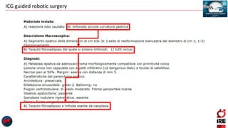
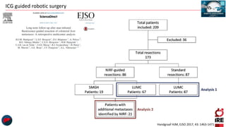
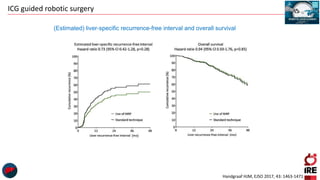
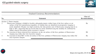
Gian Luca Grazi
Hepato-Biliary-Pancreatic Surgery
National Cancer Institute Regina Elena
Rome
Future of robotic surgery
ICG guided robotic surgery
(20 minutes)
ICG guided robotic surgery
HCC
214
Intrahepatic
Cholangiocarcinoma
79
Metastases
402
Perihilar
Cholangiocarcinoma
36
Others
93
Gallbladder
29
892 Liver Resections
(June 2010 – September 2022)
ICG guided robotic surgery
5
7
10
11
19
23
1
0
5
10
15
20
25
2011 2012 2013 2014 2015 2016 2017 2018 2019 2020 2021 2022 2023
#
procedures
Years
Per Year # of 76 Robotic Hepatectomies
ICG guided robotic surgery
HCC
38%
Metastases
18%
Other Cancers
4%
CCA
12%
Benign
28%
Diagnosis
Left lateral
sectionectomy
12%
Minor resections
42%
1-2 Segments
34%
Left hepatectomy
5%
Right hepatectomy
7%
Type of Resection
ICG guided robotic surgery
 One major drawback of minimally-invasive
surgery is the lack of haptic feedback, since
palpation with laparoscopic or robotic forceps is
limited.
 The surgeon must rely on his/her own visual
impressions, making parenchymal dissections
particularly problematic
ICG guided robotic surgery
• Indocyanine green (ICG) was approved by the Food and Drug Administration (FDA) in
1957 and has been used in various medical fields.
• Since the 1980s, ICG has been used to test liver function prior to hepatobiliary surgery.
• In this indication (LiMON test), ICG is administered intravenously days before surgery
and the blood concentration and ICG plasma disappearance rates are measured
noninvasively.
• In healthy liver tissue, ICG is fully excreted after 72 h and no remnants should be
detectable.
• In 2009 it was noticed ICG accumulation in hepatocellular carcinoma (HCC) and hepatic
metastasis (HM) of colorectal cancer up to 14 days after ICG application for liver
function evaluation
Brief History of Indocyanine Green
ICG guided robotic surgery
 In open surgery, additional near-infrared (NIR) cameras and/or monitors are
needed for ICG visualization, theater lights need to be switched off, and the
operating surgeon must remove their focus from the operation field while
performing crucial parts of the operation.
 In laparoscopic surgery, the NIR camera is integrated into some systems.
 The Firefly™camera (Intuitive, Sunnyvale, CA, USA) is integrated in the da
Vinci Surgical Systems (Intuitive, Sunnyvale, CA, USA) and can easily be used
to intraoperatively visualize ICG accumulation.
Usage in Surgery
ICG guided robotic surgery
Indocyanine Green Fluorescence Navigation in Liver Surgery
A Systematic Review on Dose and Timing of Administration
Wakabayashi T, Ann Surg 2022;275:1025–1034
ICG guided robotic surgery
•Time of administration
•Dose to be given
•Impaired liver function (fibrosis or cirrhosis)
•Elderly patients with slower metabolism
Open Questions in the usage of Indocyanine Green
ICG guided robotic surgery
The reported depth penetration is
limited to a maximum of ≈ 8 mm
ICG guided robotic surgery
1.Study of the anatomy, mainly biliary
2.Detection of bile leaks
3.Guiding mini-invasive liver resections
4.Intra operative tumor staging
ICG guided robotic surgery
Wang X, Ann Surg 2021, 274: 97-106
ICG guided robotic surgery
Wang X, Ann Surg 2021, 274: 97-106
ICG guided robotic surgery
1.Study of the anatomy, mainly biliary
2.Detection of bile leaks
3.Guiding mini-invasive liver resections
4.Intra operative tumor staging
ICG guided robotic surgery
On POD3, the bilirubin levels in the drainage fluid were
significantly lower in the IO-G than in the CO-G. Bars indicate
the median values and interquartile ranges.
Bilirubin levels in serum and drainage effluent on the third postoperative day (POD3).
Hanaki T, Anticancer Res 2022,42:4787-4793
ICG guided robotic surgery
1.Study of the anatomy, mainly biliary
2.Detection of bile leaks
3.Guiding mini-invasive liver resections
4.Intra operative tumor staging
ICG guided robotic surgery
Ishizawa T, Arch Surg 2012, 147: 393-394
Positive staining Negative staining
The portal branch of the segment to be resected is
punctured with a needle.
The ICG dye (0.025 mg in 10 mL of normal saline) is
injected without clamping the hepatic artery.
The liver surface of segment to be resected starts to
fluoresce following the injection of the ICG dye.
The intensity of the fluorescence on the surface of
segment to be resected is highest 10 minutes after
injection, which allows clear differentiation between
segment to be resected and adjacent.
The positive-staining technique with ICG dye does not
require hepatic artery clamping.
The root of the portal pedicle of the segment to be
resected had to be temporarily clamped and the ICG dye
(2.5 mg in 1 mL of normal saline) is intravenously
injected.
All hepatic segments, except segment to be resected,
are clearly fluorescent 1 minute after injection.
The hepatic segments with maintained portal and
arterial blood flow are illuminated with ICG demarcating
the area to be resected.
Positive and Negative Staining of Hepatic Segments by Use of Fluorescent
Imaging Techniques During Laparoscopic Hepatectomy
ICG guided robotic surgery
Wang X, Ann Surg 2021, 274: 97-106
ICG guided robotic surgery
Indocyanine Green Fluorescence Navigation in Liver Surgery
A Systematic Review on Dose and Timing of Administration
Wakabayashi T, Ann Surg 2022;275:1025–1034
ICG guided robotic surgery
Chiow AKH, HPB 2021, 23: 475-482
Robotic ICG guided anatomical liver resection in a multicenter cohort: an
evolution from “positive staining” into “negative staining” method
ICG guided robotic surgery
• Robotic ICG guided hepatectomy technique for anatomical
liver resection is safe and feasible and has the potential
benefit for improving visualization of the demarcation line
especially in living donor liver graft harvest and cirrhotic
patients with minimal complications.
• The negative staining technique was easy to perform and
is recommended in major anatomical resections.
Chiow AKH, HPB 2021, 23: 475-482
ICG guided robotic surgery
Wang X, Ann Surg 2021, 274: 97-106
ICG guided robotic surgery
1.Study of the anatomy, mainly biliary
2.Detection of bile leaks
3.Guiding mini-invasive liver resections
4.Intra operative tumor staging
ICG guided robotic surgery
ASSOCIATING INTRAOPERATIVE INDOCYANINE
GREEN FLUORESCENCE IMAGING AND
ULTRASOUND TO DETECT MICROSCOPIC CANCER
LESIONS DURING HEPATIC SURGERY
Scarinci A, Di Filippo S, Palmieri A, Police A, Marcelli ME,
Diodoro MG, Grazi GL
Abstract – Oral presentation
8 patients
Evaluated with CT/MR + IOUS
• 4 HCC
• 2 METS
• 2 CCA
Additional resection
6 patients
Planned resection
2 patients
11 more nodules
7
Tumoral
nodules
4
non tumoral nodules
2 regenerative nodule
1 fat containing cell
1 cirrhosis
ICG guided robotic surgery
Boogerd LSF, Surg Endosc 2017, 31: 952-961
Sensitivity of all imaging modalities employed.
Sensitivity and positive predictive value of computed tomography (CT), magnetic resonance imaging (MRI), visual
inspection, laparoscopic ultrasonography (LUS), near-infrared fluorescence imaging (NIRF), and combination of LUS and
NIRF.
Twenty-two patients planned
to undergo laparoscopic
staging (n = 4) or resection (n
= 19) of one or multiple
tumors confined to the liver
were included from April 2013
to November 2015.
ICG guided robotic surgery
Peyrat P, J Surg Oncol 2018;117:922-927
ICG guided robotic surgery
Wang X, Ann Surg 2021, 274: 97-106
ICG guided robotic surgery
Fluorescence
patterns
Total all tumor tissue showed
uniform fluorescence
all well-differentiated
HCCs
the expression levels of portal uptake transporters of ICG were
well preserved, but functional or morphological biliary
excretion disorders were present, leading to retention of ICG in
cancerous tissues at the time of surgery, following
preoperative intravenous injection.
Partial some tumor tissues
showed fluorescence
Rim the cancer tissues were
negative for
fluorescence, but the
surrounding liver
parenchyma showed
fluorescence
poorly differentiated
HCCs
and CRLM
the portal uptake transporters were downregulated in
cancerous tissues but biliary excretion of ICG by surrounding
non-cancerous hepatic parenchyma was also disordered,
resulting in rim-type fluorescence.
The rim-type fluorescence signal in CRLM has been reported to
be caused by immature hepatocytes with decreased bile
excretion ability that surrounds the tumor
Ishizawa, HepatoBiliary Surg Nutr 2016, 5: 322-328
ICG guided robotic surgery
Liu T, Surgical Innovation 2022, 29:532–539
Fluorescent patterns of liver cancers on surgical specimens and their images and overlay
HCC
Fluorescent
Type
Overlay
Total fluorescent type, well-
differentiated HCC
Partial fluorescent type,
moderately differentiated
HCC with haemorrhagic
necrosis
Rim fluorescent type,
poorly differentiated HCC
ICG guided robotic surgery
Marino MV, HPB 2020, 22: 422-431
ICG guided robotic surgery
 Besides being highly user-dependent, IOUS has the
problem of not detecting lesions that are just below
the surface within the first cm of the liver.
 In contrast, NIR light can only penetrate up to 1 cm
into liver parenchyma and thereby fails to detect
deeper tumors.
 The combination of IOUS and ICG therefore seems to
increase the detection rate of hepatic metastasis
ICG guided robotic surgery
Handgraaf HJM, EJSO 2017, 43: 1463-1471
ICG guided robotic surgery
Handgraaf HJM, EJSO 2017, 43: 1463-1471
Additionally identified colorectal liver metastases
ICG guided robotic surgery
(Estimated) liver-specific recurrence-free interval and overall survival
Handgraaf HJM, EJSO 2017, 43: 1463-1471
ICG guided robotic surgery
Wang X, Ann Surg 2021, 274: 97-106
ICG guided robotic surgery
Future of robotic surgery
ICG guided robotic surgery
Future of robotic surgery
ICG guided robotic surgery
Progress of NIR-II Fluorescence Technology
NIR-II (1000–1700 nm) fluorescence imaging technology has a longer
emission wavelength than NIR-I (750–900 nm) and can significantly
diminish photon scattering within biological tissues and reduce tissue
autofluorescence and light absorption, leading to significant benefits in
terms of
 deeper detection,
 higher resolution, and
 fidelity.
Liu T, Surgical Innovation 2022, 29:532–539
ICG guided robotic surgery
• Patients with liver cancer were enrolled in the study, and then received
preoperative imaging examinations, including enhanced CT, MRI,
ultrasonography and PET.
• Before surgery, the patients were injected with ICG intravenously at a
dose of 0.5 mg kg−1 body weight as a routine preoperative liver function
test.
• One to seven days later, on the day of surgery, the patients received a
laparotomy.
• The liver surface was examined by the integrated NIR-I/II and visible
multispectral imaging instrument and visible and NIR-I/II images were
obtained.
• Tumours were resected by the guidance of ultrasonography and NIR-I
imaging.
• During the resection, NIR-II images were also acquired. After the
operation, visible and NIR-I/II images of the resected specimens were
obtained.
• Pathological examination of the resected tissues was conducted.
First-in-human liver-tumour surgery guided by multispectral fluorescence imaging in the
visible and near-infrared-I/II windows
Hu Z, Nat Biomed Eng 2020;4:259-271
ICG guided robotic surgery
Hu Z, Nat Biomed Eng 2020;4:259-271
Intraoperative NIR-I/II fluorescence image-guided tumour resection
For a typical patient
with HCC, guided by
ultrasonography and
the visible light image,
the tumour was
resected and thought
to be completely
removed on the basis
of the experience of
the surgeons.
NIR-II imaging detected
fluorescence signals in
the remaining tissue
sections.
NIR-I imaging
did not reveal
any signals
The fluorescent residual
tissues were further
resected and received
histopathological
examination to verify that
the tissues were HCC.
ICG guided robotic surgery
Compared with the current preoperative imaging modalities (ultrasonography, MRI,
CT) and intraoperative imaging techniques (ultrasonography), intraoperative ICG-
based NIR-I/II fluorescence imaging can detect the tumour lesions with no obvious
imaging characteristics.
After laparotomy, intraoperative NIR-I/II fluorescence imaging was able to detect
the lesions missed by preoperative imaging modalities, which can substantially
promote the accuracy of patient staging and management.
During surgery, NIR-I/II imaging can also identify residual lesions that are difficult to
be recognized by surgeons or intraoperative ultrasonography.
Moreover, our imaging study revealed that ICG distributed quite uniformly in well-
differentiated or moderately differentiated tumours, but partial-type and rim-type
distributions were found in the poorly differentiated HCCs, which is consistent with
previously reported findings.
Hu Z, Nat Biomed Eng 2020;4:259-271
ICG guided robotic surgery
Molecular imaging is currently a hot research field in the world.
ICG fluorescence imaging technology can
• accurately identify liver tumors and enable real-time surgical navigation to ensure complete tumor resection;
• aid in the determination of tumor differentiation, creating a new target for accurate hepatobiliary surgery.
ICG technology combined with tumor-targeting nanoparticles and the emergence of novel targeted probes greatly
overcome the limitations of ICG.
With probe improvement, the fluorescence imaging system can be combined with other clinical treatment or examination
methods to improve the treatment efficiency of liver cancer.
The rapid development of NIR-II fluorescence imaging is conducive to achieving a more accurate intraoperative navigation
system.
With the advent of precision medicine and the progress of various biotechnology methods, fluorescence imaging technology
will be better developed and applied in the
• diagnosis,
• surgical navigation, and
• treatment of liver cancer.
TAKE HOME MESSAGE
ICG guided robotic surgery
As image-guided surgery solutions are quite often technically challenging.
Ethics and regulations provide a healthy translational hurdle to protect patients, while financial aspects
may also constrain development.
Lead compounds and detection device prototypes must be developed and refined in research setting and
often not within the domain of patient care.
While this helps preventing the patient exposure to potentially harmful technologies, it also means that
some approaches can become more ‘technology-driven’ than ‘clinical need-driven’. All developments
should be done with clinical translation in mind and based on real-life unmet surgical needs.
It is extremely challenging to translate laboratory findings to the clinic.
Most chemical and engineering efforts still find applications in multiple settings. The success stories in
the field of image-guided surgery are based on technologies that maximally align with innovations made
in other fields. For example, initial work on fluorescence laparoscopy presented in prostate cancer
surgery was later transferred to breast surgery and the technique is now also implemented during i.e.,
laparoscopic surgery.
FINAL CONSIDERATIONS
ICG guided robotic surgery
Gian Luca Grazi
Hepato Biliary Pancreatic Surgery
National Cancer Institute “Regina Elena”, Rome, Italy
gianluca.grazi@ifo.it
www.chirurgiadelfegato.it

ICG guided robotic liver surgery

  • 1.
    Gian Luca Grazi Hepato-Biliary-PancreaticSurgery National Cancer Institute Regina Elena Rome Future of robotic surgery ICG guided robotic surgery (20 minutes)
  • 2.
    ICG guided roboticsurgery HCC 214 Intrahepatic Cholangiocarcinoma 79 Metastases 402 Perihilar Cholangiocarcinoma 36 Others 93 Gallbladder 29 892 Liver Resections (June 2010 – September 2022)
  • 3.
    ICG guided roboticsurgery 5 7 10 11 19 23 1 0 5 10 15 20 25 2011 2012 2013 2014 2015 2016 2017 2018 2019 2020 2021 2022 2023 # procedures Years Per Year # of 76 Robotic Hepatectomies
  • 4.
  • 5.
    ICG guided roboticsurgery HCC 38% Metastases 18% Other Cancers 4% CCA 12% Benign 28% Diagnosis Left lateral sectionectomy 12% Minor resections 42% 1-2 Segments 34% Left hepatectomy 5% Right hepatectomy 7% Type of Resection
  • 6.
    ICG guided roboticsurgery  One major drawback of minimally-invasive surgery is the lack of haptic feedback, since palpation with laparoscopic or robotic forceps is limited.  The surgeon must rely on his/her own visual impressions, making parenchymal dissections particularly problematic
  • 7.
    ICG guided roboticsurgery • Indocyanine green (ICG) was approved by the Food and Drug Administration (FDA) in 1957 and has been used in various medical fields. • Since the 1980s, ICG has been used to test liver function prior to hepatobiliary surgery. • In this indication (LiMON test), ICG is administered intravenously days before surgery and the blood concentration and ICG plasma disappearance rates are measured noninvasively. • In healthy liver tissue, ICG is fully excreted after 72 h and no remnants should be detectable. • In 2009 it was noticed ICG accumulation in hepatocellular carcinoma (HCC) and hepatic metastasis (HM) of colorectal cancer up to 14 days after ICG application for liver function evaluation Brief History of Indocyanine Green
  • 8.
    ICG guided roboticsurgery  In open surgery, additional near-infrared (NIR) cameras and/or monitors are needed for ICG visualization, theater lights need to be switched off, and the operating surgeon must remove their focus from the operation field while performing crucial parts of the operation.  In laparoscopic surgery, the NIR camera is integrated into some systems.  The Firefly™camera (Intuitive, Sunnyvale, CA, USA) is integrated in the da Vinci Surgical Systems (Intuitive, Sunnyvale, CA, USA) and can easily be used to intraoperatively visualize ICG accumulation. Usage in Surgery
  • 9.
    ICG guided roboticsurgery Indocyanine Green Fluorescence Navigation in Liver Surgery A Systematic Review on Dose and Timing of Administration Wakabayashi T, Ann Surg 2022;275:1025–1034
  • 10.
    ICG guided roboticsurgery •Time of administration •Dose to be given •Impaired liver function (fibrosis or cirrhosis) •Elderly patients with slower metabolism Open Questions in the usage of Indocyanine Green
  • 11.
    ICG guided roboticsurgery The reported depth penetration is limited to a maximum of ≈ 8 mm
  • 12.
    ICG guided roboticsurgery 1.Study of the anatomy, mainly biliary 2.Detection of bile leaks 3.Guiding mini-invasive liver resections 4.Intra operative tumor staging
  • 13.
    ICG guided roboticsurgery Wang X, Ann Surg 2021, 274: 97-106
  • 14.
    ICG guided roboticsurgery Wang X, Ann Surg 2021, 274: 97-106
  • 15.
    ICG guided roboticsurgery 1.Study of the anatomy, mainly biliary 2.Detection of bile leaks 3.Guiding mini-invasive liver resections 4.Intra operative tumor staging
  • 16.
    ICG guided roboticsurgery On POD3, the bilirubin levels in the drainage fluid were significantly lower in the IO-G than in the CO-G. Bars indicate the median values and interquartile ranges. Bilirubin levels in serum and drainage effluent on the third postoperative day (POD3). Hanaki T, Anticancer Res 2022,42:4787-4793
  • 17.
    ICG guided roboticsurgery 1.Study of the anatomy, mainly biliary 2.Detection of bile leaks 3.Guiding mini-invasive liver resections 4.Intra operative tumor staging
  • 18.
    ICG guided roboticsurgery Ishizawa T, Arch Surg 2012, 147: 393-394 Positive staining Negative staining The portal branch of the segment to be resected is punctured with a needle. The ICG dye (0.025 mg in 10 mL of normal saline) is injected without clamping the hepatic artery. The liver surface of segment to be resected starts to fluoresce following the injection of the ICG dye. The intensity of the fluorescence on the surface of segment to be resected is highest 10 minutes after injection, which allows clear differentiation between segment to be resected and adjacent. The positive-staining technique with ICG dye does not require hepatic artery clamping. The root of the portal pedicle of the segment to be resected had to be temporarily clamped and the ICG dye (2.5 mg in 1 mL of normal saline) is intravenously injected. All hepatic segments, except segment to be resected, are clearly fluorescent 1 minute after injection. The hepatic segments with maintained portal and arterial blood flow are illuminated with ICG demarcating the area to be resected. Positive and Negative Staining of Hepatic Segments by Use of Fluorescent Imaging Techniques During Laparoscopic Hepatectomy
  • 19.
    ICG guided roboticsurgery Wang X, Ann Surg 2021, 274: 97-106
  • 20.
    ICG guided roboticsurgery Indocyanine Green Fluorescence Navigation in Liver Surgery A Systematic Review on Dose and Timing of Administration Wakabayashi T, Ann Surg 2022;275:1025–1034
  • 21.
  • 22.
  • 23.
    ICG guided roboticsurgery Chiow AKH, HPB 2021, 23: 475-482 Robotic ICG guided anatomical liver resection in a multicenter cohort: an evolution from “positive staining” into “negative staining” method
  • 24.
    ICG guided roboticsurgery • Robotic ICG guided hepatectomy technique for anatomical liver resection is safe and feasible and has the potential benefit for improving visualization of the demarcation line especially in living donor liver graft harvest and cirrhotic patients with minimal complications. • The negative staining technique was easy to perform and is recommended in major anatomical resections. Chiow AKH, HPB 2021, 23: 475-482
  • 25.
    ICG guided roboticsurgery Wang X, Ann Surg 2021, 274: 97-106
  • 26.
    ICG guided roboticsurgery 1.Study of the anatomy, mainly biliary 2.Detection of bile leaks 3.Guiding mini-invasive liver resections 4.Intra operative tumor staging
  • 27.
  • 28.
  • 29.
    ICG guided roboticsurgery ASSOCIATING INTRAOPERATIVE INDOCYANINE GREEN FLUORESCENCE IMAGING AND ULTRASOUND TO DETECT MICROSCOPIC CANCER LESIONS DURING HEPATIC SURGERY Scarinci A, Di Filippo S, Palmieri A, Police A, Marcelli ME, Diodoro MG, Grazi GL Abstract – Oral presentation 8 patients Evaluated with CT/MR + IOUS • 4 HCC • 2 METS • 2 CCA Additional resection 6 patients Planned resection 2 patients 11 more nodules 7 Tumoral nodules 4 non tumoral nodules 2 regenerative nodule 1 fat containing cell 1 cirrhosis
  • 30.
    ICG guided roboticsurgery Boogerd LSF, Surg Endosc 2017, 31: 952-961 Sensitivity of all imaging modalities employed. Sensitivity and positive predictive value of computed tomography (CT), magnetic resonance imaging (MRI), visual inspection, laparoscopic ultrasonography (LUS), near-infrared fluorescence imaging (NIRF), and combination of LUS and NIRF. Twenty-two patients planned to undergo laparoscopic staging (n = 4) or resection (n = 19) of one or multiple tumors confined to the liver were included from April 2013 to November 2015.
  • 31.
    ICG guided roboticsurgery Peyrat P, J Surg Oncol 2018;117:922-927
  • 32.
    ICG guided roboticsurgery Wang X, Ann Surg 2021, 274: 97-106
  • 33.
    ICG guided roboticsurgery Fluorescence patterns Total all tumor tissue showed uniform fluorescence all well-differentiated HCCs the expression levels of portal uptake transporters of ICG were well preserved, but functional or morphological biliary excretion disorders were present, leading to retention of ICG in cancerous tissues at the time of surgery, following preoperative intravenous injection. Partial some tumor tissues showed fluorescence Rim the cancer tissues were negative for fluorescence, but the surrounding liver parenchyma showed fluorescence poorly differentiated HCCs and CRLM the portal uptake transporters were downregulated in cancerous tissues but biliary excretion of ICG by surrounding non-cancerous hepatic parenchyma was also disordered, resulting in rim-type fluorescence. The rim-type fluorescence signal in CRLM has been reported to be caused by immature hepatocytes with decreased bile excretion ability that surrounds the tumor Ishizawa, HepatoBiliary Surg Nutr 2016, 5: 322-328
  • 34.
    ICG guided roboticsurgery Liu T, Surgical Innovation 2022, 29:532–539 Fluorescent patterns of liver cancers on surgical specimens and their images and overlay HCC Fluorescent Type Overlay Total fluorescent type, well- differentiated HCC Partial fluorescent type, moderately differentiated HCC with haemorrhagic necrosis Rim fluorescent type, poorly differentiated HCC
  • 35.
    ICG guided roboticsurgery Marino MV, HPB 2020, 22: 422-431
  • 36.
    ICG guided roboticsurgery  Besides being highly user-dependent, IOUS has the problem of not detecting lesions that are just below the surface within the first cm of the liver.  In contrast, NIR light can only penetrate up to 1 cm into liver parenchyma and thereby fails to detect deeper tumors.  The combination of IOUS and ICG therefore seems to increase the detection rate of hepatic metastasis
  • 37.
  • 38.
  • 39.
  • 40.
    ICG guided roboticsurgery Handgraaf HJM, EJSO 2017, 43: 1463-1471
  • 41.
    ICG guided roboticsurgery Handgraaf HJM, EJSO 2017, 43: 1463-1471 Additionally identified colorectal liver metastases
  • 42.
    ICG guided roboticsurgery (Estimated) liver-specific recurrence-free interval and overall survival Handgraaf HJM, EJSO 2017, 43: 1463-1471
  • 43.
    ICG guided roboticsurgery Wang X, Ann Surg 2021, 274: 97-106
  • 44.
    ICG guided roboticsurgery Future of robotic surgery ICG guided robotic surgery Future of robotic surgery ICG guided robotic surgery
  • 45.
    ICG guided roboticsurgery Progress of NIR-II Fluorescence Technology NIR-II (1000–1700 nm) fluorescence imaging technology has a longer emission wavelength than NIR-I (750–900 nm) and can significantly diminish photon scattering within biological tissues and reduce tissue autofluorescence and light absorption, leading to significant benefits in terms of  deeper detection,  higher resolution, and  fidelity. Liu T, Surgical Innovation 2022, 29:532–539
  • 46.
    ICG guided roboticsurgery • Patients with liver cancer were enrolled in the study, and then received preoperative imaging examinations, including enhanced CT, MRI, ultrasonography and PET. • Before surgery, the patients were injected with ICG intravenously at a dose of 0.5 mg kg−1 body weight as a routine preoperative liver function test. • One to seven days later, on the day of surgery, the patients received a laparotomy. • The liver surface was examined by the integrated NIR-I/II and visible multispectral imaging instrument and visible and NIR-I/II images were obtained. • Tumours were resected by the guidance of ultrasonography and NIR-I imaging. • During the resection, NIR-II images were also acquired. After the operation, visible and NIR-I/II images of the resected specimens were obtained. • Pathological examination of the resected tissues was conducted. First-in-human liver-tumour surgery guided by multispectral fluorescence imaging in the visible and near-infrared-I/II windows Hu Z, Nat Biomed Eng 2020;4:259-271
  • 47.
    ICG guided roboticsurgery Hu Z, Nat Biomed Eng 2020;4:259-271 Intraoperative NIR-I/II fluorescence image-guided tumour resection For a typical patient with HCC, guided by ultrasonography and the visible light image, the tumour was resected and thought to be completely removed on the basis of the experience of the surgeons. NIR-II imaging detected fluorescence signals in the remaining tissue sections. NIR-I imaging did not reveal any signals The fluorescent residual tissues were further resected and received histopathological examination to verify that the tissues were HCC.
  • 48.
    ICG guided roboticsurgery Compared with the current preoperative imaging modalities (ultrasonography, MRI, CT) and intraoperative imaging techniques (ultrasonography), intraoperative ICG- based NIR-I/II fluorescence imaging can detect the tumour lesions with no obvious imaging characteristics. After laparotomy, intraoperative NIR-I/II fluorescence imaging was able to detect the lesions missed by preoperative imaging modalities, which can substantially promote the accuracy of patient staging and management. During surgery, NIR-I/II imaging can also identify residual lesions that are difficult to be recognized by surgeons or intraoperative ultrasonography. Moreover, our imaging study revealed that ICG distributed quite uniformly in well- differentiated or moderately differentiated tumours, but partial-type and rim-type distributions were found in the poorly differentiated HCCs, which is consistent with previously reported findings. Hu Z, Nat Biomed Eng 2020;4:259-271
  • 49.
  • 50.
    ICG guided roboticsurgery Molecular imaging is currently a hot research field in the world. ICG fluorescence imaging technology can • accurately identify liver tumors and enable real-time surgical navigation to ensure complete tumor resection; • aid in the determination of tumor differentiation, creating a new target for accurate hepatobiliary surgery. ICG technology combined with tumor-targeting nanoparticles and the emergence of novel targeted probes greatly overcome the limitations of ICG. With probe improvement, the fluorescence imaging system can be combined with other clinical treatment or examination methods to improve the treatment efficiency of liver cancer. The rapid development of NIR-II fluorescence imaging is conducive to achieving a more accurate intraoperative navigation system. With the advent of precision medicine and the progress of various biotechnology methods, fluorescence imaging technology will be better developed and applied in the • diagnosis, • surgical navigation, and • treatment of liver cancer. TAKE HOME MESSAGE
  • 51.
    ICG guided roboticsurgery As image-guided surgery solutions are quite often technically challenging. Ethics and regulations provide a healthy translational hurdle to protect patients, while financial aspects may also constrain development. Lead compounds and detection device prototypes must be developed and refined in research setting and often not within the domain of patient care. While this helps preventing the patient exposure to potentially harmful technologies, it also means that some approaches can become more ‘technology-driven’ than ‘clinical need-driven’. All developments should be done with clinical translation in mind and based on real-life unmet surgical needs. It is extremely challenging to translate laboratory findings to the clinic. Most chemical and engineering efforts still find applications in multiple settings. The success stories in the field of image-guided surgery are based on technologies that maximally align with innovations made in other fields. For example, initial work on fluorescence laparoscopy presented in prostate cancer surgery was later transferred to breast surgery and the technique is now also implemented during i.e., laparoscopic surgery. FINAL CONSIDERATIONS
  • 52.
    ICG guided roboticsurgery Gian Luca Grazi Hepato Biliary Pancreatic Surgery National Cancer Institute “Regina Elena”, Rome, Italy gianluca.grazi@ifo.it www.chirurgiadelfegato.it