
Recommended
More Related Content
Similar to Heterozygous 8q24.3 Duplication in a Neonate with Jarcho-Levin Syndrome Presenting as Severe Scoliosis, Respiratory Distress, Spondylo-Costal Abnormality, Two Vessel Umbilical Cord and Mild Bilateral Hydronephrosis
Similar to Heterozygous 8q24.3 Duplication in a Neonate with Jarcho-Levin Syndrome Presenting as Severe Scoliosis, Respiratory Distress, Spondylo-Costal Abnormality, Two Vessel Umbilical Cord and Mild Bilateral Hydronephrosis (20)
More from semualkaira
More from semualkaira (20)
Recently uploaded
Recently uploaded (20)
Heterozygous 8q24.3 Duplication in a Neonate with Jarcho-Levin Syndrome Presenting as Severe Scoliosis, Respiratory Distress, Spondylo-Costal Abnormality, Two Vessel Umbilical Cord and Mild Bilateral Hydronephrosis
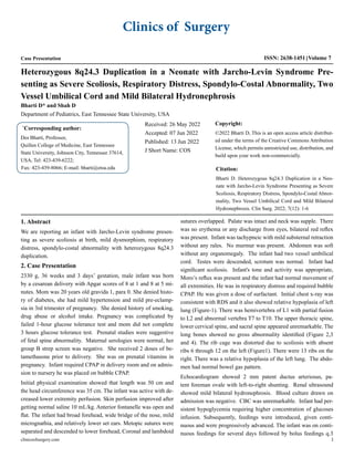
- 1. Bharti D* and Shah D Department of Pediatrics, East Tennessee State University, USA * Corresponding author: Des Bharti, Professor, Quillen College of Medicine, East Tennessee State University, Johnson City, Tennessee 37614, USA, Tel: 423-439-6222; Fax: 423-439-8066; E-mail: bharti@etsu.edu Received: 26 May 2022 Accepted: 07 Jun 2022 Published: 13 Jun 2022 J Short Name: COS Copyright: ©2022 Bharti D, This is an open access article distribut- ed under the terms of the Creative Commons Attribution License, which permits unrestricted use, distribution, and build upon your work non-commercially. Citation: Bharti D. Heterozygous 8q24.3 Duplication in a Neo- nate with Jarcho-Levin Syndrome Presenting as Severe Scoliosis, Respiratory Distress, Spondylo-Costal Abnor- mality, Two Vessel Umbilical Cord and Mild Bilateral Hydronephrosis. Clin Surg. 2022; 7(12): 1-6 Heterozygous 8q24.3 Duplication in a Neonate with Jarcho-Levin Syndrome Pre- senting as Severe Scoliosis, Respiratory Distress, Spondylo-Costal Abnormality, Two Vessel Umbilical Cord and Mild Bilateral Hydronephrosis Clinics of Surgery Case Presentation ISSN: 2638-1451 Volume 7 clinicsofsurgery.com 1 1. Abstract We are reporting an infant with Jarcho-Levin syndrome presen- ting as severe scoliosis at birth, mild dysmorphism, respiratory distress, spondylo-costal abnormality with heterozygous 8q24.3 duplication. 2. Case Presentation 2330 g, 36 weeks and 3 days’ gestation, male infant was born by a cesarean delivery with Apgar scores of 8 at 1 and 8 at 5 mi- nutes. Mom was 20 years old gravida 1, para 0. She denied histo- ry of diabetes, she had mild hypertension and mild pre-eclamp- sia in 3rd trimester of pregnancy. She denied history of smoking, drug abuse or alcohol intake. Pregnancy was complicated by failed 1-hour glucose tolerance test and mom did not complete 3 hours glucose tolerance test. Prenatal studies were suggestive of fetal spine abnormality. Maternal serologies were normal, her group B strep screen was negative. She received 2 doses of be- tamethasone prior to delivery. She was on prenatal vitamins in pregnancy. Infant required CPAP in delivery room and on admis- sion to nursery he was placed on bubble CPAP. Initial physical examination showed that length was 50 cm and the head circumference was 35 cm. The infant was active with de- creased lower extremity perfusion. Skin perfusion improved after getting normal saline 10 mL/kg. Anterior fontanelle was open and flat. The infant had broad forehead, wide bridge of the nose, mild micrognathia, and relatively lower set ears. Metopic sutures were separated and descended to lower forehead, Coronal and lambdoid sutures overlapped. Palate was intact and neck was supple. There was no erythema or any discharge from eyes, bilateral red reflex was present. Infant was tachypneic with mild substernal retraction without any rales. No murmur was present. Abdomen was soft without any organomegaly. The infant had two vessel umbilical cord. Testes were descended, scrotum was normal. Infant had significant scoliosis. Infant's tone and activity was appropriate, Moro’s reflux was present and the infant had normal movement of all extremities. He was in respiratory distress and required bubble CPAP. He was given a dose of surfactant. Initial chest x-ray was consistent with RDS and it also showed relative hypoplasia of left lung (Figure-1). There was hemivertebra of L1 with partial fusion to L2 and abnormal vertebra T7 to T10. The upper thoracic spine, lower cervical spine, and sacral spine appeared unremarkable. The long bones showed no gross abnormality identified (Figure 2,3 and 4). The rib cage was distorted due to scoliosis with absent ribs 6 through 12 on the left (Figure1). There were 13 ribs on the right. There was a relative hypoplasia of the left lung. The abdo- men had normal bowel gas pattern. Echocardiogram showed 2 mm patent ductus arteriosus, pa- tent foreman ovale with left-to-right shunting. Renal ultrasound showed mild bilateral hydronephrosis. Blood culture drawn on admission was negative. CBC was unremarkable. Infant had per- sistent hypoglycemia requiring higher concentration of glucoses infusion. Subsequently, feedings were introduced, given conti- nuous and were progressively advanced. The infant was on conti- nuous feedings for several days followed by bolus feedings q.3
- 2. clinicsofsurgery.com 2 Volume 7 Issue 11-2022 Case Presentation hours. Initial insulin level was 4.2 mIU/ml which was thought to be elevated in the settings of hypoglycemia, cortisol was 4.9 mcg/ ml, TSH was 6.49 mIU/ml and free T4 was 3. 3 ug/dl. Blood su- gars stabilized by day 9 of life. At that time total parenteral nutri- tion was discontinued and infant was weaned off IV fluids. Fee- dings at this point had been changed to q.3 hours’ feedings. He was discharged home on Enfamil neuropro 60 mL q.3 hours. The endocrinologists were of the opinion that the infant had transient hypoglycemia of unknown significance. There was a mild thrombocytopenia with a platelet count of 50,000 per mL on admission. Maternal platelets were normal. Mom had hypertension/preeclampsia that probably contributed to mild thrombocytopenia. Platelet count prior to discharge was 209,000/ ml. Urine for CMV was negative. While the infant was admitted in the neonatal ICU and when he was 2 weeks of age, his mom tested positive for COVID with mild symptoms. Infant's CO- VID test was negative. Infant was on isolation precautions for 1 week. The infant required antibiotics for less than 36 hours pending a report of blood culture. Microarray analysis showed a single copy number increase of 110.77 kb involving chromosome 8q24.3. Results represented a male with a heterozygous 8q24.3 duplications. Skeletal survey showed marked thoracic congenital vertebral body anomalies and chest wall deformity with resultant congenital scoliosis. No additional bony abnormality was noted. No definite definable bony dysplasia was identified from the stu- dy. Initially, we were considering possibilities of developmental defect, maternal exposure, or chromosomal abnormality. Karyo- type was reported normal. Skeletal disorders panel and Kabuki syndrome panel showed C2CD3 heterozygous, HSPG2 hetero- zygous, TCTN3 heterozygous, and TUBGCP6 heterozygous. All of these were of unclear significance. Head ultrasound did not show any abnormality. MRI of the head was negative for Chari malfor- mation. Spinal cord MRI is recommended after discharge to rule out a possibility of myelodysplasia. Ophthalmology consultation reported normal eye examination. Based on the clinical picture we considered a diagnosis of Jarcho-Levin syndrome with spon- dylo-costal dysplasia. Arrangement was made to have infant to get follow-up by Shriners Children Hospital for further investiga- tions of the spine and spinal cord and corrective surgical proce- dures. Genetic consultation was suggested to the parents. Figure 1: Hemivertebra with severe scoliosis: There was hemivertebra of L1 with partial fusion to L2. Abnormal vertebra T7-T10. 5 ribs on the left and 13 ribs on the right.
- 3. clinicsofsurgery.com 3 Volume 7 Issue 11-2022 Case Presentation Figures 2: Left arm. No abnormality noted Figure 3: Left foot. No abnormality noted
- 4. clinicsofsurgery.com 4 Volume 7 Issue 11-2022 Case Presentation Figure 4: Left hand. No abnormality noted 3. Case Discussion Congenital scoliosis is a malformation of the spine. It may be due to failure of development, failure of segmentation, or a combina- tion. The extent of the vertebral involvement and associated rib abnormalities will determine the severity of the disease manifesta- tion. Congenital scoliosis is usually progressive and, if untreated, results in severe spinal deformity. The ultimate severity of the curve depends on both the type of anomaly and the site at which it oc- curs. The most progressive type is associated with hemi-vertebrae on the contralateral side with or without associated fused ribs. It could be painless in earlier phases and may not even have signifi- cant respiratory distress associated with. With progression of the scoliosis, there may be associated pain and deformity and a risk of restrictive lung disease. Cardiorespiratory failure may eventually occur with a potential for neurological involvement that may in- clude Chiari malformation, tethered cord syndrome, paresthesia, and motor weakness. There may be a delay in acquisition of mo- tor milestones. There may be associated torticollis. It is important to make sure there is no associated abnormality of the kidneys, heart or other organs. Presence of midline lesions like sacral di- mples, hair patches, or skin discoloration may indicate associated neurological conditions. Neurological findings on examination may include clubfeet, pes cavus, asymmetry of the lower leg or foot. There could be potentially an associated myelomeningocele [1]. If the disc space above and below the hemivertebra are normal it allows near-normal longitudinal growth. An absence of a por- tion of the vertebral body and growth plates on the side of the unformed vertebra results in limited growth potential. Associat- ed myelodysplasia occurs in 15% to 20% of cases of scoliosis or kyphosis manifesting as hydrosyringomyelia, diastematomye- lia, neurenteric cyst, or a tethered cord. In childhood other caus- es of kyphoscoliosis are abnormal posture, Scheuermann disease, neuromuscular disorder, trauma, inflammation, surgery, radiation therapy, metabolic disorders, chondrodysplasia, arthritis, and tu- mors [2]. Diastematomyelia is believed to be the most common associated defect. It is a result of sagittal split in the spinal cord or cauda equina by a bony or cartilage spur extending posteriorly from the vertebral body. Abnormalities of the gastrointestinal system can also be seenwith VATER syndrome with esophageal atresia and imperforate in ad- dition to vertebral defects and radial and renal dysplasia [3]. Devel- opmental scoliosis can be seen in patients with neurofibromatosis, Marfan's syndrome, Ehlers-Danlos syndrome, and mucopolysac-
- 5. clinicsofsurgery.com 5 Volume 7 Issue 11-2022 Case Presentation charidoses, in whom additional clinical and rheumatologic stig- mata may be encountered. Neurofibromatosis type 1 is associated with scoliosis presenting as most common skeletal abnormality. Bonaglia and associates reported duplication of chromosome 8q24.3 and severe mental retardation and epilepsy detected by standard karyotype. 2.3 Mb inverted duplication of 8q24.3 with- out apparently associated deletion was associated with profound psychomotor retardation, idiopathic epilepsy and growth delay [4]. A retrospective study of patients with spondylo-costal dysostosis revealed that anomalies involved the thoracic region in all cases; many also involved the cervical spine; most patients had equal or more than four vertebral anomalies; frequent vertebral anomalies were butterfly vertebra, hemivertebra, complete block, and unilat- eral bar, which were associated with both rib absence and fusion; short stature was not always present at birth [5]. The Jarcho-Levin syndrome is a condition manifested by vertebral body and rib malformations. There are two major subtypes spon- dylo-costal dysostosis and spondylo-thoracic dysostosis. Patients with spondylo-thoracic dysostosis have vertebral body malforma- tions and ribs which flare in a fanlike pattern but which are not sig- nificantly malformed. This is an autosomal recessive trait, and the patients have a higher mortality rate and greater incidence of neu- ral tube defects. Individuals with spondylo-costal dysostosis have vertebral malformations, frequent dramatic rib malformations, and short stature, but do not have a fanlike thoracic configuration. Most cases of spondylo-costal dysostosis are inherited in an auto- somal recessive fashion. There have been few autosomal dominant reports with better survivals. Respiratory compromise was previ- ously accounted for the high mortality in these conditions. With recent technological advances in respiratory management more and more of these patients are surviving [6]. Concolino et al. described a “pure” de novo duplication of chro- mosome 8q22.2-8q24.3 and suggested that mental retardation and facial dysmorphism such as hypertelorism, microretrognathia, and telecanthus are key features of 8q22.2-q24.3 duplication syn- drome [7]. Most of them have growth retardation and short stat- ure. Main facial features were prominent forehead, hypertelorism, large nasal bridge and low-set ears. Other associations could be microretrognathia, anteverted nares, broad mouth, and long phil- trum. There may be associated abnormalities that include congen- ital heart disease, anal atresia, cleft lip and palate, and cryptorchi- dism. Hilger et al. found that a male patient of VACTERL associa- tion who had a 120 kb microduplication of 8q24.3 [8].His pheno- types included butterfly vertebrae, anal atresia, congenital subval- vularaortic stenosis and ventricular septal defect, esophageal atre- sia,high-grade bilateral vesico-ureteral reflux, and bilateral crypt- orchidism. Velinov et al [9] found a female infant with hypoplastic left heart with aortic atresia and hypoplastic aortic arch, ventricu- lar septal defect, and a nonrestrictive atrial communication. Cioc- ca et al reported a syndromic female newborn of 21q22.3 deletion with severe stenosis of the aortic valve and ascending aorta, mitral valve atresia, and hypoplastic left ventricle, who died of cardiopul- monary insufficiency after birth [10]. Kabuki syndrome is a multi-system disorder presenting at birth with symptoms pertaining to facial dysmorphism, skeletal ab- normalities, short stature, cardiac defects and intellectual delay, microcephaly. Others have reported seizures, hypotonia, nystag- mus, cleft lip and cleft palate and abnormal dentition. Skeletal ab- normalities may include a clinodactyly, spine abnormalities, and joint dislocations. Facial features show high arched eyebrows; hypertelorism with upwards slant; long and thick eyelashes; blue sclerae; prominent ears; downward slanting corners of the mouth; and a depressed tip of the nose [11]. Kabuki syndrome is most often caused by a mutation in the KMT2D gene, and is usually autosomal dominant [12]. Some cases are due to a mutation in the KDM6A gene and are inherited in an X-linked dominant manner. Treatment is focused on the associated cardiac and skeletal abnor- malities. Jarcho-Levin syndrome is an uncommon condition presenting in the neonate. Congenital severe scoliosis and spondylo-costal ab- normalities are commonly associated with this disorder. The dis- order is usually autosomal recessive and generally has a better out- come. A follow-up and management of severe congenital scoliosis requires multidisciplinary care. Since congenital scoliosis can be associated with genitourinary, heart, kidney and spina bifida dis- orders, these infants need to be investigated carefully. Multi stage repair of the spine may require spinal fusion, spinal osteotomy or vertebrectomy in severe cases. References 1. Shaffrey CI, Wiggins GC. Anatomy and Physiology of Congenital Spinal Lesions, in Spine Surgery 3rd Ed, 2005. 2. Major Congenital Orthopedic Deformities, Kosmas Kayes, Wil- liam Didelot, in Pediatric Surgery. 7th ED, 2012. 3. Orphanet J Rare Dis. 2011; 6: 56. 4. Bonaglia MC,Giorda R.A2.3 Mb duplication of chromosome 8q24.3 associated with severe mental retardation and epilepsy detected by standard karyotype. Eur J Hum Genet. 2005; 13(5): 586-591. 5. Takikawa K, Haga N. Spine and rib abnormalities and stature in spondylocostal dysostosis. Spine (Phila Pa 1976). 2006; 31(7): E192-197. 6. Karnes PS, Berry SA. Jarcho-Levin syndrome: four new cases and classification of subtypes. Am J Med Genet. 1991; 40(3): 264-270. 7. Concolino D, Iembo MA, Moricca MT, Rapsomaniki M, Marotta R, Galesi O, et al. A de novo 8q22.2-24.3 duplication in a patient with mild phenotype. Eur J Med Genet. 2012; 55(1): 67-70.
- 6. clinicsofsurgery.com 6 Volume 7 Issue 11-2022 Case Presentation 8. Hilger A, Schramm C, Draaken M. Familial occurrence of the vater/ vacterl association. PediatrSurg Int. 2012; 28(7): 725-729. 9. Velinov M, Gu H. Hypoplastic left heart in a female infant with par- tial trisomy 4q due to de novo 4;21 translocation American journal of medical genetics. 2002; 107(4): 330-333. 10. 10.Ciocca L, Digilio MC, Lombardo A, D’Elia G, Baban A, Capoli- no R, et al. Hypoplastic left heart syndrome and 21q22.3 deletion. Am J Med Genet A. 2015; 167A(3): 579-586. 11. Adam M. Kabuki syndrome. Orphanet. 2012. 12. Kabuki syndrome. Genetics Home Reference.2013.