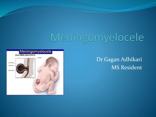
SlideShare a Scribd company logo
1 of 47
Dr Gagan Adhikari
MS Resident
Embryology
 NTDs are the results of an abnormality in the process of neurulation,
 conversion of the neural plate into a neural tube by a process of
folding,
 which occurs in the fourth week.
 Failure of part of the neural tube to close disrupts both
 The differentiation of the central nervous system
 and the induction of the vertebral arches
 which can result in a number of developmental anomalies.
Embryology
Embryology
Malformations usually involve part of the cranial or
caudal neuropore,
• the unfused anterior/rostral neural folds
results in a defect of the cranial regions
• and unfused posterior/caudal neural folds
results in defect in lower lumbar and sacral
regions
Cranial and caudal neuropore close on day 24 and on
day 26 respectively (Larsen 1993).
History
 The first reports of fetuses and infants with anencephaly, myelomeningocele,
and craniorachischisis originate in ancient Egypt
 Caspar Baulinin is credited with the first accurate description of spina bifida in
the early seventeenth century (Morgagni 1762).
 The term “spina dorsi bifida” was coined by Nicholas Talpius (Tulp) in 1641
(Doran and Guthkelch 1961; Tulp 1672)
 Virchow introduced the term “spina bifida occulta” in 1875 (Virchow 1875).
 In 1963, Sharrard proposed emergency operative closure of the back lesion to
decrease mortality and improve muscle function (Sharrard et al. 1967).
History
 The association of hydrocephalus and spina bifida was recognized by
Morgagni in 1761
 attributed bladder, rectal, and limb abnormalities to the neuronal damage
in the defective spinal cord (Morgagni 1762).
 Lorber reviewed 524 cases of myelomeningocele treated actively and
concluded that there were four main criteria associated with a poor
prognosis:
 gross hydrocephalus,
 severe paraplegia,
 kyphosis,
 associated gross congenital anomalies, or major birth injury (Lorber 1971).
Neural tube defects
Introduction: Meningomyelocele
 The most common open neural tube defect.
 It is characterized by failure of the neural tube to close in the
lumbosacral region during embryonic development
 leading to the herniation of the meninges and spinal cord through a
vertebral defect
 The neural tube fusion starts at the level of the hindbrain (medulla
and pons) and progresses rostrally and caudally.
 Incomplete fusion caudally leads to the formation of
meningomyelocele around day 26 of gestation
Copp AJ, Adzick NS, Chitty LS, Fletcher JM, Holmbeck GN, Shaw GM. Spina bifida. Nat Rev Dis
Primers. 2015 Apr 30;1:15007.
Brody BA, Kinney HC, Kloman AS, Gilles FH. Sequence of central nervous system myelination in human
infancy. I. An autopsy study of myelination. J Neuropathol Exp Neurol. 1987 May;46(3):283-301.
Etiology
 Most occur sporadically. however, several risk factors have been linked
 Chromosomal and genetic conditions:
 Parent or sibling with neural tube defect
 Trisomies 18 and 13
 Meckel-Gruber syndrome ( renal cysts, neural tube defects, and
polydactyly).
 Roberts, Jarcho-Levin (present with multiple defects in the spine and fan-
like ribs anomalies)
 HARD syndrome (Hydrocephalus, agyria and retinal dysplasia),
 VACTERAL and VATER associations
 X-linked neural tube defects, among others.
Sepulveda W, Corral E, Ayala C, Be C, Gutierrez J, Vasquez P. Chromosomal abnormalities in
fetuses with open neural tube defects: prenatal identification with ultrasound. Ultrasound Obstet
Gynecol. 2004 Apr;23(4):352-6.
Etiology
 Incidence is 1–2/1000 live births (0.1– 0.2%).
 Risk increases to 2–3% if there is one previous birth with MM,
 and 6–8% after two affected children.
 The risk is also increased in families where close relatives (e.g.
siblings) have given birth to MM children, especially when on the
mother’s side of the family.
 Transmission follows non-Mendelian genetics, and is probably
multifactorial.
 Prenatal folate (in the form of folic acid) lowers the incidence of MM
Au KS, Ashley-Koch A, Northrup H. Epidemiologic and genetic aspects of spina bifida and other neural
tube defects. Dev Disabil Res Rev. 2010;16(1):6-15.
Shimoji K, Kimura T, Kondo A, Tange Y, Miyajima M, Arai H. Genetic studies of
myelomeningocele. Childs Nerv Syst. 2013 Sep;29(9):1417-25.
Etiology
 Maternal environmental factors and exposure:
 alcohol use, caffeine intake,
 smoking, air pollution,
 disinfectant byproducts in drinking water,
 exposure to organic solvents, pesticides, nitrate-related compounds,
polycyclic aromatic hydrocarbons,
 maternal fever or hyperthermia ( especially in the first trimester)
from febrile illness or external sources like sauna, hot tub.
 Maternal medical conditions:
 elevated glycemic index, and gestational diabetes mellitus
 infections,
 obesity,
 and stress
Etiology
 Amniotic bands
 that disrupt normal neural tube formation.
 Maternal nutritional deficiencies
 folate, methionine, zinc, vitamin C, vitamin B12, and choline.
 Maternal medications:
 various folic acid antagonists like valproic acid, carbamazepine, and
methotrexate.
Pathophysiology
 Failure of the closure of the neural tube leads to exposure of the neural
tube to amniotic fluid.
 Although the neuroepithelium, neuronal differentiation, and function
develop normally in the beginning,
 these neurons die over time because of toxicity from exposure to
amniotic fluid.
 The failed neural tube closure and neurodegeneration in utero is
described as a "Two-hit" process.
Greene ND, Massa V, Copp AJ. Understanding the causes and prevention of neural tube defects:
Insights from the splotch mouse model. Birth Defects Res A Clin Mol Teratol. 2009 Apr;85(4):322-30
Hydrocephalus in myelomeningocele
 Hydrocephalus (HCP) develops in 65–85% of patients with MM, and 5–
10% of MM patients have clinically overt HCP at birth.
 Over 80% of MM patients who will develop HCP do so before age 6
mos.
 Most MM patients will have an associated Chiari type 2 malformation.
 Closure of the MM defect may convert a latent HCP to active HCP by
eliminating a route of egress of CSF.
Stein SC, Schut L. Hydrocephalus in Myelomeningocele Childs Brain. 1979; 5:413–419
Latex allergy in myelomeningocele
 Up to 73% of MM patients are allergic to proteins present in latex (the
milky sap from the rubber tree Hevea brasiliensis),
 found only in naturally occurring rubber products
 which are not present in synthetics such as: silicone, vinyl, plastic,
neoprene, nitrile
 The allergy is thought to arise from early and frequent exposure to latex
products during medical care
 There is a suggestion that latex-free surgery on these infants may
reduce the risk of the development of latex allergy.
Cremer R, Kleine-Diepenbruck U, Hoppe A, et al. Latex allergy in spina bifida patients–prevention by
primary prophylaxis. Allergy. 1998; 53:709–711
History and examination
The diagnosis in a newborn is usually
apparent because of the grossly visible
lesion in the back.
Protruding membrane-covered sac-
containing meninges, cerebrospinal
fluid (CSF), and nerve tissue are seen
through a vertebral column defect.
History and examination
 The clinical features of myelomeningocele depend on the:
 Level of involvement
 The presence of hydrocephalus
 Associated brain abnormalities
 Impairment in sensory, motor and sphincter function depends on
the lesion level.
 Bowel and bladder function is impaired in almost 97% of the
population with spina bifida.
History and Physical examination
 Newborns may remain asymptomatic up to 6 weeks of age.
 In the presence of hydrocephalus
 Clinical signs of increased ICP(increase in head circumference,
irritability, lethargy, and limited upward gaze) may be present.
 Due to loss of function in antigravity muscles like iliopsoas and
quadriceps,
 ambulation problems are common
 and usually progress with age.
 Most individuals have complete paralysis and loss of sensation in their
lower extremities and trunk, below the lesion level.
Avagliano L, Massa V, George TM, Qureshy S, Bulfamante GP, Finnell RH. Overview on neural tube
defects: From development to physical characteristics. Birth Defects Res. 2019 Nov 15;111(19):1455-1467.
Williams EN, Broughton NS, Menelaus MB. Age-related walking in children with spina bifida. Dev Med
Child Neurol. 1999 Jul;41(7):446-9.
History and Physical examination
 Spina bifida can also be associated with Chiari-II malformation,
 characterized by downward displacement of the cerebellar tonsils and
medulla.
 This malformation leads to obstruction of the CSF flow through the
posterior fossa leading to hydrocephalus.
 If brainstem dysfunction is present, these patients can have swallowing
difficulties, vocal cord paresis leading to apnea and stridor.
Naidich TP, McLone DG, Fulling KH. The Chiari II malformation: Part IV. The hindbrain
deformity. Neuroradiology. 1983;25(4):179-97.
Nagler J, Levy JA, Bachur RG. Stridor in an infant with myelomeningocele. Pediatr Emerg
Care. 2007 Jul;23(7):478-81
Associated symptoms
 Learning disabilities and cognitive impairments
 Seizures
 Paralysis and loss of sensation below the site of the lesion,
 Decreased mobility due to associated muscle weakness
 Neurogenic bladder and frequent urinary tract infections
 Bowel dysfunction
 Pressure ulcers due to sensory loss
 Orthopedic problems associated with paralysis, for example, scoliosis,
contractures, hip dislocation, among others
Mummareddy N, Dewan MC, Mercier MR, Naftel RP, Wellons JC, Bonfield CM. Scoliosis in
myelomeningocele: epidemiology, management, and functional outcome. J Neurosurg Pediatr. 2017
Jul;20(1):99-108
Prenatal Diagnosis
 Allows for parental informed decision
 To continue the pregnancy or terminate if desired,
 and also improved obstetric and neonatal care of the affected infant
(White-Van Mourik et al. 1990).
 Maternal serum -fetoprotein and ultrasound are now routinely used
 Positive findings from either of these two screens can be followed by
amniocentesis or detailed sonography, or both.
 When amniocentesis is done, amniotic fluid -fetoprotein and
acetylcholinesterase concentrations can be used
 to confirm the presence of an open fetal malformation and
Prenatal Diagnosis
 Fetal karyotype can be examined to rule out chromosomal anomalies.
 Sonography:
 to differentiate between ventral wall and neural tube defects,
 to identify additional structural malformations that are
characteristic of fetuses with chromosomal abnormalities.
 When a diagnosis of spina bifida is confirmed, ultrasound is used to
assess
 spontaneous leg and foot motion,
 leg and spine deformities,
 the presence of a Chiari II malformation and other physical defects.
Prenatal Diagnosis
Prenatal MRI, with
ultrafast T2-weighted
sequences are helpful
MRI do characterise the
Chiari II and other
malformations.
Prevention
 Administration of a folic acid-containing multivitamin supplement
 reduced the risk of NTD recurrence in women with a previously affected
pregnancy
 UK Medical Research Council randomized clinical trial of NTD
recurrence, a randomized trial of NTD first occurrence and a number
of observational epidemiological studies
 provided evidence that folic acid supplements can prevent NTDs from
occurring during pregnancy.
Smithells, R. W. et al. Apparent prevention of neural tube defects by periconceptional vitamin
supplementation. Arch. Dis. Child. 56, 911–918 (1981).
Prevention
 Women at high risk while planning a pregnancy (including those with
a previous history of an NTD-affected pregnancy)
 are recommended to take 4 mg of folic acid per day
 Whereas those at low risk
 are advised to take 0.4 mg per day.
Obican, S. G., Finnell, R. H., Mills, J. L., Shaw, G. M. & Scialli, A. R. Folic acid in early pregnancy: a
public health success story. FASEB J. 24, 4167–4174 (2010).
Modes of delivery
 Most fetuses with spina bifida that are not electively terminated
 receive no treatment until after birth.
 Several studies have investigated whether method of delivery
influences the outcome for infants with the disorder. Anteby and Yagel
concluded that,
 no conclusive evidence that caesarean section improves the
outcome in children with spina bifida compared to vaginal delivery.
 caesarean section might be justified for large lesions, to reduce the
risk of trauma,
 caesarean section is done after inutero treatment of spina bifida
because the forces of labour are likely to produce a dehiscence.
Anteby EY, Yagel S. Route of delivery of fetuses with structural anomalies. Eur J Obstet Gynecol Reprod
Biol 2003; 106: 5–9.
Fetal surgery
 The rationale for fetal surgery is that damage to the exposed spinal cord
progresses during gestation.
 Hence, early repair of the lesion in utero might prevent continuing
damage and improve clinical outcome.
 Additionally, myelomeningocele repair arrests the leak of cerebrospinal
fluid from the lesion, enabling the reversal or resolution of hindbrain
herniation
 Successful in utero spina bifida repair was first reported in 1998
Adzick, N. S., Sutton, L. N., Crombleholme, T. M. & Flake, A. W. Successful fetal surgery for spina
bifida. Lancet 352, 1675–1676 (1998).
Closure of the defect follows a standard procedure similar to that used postnatally:
• the cystic membrane is excised,
• meningeal attachments to skin and soft tissues are mobilized,
• and the neural placode is separated from surrounding tissue and positioned in the
spinal canal.
• If possible, the dura is identified, reflected over the placode and closed with
sutures.
• Paraspinal myofascial flaps are created and closed in the midline.
• Skin flaps are then used to complete the repair, but if the skin cannot be closed
primarily, the procedure is completed using an acellular human dermis graft.
MOMS
trial
Adzick, N. S. et al. A randomized trial of prenatal versus postnatal repair of myelomeningocele. N. Engl.
J. Med. 364, 993–1004 (2011).
MOMS trial
 A randomized control trial comparing prenatal surgery's efficacy vs. postnatal
repair was done
 And was stopped early due to the evident better outcomes with prenatal
surgery.
 The trial showed that prenatal surgery was associated with a decreased need for
shunt placement (40% in the prenatal group and 82% in the postnatal surgery
group).
 It also showed that children who underwent prenatal surgery had improved
mental development and motor function at 30 months.
 Complications associated with prenatal surgery were increased risk of prenatal
delivery and uterine dehiscence at the time of delivery
Adzick, N. S. et al. A randomized trial of prenatal versus postnatal repair of myelomeningocele. N. Engl.
J. Med. 364, 993–1004 (2011).
Management
 The management of myelomeningocele traditionally involves surgery
 The child’s back is closed to minimize the risk of ascending infection,
 which can otherwise result in meningitis.
 Assessment and management of lesion
 measure size of defect
 assess whether lesion is ruptured or unruptured
 ruptured: start antibiotics (e.g. nafcillin and gentamicin;6 hrs
after MM closure, or continue if shunt anticipated in next 5 or 6
days)
 unruptured: no antibiotics necessary
General management
 Cover lesion with telfa, then sponges soaked in lactated ringers or
normal saline (form a sterile gauze ring around the lesion if it is cystic
and protruding)
 to prevent desiccation
 Trendelenburg position, patient on stomach
 (keeps pressure off lesion)
 Perform surgical closure within 48 hrs
 unless there is a contraindication to surgery (simultaneous shunt is
not usually done except if overt hydrocephalus (HCP) at birth)
Timing of MM closure
 Early closure:
 is not associated with improvement of neurologic function,
 but evidence supports lower infection rate with early closure.
 MM should be closed within 48 hrs
 whether or not membrane is intact
 after ≈ 36 hrs, the back lesion is colonized and there is
increased risk of postoperative infection.
MMC repair and VP shunting
 In patients without hydrocephalus, most surgeons wait at least ≈ 3 days
after MM repair before shunting.
 In MM patients with clinically overt HCP at birth (ventriculomegaly
with enlarged OFC and/or symptoms),MM repair and shunting may be
performed in the same sitting
 without increased incidence of infection, and with shorter
hospitalization.
 It may also reduce the risk of MM repair breakdown previously seen
during the interval before shunting.
MMC repair: Key concepts
 critical goals:
 1) free placode from dura (to avoid tethering),
 2) water-tight dural closure,
 3) skin closure (can be accomplished in all cases).
 Closure does not restore any neurologic function
 timing goal: surgical closure with latex-free setup ideally ≤ 36 hours after
birth
 helpful tips:
 start at normal dura,
 open as wide as the defect,
 trim placode if necessary to close dura,
 undermine skin to achieve closure (avoid trapping skin → dermoid
tumor)
 post-op CSF leak usually means a shunt is required
General principles
 Prevent desiccation—keep the exposed neural tissue moist.
 Use latex-free environment (reduces development of latex allergy, as well as
attack by maternal antibodies that may have crossed through the placenta).
 Do not allow scrub solutions or chemical antimicrobials to contact neural
placode.
 Do not use monopolar cautery.
 At every point during the closure, avoid placing tension on the neural
placode.
 Multiple layer closure is advocated, 5 layers should be attempted, although
occasionally only 2 or so layers may be closed.
 There is no evidence that multiple layer closure either improves
neurologic function or prevents later tethering,
 but there is a suggestion that when tethering does occur, it may be
easier to release when a previous multilayered closure was performed.
Patient position on
operating table
Steps
 Begin by dividing the abnormal epithelial covering from the normal skin.
 The pia-arachnoid may be separated from the neural tissue.
 The placode is folded into a tube and the pia-arachnoid is then approximated
around it with 7–0 suture (absorbable suture, e.g. PDS, may make future
reoperation easier).
 It often helps to start with normal dura above, and then work down.
 The dura can then be isolated around the periphery and followed deep to the
spinal canal superiorly.
 The dura is then also formed into a tube and approximated in a water-tight
closure.
 If the dura cannot be closed, the placode may be judiciously trimmed.
 The filum terminale should be divided if it can be located.
 The skin is then mobilized and closed. Dermoid tumors may result from
retained skin during the closure, but alternatively dermoids may also be
present congenitally.
Late problems/issues
• hydrocephalus:
• may mimic other complications.
• ALWAYS RULE OUT SHUNT MALFUNCTION when an MM patient
deteriorates
• syringomyelia
• Tethered cord syndrome
• as many as 70% of MM patients have a tethered cord
radiographically(some quote 10–20%),
• but only a minority are symptomatic.
• scoliosis: early untethering of cord may improve scoliosis
• symptomatic tethering may manifest as delayed neurological deterioration
• dermoid tumor at the MM site: incidence ≈ 16%
• medullary compression at foramen magnum, symptomatic Chiari II
malformation
45-day-old male child was brought to Neurosurgical Out-Patient Clinic by
his parents, presenting with an enlarging bilobed cystic swelling over the
upper back since birth.
The right lobe of the swelling was slightly smaller (2.5 x 3.0 x 2.5 cm3 )
compared to the left lobe which was slightly larger (3.0 x 3.0 x 2.5 cm3 )
Meningiomyleocele

More Related Content

What's hot

Cerebellar syndromes
Cerebellar syndromesCerebellar syndromes
Cerebellar syndromes
PS Deb
 
Posterior reversible encephalopathy syndrome
Posterior reversible encephalopathy syndromePosterior reversible encephalopathy syndrome
Posterior reversible encephalopathy syndrome
Prashant Makhija
 
Localization in neurology 2
Localization in neurology 2Localization in neurology 2
Localization in neurology 2
Puneet Shukla
 
Demyelinating diseases
Demyelinating diseasesDemyelinating diseases
Demyelinating diseases
Puneet Shukla
 

What's hot (20)

Multifocal motor neuropathy
Multifocal motor neuropathyMultifocal motor neuropathy
Multifocal motor neuropathy
 
Neuro degenerative disorders
Neuro degenerative disordersNeuro degenerative disorders
Neuro degenerative disorders
 
Cerebellar syndromes
Cerebellar syndromesCerebellar syndromes
Cerebellar syndromes
 
Progressive Supranuclear Palsy
Progressive Supranuclear Palsy Progressive Supranuclear Palsy
Progressive Supranuclear Palsy
 
Complicated Migraine
Complicated Migraine Complicated Migraine
Complicated Migraine
 
Approach to Vertigo
Approach to VertigoApproach to Vertigo
Approach to Vertigo
 
Progressive Myoclonic Epilepsy
Progressive Myoclonic Epilepsy Progressive Myoclonic Epilepsy
Progressive Myoclonic Epilepsy
 
Clinical Approach to Paraplegia
Clinical Approach to ParaplegiaClinical Approach to Paraplegia
Clinical Approach to Paraplegia
 
Posterior reversible encephalopathy syndrome
Posterior reversible encephalopathy syndromePosterior reversible encephalopathy syndrome
Posterior reversible encephalopathy syndrome
 
Neurofibromatosis abhijeet
Neurofibromatosis abhijeetNeurofibromatosis abhijeet
Neurofibromatosis abhijeet
 
Posterior reversible encephalopathy syndrome
Posterior reversible encephalopathy syndromePosterior reversible encephalopathy syndrome
Posterior reversible encephalopathy syndrome
 
Stroke localization
Stroke localizationStroke localization
Stroke localization
 
Stiff Person Syndrome
Stiff Person Syndrome Stiff Person Syndrome
Stiff Person Syndrome
 
Dysmyelination syndromes
Dysmyelination syndromesDysmyelination syndromes
Dysmyelination syndromes
 
Localization in neurology 2
Localization in neurology 2Localization in neurology 2
Localization in neurology 2
 
PRIMARY TUMOUR OF CNS IN ADULT
PRIMARY TUMOUR OF CNS IN ADULTPRIMARY TUMOUR OF CNS IN ADULT
PRIMARY TUMOUR OF CNS IN ADULT
 
Demyelinating diseases
Demyelinating diseasesDemyelinating diseases
Demyelinating diseases
 
Genetics in Epilepsy.pptx
Genetics in Epilepsy.pptxGenetics in Epilepsy.pptx
Genetics in Epilepsy.pptx
 
CMT and other hereditory neuropathies
CMT and other hereditory neuropathiesCMT and other hereditory neuropathies
CMT and other hereditory neuropathies
 
Forgotten reflex testing
Forgotten reflex testingForgotten reflex testing
Forgotten reflex testing
 

Similar to Meningiomyleocele

Dementia-final+++++++++++++++
Dementia-final+++++++++++++++Dementia-final+++++++++++++++
Dementia-final+++++++++++++++
dhavalshah4424
 
Menkes Disease A Differential Diagnosis To Child Abuse
Menkes Disease A Differential Diagnosis To Child AbuseMenkes Disease A Differential Diagnosis To Child Abuse
Menkes Disease A Differential Diagnosis To Child Abuse
alisonegypt
 
Menkes disease a differential diagnosis to child abuse
Menkes disease a differential diagnosis to child abuseMenkes disease a differential diagnosis to child abuse
Menkes disease a differential diagnosis to child abuse
Alison Stevens
 

Similar to Meningiomyleocele (20)

publication
publicationpublication
publication
 
Neural tube defects
Neural tube defectsNeural tube defects
Neural tube defects
 
Dementia-final+++++++++++++++
Dementia-final+++++++++++++++Dementia-final+++++++++++++++
Dementia-final+++++++++++++++
 
Meningomyelocoele
MeningomyelocoeleMeningomyelocoele
Meningomyelocoele
 
Meningomyelocoele
MeningomyelocoeleMeningomyelocoele
Meningomyelocoele
 
Menkes Disease A Differential Diagnosis To Child Abuse
Menkes Disease A Differential Diagnosis To Child AbuseMenkes Disease A Differential Diagnosis To Child Abuse
Menkes Disease A Differential Diagnosis To Child Abuse
 
Menkes disease a differential diagnosis to child abuse
Menkes disease a differential diagnosis to child abuseMenkes disease a differential diagnosis to child abuse
Menkes disease a differential diagnosis to child abuse
 
Case record...Cerebral palsy
Case record...Cerebral palsyCase record...Cerebral palsy
Case record...Cerebral palsy
 
Neural tube defects
Neural tube defectsNeural tube defects
Neural tube defects
 
Seminar on cerebral palsy ( akshay )
Seminar on cerebral palsy   ( akshay )Seminar on cerebral palsy   ( akshay )
Seminar on cerebral palsy ( akshay )
 
Neural tube defects
Neural tube defectsNeural tube defects
Neural tube defects
 
Case record...Progressive supranuclear palsy
Case record...Progressive supranuclear palsyCase record...Progressive supranuclear palsy
Case record...Progressive supranuclear palsy
 
Transtornos de desarrollo cortical 2016
Transtornos de desarrollo cortical 2016Transtornos de desarrollo cortical 2016
Transtornos de desarrollo cortical 2016
 
Fetal central nervous system anomalies
Fetal central nervous system anomaliesFetal central nervous system anomalies
Fetal central nervous system anomalies
 
Neural Tube Defects
Neural Tube DefectsNeural Tube Defects
Neural Tube Defects
 
Jhy paper Himys publication
Jhy paper Himys publication Jhy paper Himys publication
Jhy paper Himys publication
 
Approach to hypotonia
Approach to hypotoniaApproach to hypotonia
Approach to hypotonia
 
Radiological findings of congenital anomalies of the spine and spinal cord
Radiological findings of congenital anomalies of the spine and spinal cordRadiological findings of congenital anomalies of the spine and spinal cord
Radiological findings of congenital anomalies of the spine and spinal cord
 
Fetal surgery for neural tube defects
Fetal surgery for neural tube defectsFetal surgery for neural tube defects
Fetal surgery for neural tube defects
 
Displasia septooptica
Displasia septoopticaDisplasia septooptica
Displasia septooptica
 

More from Gagan Adhikari (6)

BENIGN PROSTATIC HYPERPLASIA: Epidemiology, Etiology, Pathophysiology, and ev...
BENIGN PROSTATIC HYPERPLASIA: Epidemiology, Etiology, Pathophysiology, and ev...BENIGN PROSTATIC HYPERPLASIA: Epidemiology, Etiology, Pathophysiology, and ev...
BENIGN PROSTATIC HYPERPLASIA: Epidemiology, Etiology, Pathophysiology, and ev...
 
Diffuse axonal injury
Diffuse axonal injuryDiffuse axonal injury
Diffuse axonal injury
 
malignant melanoma
malignant melanomamalignant melanoma
malignant melanoma
 
Renal cell carcinoma
Renal cell carcinomaRenal cell carcinoma
Renal cell carcinoma
 
Bph management
Bph managementBph management
Bph management
 
Choledochal cyst
Choledochal cyst Choledochal cyst
Choledochal cyst
 

Recently uploaded

Call Girl in Chennai | Whatsapp No 📞 7427069034 📞 VIP Escorts Service Availab...
Call Girl in Chennai | Whatsapp No 📞 7427069034 📞 VIP Escorts Service Availab...Call Girl in Chennai | Whatsapp No 📞 7427069034 📞 VIP Escorts Service Availab...
Call Girl in Chennai | Whatsapp No 📞 7427069034 📞 VIP Escorts Service Availab...
amritaverma53
 
❤️ Chandigarh Call Girls☎️98151-579OO☎️ Call Girl service in Chandigarh ☎️ Ch...
❤️ Chandigarh Call Girls☎️98151-579OO☎️ Call Girl service in Chandigarh ☎️ Ch...❤️ Chandigarh Call Girls☎️98151-579OO☎️ Call Girl service in Chandigarh ☎️ Ch...
❤️ Chandigarh Call Girls☎️98151-579OO☎️ Call Girl service in Chandigarh ☎️ Ch...
Rashmi Entertainment
 
👉 Chennai Sexy Aunty’s WhatsApp Number 👉📞 7427069034 👉📞 Just📲 Call Ruhi Colle...
👉 Chennai Sexy Aunty’s WhatsApp Number 👉📞 7427069034 👉📞 Just📲 Call Ruhi Colle...👉 Chennai Sexy Aunty’s WhatsApp Number 👉📞 7427069034 👉📞 Just📲 Call Ruhi Colle...
👉 Chennai Sexy Aunty’s WhatsApp Number 👉📞 7427069034 👉📞 Just📲 Call Ruhi Colle...
rajnisinghkjn
 
Russian Call Girls In Pune 👉 Just CALL ME: 9352988975 ✅❤️💯low cost unlimited ...
Russian Call Girls In Pune 👉 Just CALL ME: 9352988975 ✅❤️💯low cost unlimited ...Russian Call Girls In Pune 👉 Just CALL ME: 9352988975 ✅❤️💯low cost unlimited ...
Russian Call Girls In Pune 👉 Just CALL ME: 9352988975 ✅❤️💯low cost unlimited ...
chanderprakash5506
 

Recently uploaded (20)

Call girls Service Phullen / 9332606886 Genuine Call girls with real Photos a...
Call girls Service Phullen / 9332606886 Genuine Call girls with real Photos a...Call girls Service Phullen / 9332606886 Genuine Call girls with real Photos a...
Call girls Service Phullen / 9332606886 Genuine Call girls with real Photos a...
 
Call Girl in Chennai | Whatsapp No 📞 7427069034 📞 VIP Escorts Service Availab...
Call Girl in Chennai | Whatsapp No 📞 7427069034 📞 VIP Escorts Service Availab...Call Girl in Chennai | Whatsapp No 📞 7427069034 📞 VIP Escorts Service Availab...
Call Girl in Chennai | Whatsapp No 📞 7427069034 📞 VIP Escorts Service Availab...
 
Circulatory Shock, types and stages, compensatory mechanisms
Circulatory Shock, types and stages, compensatory mechanismsCirculatory Shock, types and stages, compensatory mechanisms
Circulatory Shock, types and stages, compensatory mechanisms
 
💰Call Girl In Bangalore☎️63788-78445💰 Call Girl service in Bangalore☎️Bangalo...
💰Call Girl In Bangalore☎️63788-78445💰 Call Girl service in Bangalore☎️Bangalo...💰Call Girl In Bangalore☎️63788-78445💰 Call Girl service in Bangalore☎️Bangalo...
💰Call Girl In Bangalore☎️63788-78445💰 Call Girl service in Bangalore☎️Bangalo...
 
❤️ Chandigarh Call Girls☎️98151-579OO☎️ Call Girl service in Chandigarh ☎️ Ch...
❤️ Chandigarh Call Girls☎️98151-579OO☎️ Call Girl service in Chandigarh ☎️ Ch...❤️ Chandigarh Call Girls☎️98151-579OO☎️ Call Girl service in Chandigarh ☎️ Ch...
❤️ Chandigarh Call Girls☎️98151-579OO☎️ Call Girl service in Chandigarh ☎️ Ch...
 
👉 Chennai Sexy Aunty’s WhatsApp Number 👉📞 7427069034 👉📞 Just📲 Call Ruhi Colle...
👉 Chennai Sexy Aunty’s WhatsApp Number 👉📞 7427069034 👉📞 Just📲 Call Ruhi Colle...👉 Chennai Sexy Aunty’s WhatsApp Number 👉📞 7427069034 👉📞 Just📲 Call Ruhi Colle...
👉 Chennai Sexy Aunty’s WhatsApp Number 👉📞 7427069034 👉📞 Just📲 Call Ruhi Colle...
 
💞 Safe And Secure Call Girls Coimbatore🧿 6378878445 🧿 High Class Coimbatore C...
💞 Safe And Secure Call Girls Coimbatore🧿 6378878445 🧿 High Class Coimbatore C...💞 Safe And Secure Call Girls Coimbatore🧿 6378878445 🧿 High Class Coimbatore C...
💞 Safe And Secure Call Girls Coimbatore🧿 6378878445 🧿 High Class Coimbatore C...
 
ANATOMY AND PHYSIOLOGY OF REPRODUCTIVE SYSTEM.pptx
ANATOMY AND PHYSIOLOGY OF REPRODUCTIVE SYSTEM.pptxANATOMY AND PHYSIOLOGY OF REPRODUCTIVE SYSTEM.pptx
ANATOMY AND PHYSIOLOGY OF REPRODUCTIVE SYSTEM.pptx
 
Indore Call Girls ❤️🍑7718850664❤️🍑 Call Girl service in Indore ☎️ Indore Call...
Indore Call Girls ❤️🍑7718850664❤️🍑 Call Girl service in Indore ☎️ Indore Call...Indore Call Girls ❤️🍑7718850664❤️🍑 Call Girl service in Indore ☎️ Indore Call...
Indore Call Girls ❤️🍑7718850664❤️🍑 Call Girl service in Indore ☎️ Indore Call...
 
Call Girls Kathua Just Call 8250077686 Top Class Call Girl Service Available
Call Girls Kathua Just Call 8250077686 Top Class Call Girl Service AvailableCall Girls Kathua Just Call 8250077686 Top Class Call Girl Service Available
Call Girls Kathua Just Call 8250077686 Top Class Call Girl Service Available
 
Chennai Call Girls Service {7857862533 } ❤️VVIP ROCKY Call Girl in Chennai
Chennai Call Girls Service {7857862533 } ❤️VVIP ROCKY Call Girl in ChennaiChennai Call Girls Service {7857862533 } ❤️VVIP ROCKY Call Girl in Chennai
Chennai Call Girls Service {7857862533 } ❤️VVIP ROCKY Call Girl in Chennai
 
Russian Call Girls In Pune 👉 Just CALL ME: 9352988975 ✅❤️💯low cost unlimited ...
Russian Call Girls In Pune 👉 Just CALL ME: 9352988975 ✅❤️💯low cost unlimited ...Russian Call Girls In Pune 👉 Just CALL ME: 9352988975 ✅❤️💯low cost unlimited ...
Russian Call Girls In Pune 👉 Just CALL ME: 9352988975 ✅❤️💯low cost unlimited ...
 
(RIYA)🎄Airhostess Call Girl Jaipur Call Now 8445551418 Premium Collection Of ...
(RIYA)🎄Airhostess Call Girl Jaipur Call Now 8445551418 Premium Collection Of ...(RIYA)🎄Airhostess Call Girl Jaipur Call Now 8445551418 Premium Collection Of ...
(RIYA)🎄Airhostess Call Girl Jaipur Call Now 8445551418 Premium Collection Of ...
 
Call Girls Mussoorie Just Call 8854095900 Top Class Call Girl Service Available
Call Girls Mussoorie Just Call 8854095900 Top Class Call Girl Service AvailableCall Girls Mussoorie Just Call 8854095900 Top Class Call Girl Service Available
Call Girls Mussoorie Just Call 8854095900 Top Class Call Girl Service Available
 
ANATOMY AND PHYSIOLOGY OF RESPIRATORY SYSTEM.pptx
ANATOMY AND PHYSIOLOGY OF RESPIRATORY SYSTEM.pptxANATOMY AND PHYSIOLOGY OF RESPIRATORY SYSTEM.pptx
ANATOMY AND PHYSIOLOGY OF RESPIRATORY SYSTEM.pptx
 
Call Girls in Lucknow Just Call 👉👉8630512678 Top Class Call Girl Service Avai...
Call Girls in Lucknow Just Call 👉👉8630512678 Top Class Call Girl Service Avai...Call Girls in Lucknow Just Call 👉👉8630512678 Top Class Call Girl Service Avai...
Call Girls in Lucknow Just Call 👉👉8630512678 Top Class Call Girl Service Avai...
 
Call Girls Service Jaipur {9521753030 } ❤️VVIP BHAWNA Call Girl in Jaipur Raj...
Call Girls Service Jaipur {9521753030 } ❤️VVIP BHAWNA Call Girl in Jaipur Raj...Call Girls Service Jaipur {9521753030 } ❤️VVIP BHAWNA Call Girl in Jaipur Raj...
Call Girls Service Jaipur {9521753030 } ❤️VVIP BHAWNA Call Girl in Jaipur Raj...
 
Call Girls Bangalore - 450+ Call Girl Cash Payment 💯Call Us 🔝 6378878445 🔝 💃 ...
Call Girls Bangalore - 450+ Call Girl Cash Payment 💯Call Us 🔝 6378878445 🔝 💃 ...Call Girls Bangalore - 450+ Call Girl Cash Payment 💯Call Us 🔝 6378878445 🔝 💃 ...
Call Girls Bangalore - 450+ Call Girl Cash Payment 💯Call Us 🔝 6378878445 🔝 💃 ...
 
7 steps How to prevent Thalassemia : Dr Sharda Jain & Vandana Gupta
7 steps How to prevent Thalassemia : Dr Sharda Jain & Vandana Gupta7 steps How to prevent Thalassemia : Dr Sharda Jain & Vandana Gupta
7 steps How to prevent Thalassemia : Dr Sharda Jain & Vandana Gupta
 
Chennai ❣️ Call Girl 6378878445 Call Girls in Chennai Escort service book now
Chennai ❣️ Call Girl 6378878445 Call Girls in Chennai Escort service book nowChennai ❣️ Call Girl 6378878445 Call Girls in Chennai Escort service book now
Chennai ❣️ Call Girl 6378878445 Call Girls in Chennai Escort service book now
 

Meningiomyleocele

  • 2. Embryology  NTDs are the results of an abnormality in the process of neurulation,  conversion of the neural plate into a neural tube by a process of folding,  which occurs in the fourth week.  Failure of part of the neural tube to close disrupts both  The differentiation of the central nervous system  and the induction of the vertebral arches  which can result in a number of developmental anomalies.
  • 4. Embryology Malformations usually involve part of the cranial or caudal neuropore, • the unfused anterior/rostral neural folds results in a defect of the cranial regions • and unfused posterior/caudal neural folds results in defect in lower lumbar and sacral regions Cranial and caudal neuropore close on day 24 and on day 26 respectively (Larsen 1993).
  • 5. History  The first reports of fetuses and infants with anencephaly, myelomeningocele, and craniorachischisis originate in ancient Egypt  Caspar Baulinin is credited with the first accurate description of spina bifida in the early seventeenth century (Morgagni 1762).  The term “spina dorsi bifida” was coined by Nicholas Talpius (Tulp) in 1641 (Doran and Guthkelch 1961; Tulp 1672)  Virchow introduced the term “spina bifida occulta” in 1875 (Virchow 1875).  In 1963, Sharrard proposed emergency operative closure of the back lesion to decrease mortality and improve muscle function (Sharrard et al. 1967).
  • 6. History  The association of hydrocephalus and spina bifida was recognized by Morgagni in 1761  attributed bladder, rectal, and limb abnormalities to the neuronal damage in the defective spinal cord (Morgagni 1762).  Lorber reviewed 524 cases of myelomeningocele treated actively and concluded that there were four main criteria associated with a poor prognosis:  gross hydrocephalus,  severe paraplegia,  kyphosis,  associated gross congenital anomalies, or major birth injury (Lorber 1971).
  • 8. Introduction: Meningomyelocele  The most common open neural tube defect.  It is characterized by failure of the neural tube to close in the lumbosacral region during embryonic development  leading to the herniation of the meninges and spinal cord through a vertebral defect  The neural tube fusion starts at the level of the hindbrain (medulla and pons) and progresses rostrally and caudally.  Incomplete fusion caudally leads to the formation of meningomyelocele around day 26 of gestation Copp AJ, Adzick NS, Chitty LS, Fletcher JM, Holmbeck GN, Shaw GM. Spina bifida. Nat Rev Dis Primers. 2015 Apr 30;1:15007. Brody BA, Kinney HC, Kloman AS, Gilles FH. Sequence of central nervous system myelination in human infancy. I. An autopsy study of myelination. J Neuropathol Exp Neurol. 1987 May;46(3):283-301.
  • 9. Etiology  Most occur sporadically. however, several risk factors have been linked  Chromosomal and genetic conditions:  Parent or sibling with neural tube defect  Trisomies 18 and 13  Meckel-Gruber syndrome ( renal cysts, neural tube defects, and polydactyly).  Roberts, Jarcho-Levin (present with multiple defects in the spine and fan- like ribs anomalies)  HARD syndrome (Hydrocephalus, agyria and retinal dysplasia),  VACTERAL and VATER associations  X-linked neural tube defects, among others. Sepulveda W, Corral E, Ayala C, Be C, Gutierrez J, Vasquez P. Chromosomal abnormalities in fetuses with open neural tube defects: prenatal identification with ultrasound. Ultrasound Obstet Gynecol. 2004 Apr;23(4):352-6.
  • 10. Etiology  Incidence is 1–2/1000 live births (0.1– 0.2%).  Risk increases to 2–3% if there is one previous birth with MM,  and 6–8% after two affected children.  The risk is also increased in families where close relatives (e.g. siblings) have given birth to MM children, especially when on the mother’s side of the family.  Transmission follows non-Mendelian genetics, and is probably multifactorial.  Prenatal folate (in the form of folic acid) lowers the incidence of MM Au KS, Ashley-Koch A, Northrup H. Epidemiologic and genetic aspects of spina bifida and other neural tube defects. Dev Disabil Res Rev. 2010;16(1):6-15. Shimoji K, Kimura T, Kondo A, Tange Y, Miyajima M, Arai H. Genetic studies of myelomeningocele. Childs Nerv Syst. 2013 Sep;29(9):1417-25.
  • 11. Etiology  Maternal environmental factors and exposure:  alcohol use, caffeine intake,  smoking, air pollution,  disinfectant byproducts in drinking water,  exposure to organic solvents, pesticides, nitrate-related compounds, polycyclic aromatic hydrocarbons,  maternal fever or hyperthermia ( especially in the first trimester) from febrile illness or external sources like sauna, hot tub.  Maternal medical conditions:  elevated glycemic index, and gestational diabetes mellitus  infections,  obesity,  and stress
  • 12. Etiology  Amniotic bands  that disrupt normal neural tube formation.  Maternal nutritional deficiencies  folate, methionine, zinc, vitamin C, vitamin B12, and choline.  Maternal medications:  various folic acid antagonists like valproic acid, carbamazepine, and methotrexate.
  • 13. Pathophysiology  Failure of the closure of the neural tube leads to exposure of the neural tube to amniotic fluid.  Although the neuroepithelium, neuronal differentiation, and function develop normally in the beginning,  these neurons die over time because of toxicity from exposure to amniotic fluid.  The failed neural tube closure and neurodegeneration in utero is described as a "Two-hit" process. Greene ND, Massa V, Copp AJ. Understanding the causes and prevention of neural tube defects: Insights from the splotch mouse model. Birth Defects Res A Clin Mol Teratol. 2009 Apr;85(4):322-30
  • 14. Hydrocephalus in myelomeningocele  Hydrocephalus (HCP) develops in 65–85% of patients with MM, and 5– 10% of MM patients have clinically overt HCP at birth.  Over 80% of MM patients who will develop HCP do so before age 6 mos.  Most MM patients will have an associated Chiari type 2 malformation.  Closure of the MM defect may convert a latent HCP to active HCP by eliminating a route of egress of CSF. Stein SC, Schut L. Hydrocephalus in Myelomeningocele Childs Brain. 1979; 5:413–419
  • 15. Latex allergy in myelomeningocele  Up to 73% of MM patients are allergic to proteins present in latex (the milky sap from the rubber tree Hevea brasiliensis),  found only in naturally occurring rubber products  which are not present in synthetics such as: silicone, vinyl, plastic, neoprene, nitrile  The allergy is thought to arise from early and frequent exposure to latex products during medical care  There is a suggestion that latex-free surgery on these infants may reduce the risk of the development of latex allergy. Cremer R, Kleine-Diepenbruck U, Hoppe A, et al. Latex allergy in spina bifida patients–prevention by primary prophylaxis. Allergy. 1998; 53:709–711
  • 16. History and examination The diagnosis in a newborn is usually apparent because of the grossly visible lesion in the back. Protruding membrane-covered sac- containing meninges, cerebrospinal fluid (CSF), and nerve tissue are seen through a vertebral column defect.
  • 17. History and examination  The clinical features of myelomeningocele depend on the:  Level of involvement  The presence of hydrocephalus  Associated brain abnormalities  Impairment in sensory, motor and sphincter function depends on the lesion level.  Bowel and bladder function is impaired in almost 97% of the population with spina bifida.
  • 18. History and Physical examination  Newborns may remain asymptomatic up to 6 weeks of age.  In the presence of hydrocephalus  Clinical signs of increased ICP(increase in head circumference, irritability, lethargy, and limited upward gaze) may be present.  Due to loss of function in antigravity muscles like iliopsoas and quadriceps,  ambulation problems are common  and usually progress with age.  Most individuals have complete paralysis and loss of sensation in their lower extremities and trunk, below the lesion level. Avagliano L, Massa V, George TM, Qureshy S, Bulfamante GP, Finnell RH. Overview on neural tube defects: From development to physical characteristics. Birth Defects Res. 2019 Nov 15;111(19):1455-1467. Williams EN, Broughton NS, Menelaus MB. Age-related walking in children with spina bifida. Dev Med Child Neurol. 1999 Jul;41(7):446-9.
  • 19. History and Physical examination  Spina bifida can also be associated with Chiari-II malformation,  characterized by downward displacement of the cerebellar tonsils and medulla.  This malformation leads to obstruction of the CSF flow through the posterior fossa leading to hydrocephalus.  If brainstem dysfunction is present, these patients can have swallowing difficulties, vocal cord paresis leading to apnea and stridor. Naidich TP, McLone DG, Fulling KH. The Chiari II malformation: Part IV. The hindbrain deformity. Neuroradiology. 1983;25(4):179-97. Nagler J, Levy JA, Bachur RG. Stridor in an infant with myelomeningocele. Pediatr Emerg Care. 2007 Jul;23(7):478-81
  • 20. Associated symptoms  Learning disabilities and cognitive impairments  Seizures  Paralysis and loss of sensation below the site of the lesion,  Decreased mobility due to associated muscle weakness  Neurogenic bladder and frequent urinary tract infections  Bowel dysfunction  Pressure ulcers due to sensory loss  Orthopedic problems associated with paralysis, for example, scoliosis, contractures, hip dislocation, among others Mummareddy N, Dewan MC, Mercier MR, Naftel RP, Wellons JC, Bonfield CM. Scoliosis in myelomeningocele: epidemiology, management, and functional outcome. J Neurosurg Pediatr. 2017 Jul;20(1):99-108
  • 21. Prenatal Diagnosis  Allows for parental informed decision  To continue the pregnancy or terminate if desired,  and also improved obstetric and neonatal care of the affected infant (White-Van Mourik et al. 1990).  Maternal serum -fetoprotein and ultrasound are now routinely used  Positive findings from either of these two screens can be followed by amniocentesis or detailed sonography, or both.  When amniocentesis is done, amniotic fluid -fetoprotein and acetylcholinesterase concentrations can be used  to confirm the presence of an open fetal malformation and
  • 22.
  • 23. Prenatal Diagnosis  Fetal karyotype can be examined to rule out chromosomal anomalies.  Sonography:  to differentiate between ventral wall and neural tube defects,  to identify additional structural malformations that are characteristic of fetuses with chromosomal abnormalities.  When a diagnosis of spina bifida is confirmed, ultrasound is used to assess  spontaneous leg and foot motion,  leg and spine deformities,  the presence of a Chiari II malformation and other physical defects.
  • 24. Prenatal Diagnosis Prenatal MRI, with ultrafast T2-weighted sequences are helpful MRI do characterise the Chiari II and other malformations.
  • 25. Prevention  Administration of a folic acid-containing multivitamin supplement  reduced the risk of NTD recurrence in women with a previously affected pregnancy  UK Medical Research Council randomized clinical trial of NTD recurrence, a randomized trial of NTD first occurrence and a number of observational epidemiological studies  provided evidence that folic acid supplements can prevent NTDs from occurring during pregnancy. Smithells, R. W. et al. Apparent prevention of neural tube defects by periconceptional vitamin supplementation. Arch. Dis. Child. 56, 911–918 (1981).
  • 26. Prevention  Women at high risk while planning a pregnancy (including those with a previous history of an NTD-affected pregnancy)  are recommended to take 4 mg of folic acid per day  Whereas those at low risk  are advised to take 0.4 mg per day. Obican, S. G., Finnell, R. H., Mills, J. L., Shaw, G. M. & Scialli, A. R. Folic acid in early pregnancy: a public health success story. FASEB J. 24, 4167–4174 (2010).
  • 27. Modes of delivery  Most fetuses with spina bifida that are not electively terminated  receive no treatment until after birth.  Several studies have investigated whether method of delivery influences the outcome for infants with the disorder. Anteby and Yagel concluded that,  no conclusive evidence that caesarean section improves the outcome in children with spina bifida compared to vaginal delivery.  caesarean section might be justified for large lesions, to reduce the risk of trauma,  caesarean section is done after inutero treatment of spina bifida because the forces of labour are likely to produce a dehiscence. Anteby EY, Yagel S. Route of delivery of fetuses with structural anomalies. Eur J Obstet Gynecol Reprod Biol 2003; 106: 5–9.
  • 28. Fetal surgery  The rationale for fetal surgery is that damage to the exposed spinal cord progresses during gestation.  Hence, early repair of the lesion in utero might prevent continuing damage and improve clinical outcome.  Additionally, myelomeningocele repair arrests the leak of cerebrospinal fluid from the lesion, enabling the reversal or resolution of hindbrain herniation  Successful in utero spina bifida repair was first reported in 1998 Adzick, N. S., Sutton, L. N., Crombleholme, T. M. & Flake, A. W. Successful fetal surgery for spina bifida. Lancet 352, 1675–1676 (1998).
  • 29. Closure of the defect follows a standard procedure similar to that used postnatally: • the cystic membrane is excised, • meningeal attachments to skin and soft tissues are mobilized, • and the neural placode is separated from surrounding tissue and positioned in the spinal canal. • If possible, the dura is identified, reflected over the placode and closed with sutures. • Paraspinal myofascial flaps are created and closed in the midline. • Skin flaps are then used to complete the repair, but if the skin cannot be closed primarily, the procedure is completed using an acellular human dermis graft.
  • 31.
  • 32.
  • 33. Adzick, N. S. et al. A randomized trial of prenatal versus postnatal repair of myelomeningocele. N. Engl. J. Med. 364, 993–1004 (2011).
  • 34. MOMS trial  A randomized control trial comparing prenatal surgery's efficacy vs. postnatal repair was done  And was stopped early due to the evident better outcomes with prenatal surgery.  The trial showed that prenatal surgery was associated with a decreased need for shunt placement (40% in the prenatal group and 82% in the postnatal surgery group).  It also showed that children who underwent prenatal surgery had improved mental development and motor function at 30 months.  Complications associated with prenatal surgery were increased risk of prenatal delivery and uterine dehiscence at the time of delivery Adzick, N. S. et al. A randomized trial of prenatal versus postnatal repair of myelomeningocele. N. Engl. J. Med. 364, 993–1004 (2011).
  • 35. Management  The management of myelomeningocele traditionally involves surgery  The child’s back is closed to minimize the risk of ascending infection,  which can otherwise result in meningitis.  Assessment and management of lesion  measure size of defect  assess whether lesion is ruptured or unruptured  ruptured: start antibiotics (e.g. nafcillin and gentamicin;6 hrs after MM closure, or continue if shunt anticipated in next 5 or 6 days)  unruptured: no antibiotics necessary
  • 36. General management  Cover lesion with telfa, then sponges soaked in lactated ringers or normal saline (form a sterile gauze ring around the lesion if it is cystic and protruding)  to prevent desiccation  Trendelenburg position, patient on stomach  (keeps pressure off lesion)  Perform surgical closure within 48 hrs  unless there is a contraindication to surgery (simultaneous shunt is not usually done except if overt hydrocephalus (HCP) at birth)
  • 37. Timing of MM closure  Early closure:  is not associated with improvement of neurologic function,  but evidence supports lower infection rate with early closure.  MM should be closed within 48 hrs  whether or not membrane is intact  after ≈ 36 hrs, the back lesion is colonized and there is increased risk of postoperative infection.
  • 38.
  • 39. MMC repair and VP shunting  In patients without hydrocephalus, most surgeons wait at least ≈ 3 days after MM repair before shunting.  In MM patients with clinically overt HCP at birth (ventriculomegaly with enlarged OFC and/or symptoms),MM repair and shunting may be performed in the same sitting  without increased incidence of infection, and with shorter hospitalization.  It may also reduce the risk of MM repair breakdown previously seen during the interval before shunting.
  • 40.
  • 41. MMC repair: Key concepts  critical goals:  1) free placode from dura (to avoid tethering),  2) water-tight dural closure,  3) skin closure (can be accomplished in all cases).  Closure does not restore any neurologic function  timing goal: surgical closure with latex-free setup ideally ≤ 36 hours after birth  helpful tips:  start at normal dura,  open as wide as the defect,  trim placode if necessary to close dura,  undermine skin to achieve closure (avoid trapping skin → dermoid tumor)  post-op CSF leak usually means a shunt is required
  • 42. General principles  Prevent desiccation—keep the exposed neural tissue moist.  Use latex-free environment (reduces development of latex allergy, as well as attack by maternal antibodies that may have crossed through the placenta).  Do not allow scrub solutions or chemical antimicrobials to contact neural placode.  Do not use monopolar cautery.  At every point during the closure, avoid placing tension on the neural placode.  Multiple layer closure is advocated, 5 layers should be attempted, although occasionally only 2 or so layers may be closed.  There is no evidence that multiple layer closure either improves neurologic function or prevents later tethering,  but there is a suggestion that when tethering does occur, it may be easier to release when a previous multilayered closure was performed.
  • 44. Steps  Begin by dividing the abnormal epithelial covering from the normal skin.  The pia-arachnoid may be separated from the neural tissue.  The placode is folded into a tube and the pia-arachnoid is then approximated around it with 7–0 suture (absorbable suture, e.g. PDS, may make future reoperation easier).  It often helps to start with normal dura above, and then work down.  The dura can then be isolated around the periphery and followed deep to the spinal canal superiorly.  The dura is then also formed into a tube and approximated in a water-tight closure.  If the dura cannot be closed, the placode may be judiciously trimmed.  The filum terminale should be divided if it can be located.  The skin is then mobilized and closed. Dermoid tumors may result from retained skin during the closure, but alternatively dermoids may also be present congenitally.
  • 45. Late problems/issues • hydrocephalus: • may mimic other complications. • ALWAYS RULE OUT SHUNT MALFUNCTION when an MM patient deteriorates • syringomyelia • Tethered cord syndrome • as many as 70% of MM patients have a tethered cord radiographically(some quote 10–20%), • but only a minority are symptomatic. • scoliosis: early untethering of cord may improve scoliosis • symptomatic tethering may manifest as delayed neurological deterioration • dermoid tumor at the MM site: incidence ≈ 16% • medullary compression at foramen magnum, symptomatic Chiari II malformation
  • 46. 45-day-old male child was brought to Neurosurgical Out-Patient Clinic by his parents, presenting with an enlarging bilobed cystic swelling over the upper back since birth. The right lobe of the swelling was slightly smaller (2.5 x 3.0 x 2.5 cm3 ) compared to the left lobe which was slightly larger (3.0 x 3.0 x 2.5 cm3 )

Editor's Notes

  1. Figure 4 | Myelomeningocele and associated cranial signs on ultrasonography. Diagnostic ultrasonography images of normally developing fetuses and fetuses with myelomeningocele. Compared with the regular, parallel vertebrae covered with skin in a normal fetus (part a), the spine is protruding from the vertebral column in myelomeningocele (arrow, part b). The low spinal view of a normal fetus (part c) shows the cauda equina within the vertebral canal, whereas in spina bifida, a protruding meningeal cyst is visible (arrow, part d). In a typically developing fetus, the skull has a regular, smooth frontal appearance (part e). By contrast, cranial signs that accompany myelomeningocele include the lemon sign, which is due to scalloping of the frontal bones (arrows, part f). Of note, the size of the anterior horn is also marked in part f. Compared with the dumb-bell shape of the unaffected fetal cerebellum (part g), the banana sign seen in myelomenigocele is characterized by a convex-shaped cerebellum (arrows, part h).