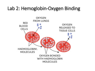
This document discusses hemoglobin and its ability to bind and transport oxygen in the blood. It contains the following key points:
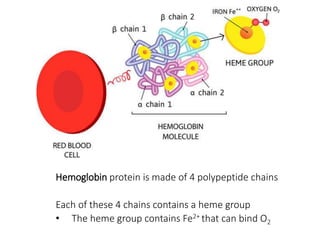
1) Hemoglobin is a protein made of four polypeptide chains, each containing a heme group with an Fe2+ ion that can bind oxygen.
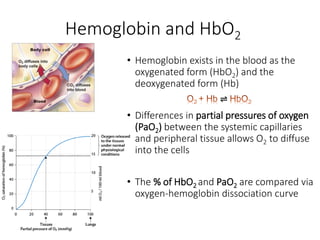
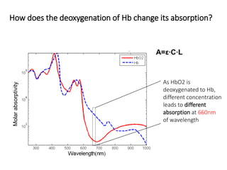
2) Hemoglobin exists in the blood in oxygenated (HbO2) and deoxygenated (Hb) forms. The ratio of these forms depends on partial pressures of oxygen in tissues.
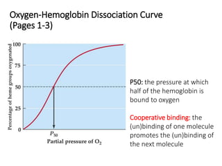
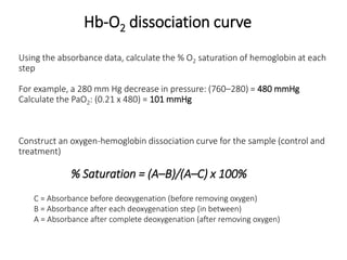
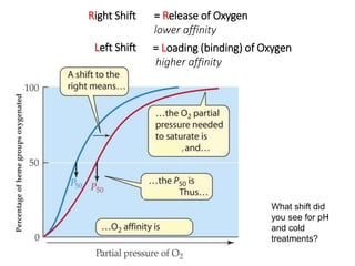
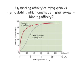
3) An oxygen-hemoglobin dissociation curve shows the relationship between Hb oxygen saturation and partial oxygen pressure. It is affected by factors like pH, temperature, and DPG levels.
This a presentation on regulation of respiration, control of the rate of increase of the the ramp signal, control of the limiting point at which ramp suddenly ceases
understanding spinal cord, its bransches, lesions, functions and anatomy.
hope to give you better knowledge of spinal cord by the end of it.
plese review ans comment for my future updates and corrections that iw ill be needing in this.
1st year medical school physiology essay:
Describe the effects between the action potential arriving at the axon terminal and skeletal muscle contraction.
Regulation of arterial blood pressure (The Guyton and Hall Physiology)Maryam Fida
BLOOD PRESSURE
The pressure exerted by the blood on vessel wall is known as blood pressure.
SYSTOLIC BLOOD PRESSURE
The maximum pressure exerted in the arteries during systole of heart.
Normal systolic pressure: 120 mm Hg.
DIASTOLIC BLOOD PRESSURE
The minimum pressure exerted in the arteries during diastole of heart.
Normal diastolic pressure: 80 mm Hg.
PULSE PRESSURE
The difference between the systolic pressure and diastolic pressure.
Normal pulse pressure: 40 mm Hg (120 – 80 = 40).
MEAN ARTERIAL BLOOD PRESSURE
The average pressure existing in the arteries.
Mean Arterial Blood Pressure = Diastolic Pressure + 1/3 Pulse Pressure
Pulse Pressure = (Systolic – Diastolic)
Mean Arterial Blood Pressure =Diastolic Pressure+1/3(Systolic – Diastolic)
HOT NEW PRODUCT! BIG SALES FAST SHIPPING NOW FROM CHINA!! EU KU DB BK substit...GL Anaacs
Contact us if you are interested:
Email / Skype : kefaya1771@gmail.com
Threema: PXHY5PDH
New BATCH Ku !!! MUCH IN DEMAND FAST SALE EVERY BATCH HAPPY GOOD EFFECT BIG BATCH !
Contact me on Threema or skype to start big business!!
Hot-sale products:
NEW HOT EUTYLONE WHITE CRYSTAL!!
5cl-adba precursor (semi finished )
5cl-adba raw materials
ADBB precursor (semi finished )
ADBB raw materials
APVP powder
5fadb/4f-adb
Jwh018 / Jwh210
Eutylone crystal
Protonitazene (hydrochloride) CAS: 119276-01-6
Flubrotizolam CAS: 57801-95-3
Metonitazene CAS: 14680-51-4
Payment terms: Western Union,MoneyGram,Bitcoin or USDT.
Deliver Time: Usually 7-15days
Shipping method: FedEx, TNT, DHL,UPS etc.Our deliveries are 100% safe, fast, reliable and discreet.
Samples will be sent for your evaluation!If you are interested in, please contact me, let's talk details.
We specializes in exporting high quality Research chemical, medical intermediate, Pharmaceutical chemicals and so on. Products are exported to USA, Canada, France, Korea, Japan,Russia, Southeast Asia and other countries.
New Drug Discovery and Development .....NEHA GUPTA
The "New Drug Discovery and Development" process involves the identification, design, testing, and manufacturing of novel pharmaceutical compounds with the aim of introducing new and improved treatments for various medical conditions. This comprehensive endeavor encompasses various stages, including target identification, preclinical studies, clinical trials, regulatory approval, and post-market surveillance. It involves multidisciplinary collaboration among scientists, researchers, clinicians, regulatory experts, and pharmaceutical companies to bring innovative therapies to market and address unmet medical needs.
These simplified slides by Dr. Sidra Arshad present an overview of the non-respiratory functions of the respiratory tract.
Learning objectives:
1. Enlist the non-respiratory functions of the respiratory tract
2. Briefly explain how these functions are carried out
3. Discuss the significance of dead space
4. Differentiate between minute ventilation and alveolar ventilation
5. Describe the cough and sneeze reflexes
Study Resources:
1. Chapter 39, Guyton and Hall Textbook of Medical Physiology, 14th edition
2. Chapter 34, Ganong’s Review of Medical Physiology, 26th edition
3. Chapter 17, Human Physiology by Lauralee Sherwood, 9th edition
4. Non-respiratory functions of the lungs https://academic.oup.com/bjaed/article/13/3/98/278874
Knee anatomy and clinical tests 2024.pdfvimalpl1234
This includes all relevant anatomy and clinical tests compiled from standard textbooks, Campbell,netter etc..It is comprehensive and best suited for orthopaedicians and orthopaedic residents.
These lecture slides, by Dr Sidra Arshad, offer a quick overview of physiological basis of a normal electrocardiogram.
Learning objectives:
1. Define an electrocardiogram (ECG) and electrocardiography
2. Describe how dipoles generated by the heart produce the waveforms of the ECG
3. Describe the components of a normal electrocardiogram of a typical bipolar leads (limb II)
4. Differentiate between intervals and segments
5. Enlist some common indications for obtaining an ECG
Study Resources:
1. Chapter 11, Guyton and Hall Textbook of Medical Physiology, 14th edition
2. Chapter 9, Human Physiology - From Cells to Systems, Lauralee Sherwood, 9th edition
3. Chapter 29, Ganong’s Review of Medical Physiology, 26th edition
4. Electrocardiogram, StatPearls - https://www.ncbi.nlm.nih.gov/books/NBK549803/
5. ECG in Medical Practice by ABM Abdullah, 4th edition
6. ECG Basics, http://www.nataliescasebook.com/tag/e-c-g-basics
Ethanol (CH3CH2OH), or beverage alcohol, is a two-carbon alcohol
that is rapidly distributed in the body and brain. Ethanol alters many
neurochemical systems and has rewarding and addictive properties. It
is the oldest recreational drug and likely contributes to more morbidity,
mortality, and public health costs than all illicit drugs combined. The
5th edition of the Diagnostic and Statistical Manual of Mental Disorders
(DSM-5) integrates alcohol abuse and alcohol dependence into a single
disorder called alcohol use disorder (AUD), with mild, moderate,
and severe subclassifications (American Psychiatric Association, 2013).
In the DSM-5, all types of substance abuse and dependence have been
combined into a single substance use disorder (SUD) on a continuum
from mild to severe. A diagnosis of AUD requires that at least two of
the 11 DSM-5 behaviors be present within a 12-month period (mild
AUD: 2–3 criteria; moderate AUD: 4–5 criteria; severe AUD: 6–11 criteria).
The four main behavioral effects of AUD are impaired control over
drinking, negative social consequences, risky use, and altered physiological
effects (tolerance, withdrawal). This chapter presents an overview
of the prevalence and harmful consequences of AUD in the U.S.,
the systemic nature of the disease, neurocircuitry and stages of AUD,
comorbidities, fetal alcohol spectrum disorders, genetic risk factors, and
pharmacotherapies for AUD.
Pulmonary Thromboembolism - etilogy, types, medical- Surgical and nursing man...VarunMahajani
Disruption of blood supply to lung alveoli due to blockage of one or more pulmonary blood vessels is called as Pulmonary thromboembolism. In this presentation we will discuss its causes, types and its management in depth.
micro teaching on communication m.sc nursing.pdfAnurag Sharma
Microteaching is a unique model of practice teaching. It is a viable instrument for the. desired change in the teaching behavior or the behavior potential which, in specified types of real. classroom situations, tends to facilitate the achievement of specified types of objectives.
2. Hemoglobin protein is made of 4 polypeptide chains
Each of these 4 chains contains a heme group
• The heme group contains Fe2+ that can bind O2
3. Hemoglobin and HbO2
• Hemoglobin exists in the blood as the
oxygenated form (HbO2) and the
deoxygenated form (Hb)
• Differences in partial pressures of oxygen
(PaO2) between the systemic capillaries
and peripheral tissue allows O2 to diffuse
into the cells
• The % of HbO2 and PaO2 are compared via
oxygen-hemoglobin dissociation curve
⇌
4. Oxygen-Hemoglobin Dissociation Curve
(Pages 1-3)
P50: the pressure at which
half of the hemoglobin is
bound to oxygen
Cooperative binding: the
(un)binding of one molecule
promotes the (un)binding of
the next molecule
6. As HbO2 is
deoxygenated to Hb,
different concentration
leads to different
absorption at 660nm
of wavelength
Molar
absorptivity
How does the deoxygenation of Hb change its absorption?
7. Be sure to set the
spectrophotometer to
Absorbance mode
Zero the
spectrophotometer with
a blank tube between
samples and different
experimental conditions
Reminders for operation of spectrophotometer
8. Hb-O2 dissociation curve
Deoxygenate the oxygen
from PaO2 760 mmHg in
140mmHg intervals
Measure absorbance of
Hb-O2 after every
decrement until PaO2
reaches 0
Spectrophotometer
9. Using the absorbance data, calculate the % O2 saturation of hemoglobin at each
step
For example, a 280 mm Hg decrease in pressure: (760–280) = 480 mmHg
Calculate the PaO2: (0.21 x 480) = 101 mmHg
Construct an oxygen-hemoglobin dissociation curve for the sample (control and
treatment)
% Saturation = (A–B)/(A–C) x 100%
Hb-O2 dissociation curve
C = Absorbance before deoxygenation (before removing oxygen)
B = Absorbance after each deoxygenation step (in between)
A = Absorbance after complete deoxygenation (after removing oxygen)
10. Right Shift = Release of Oxygen
lower affinity
Left Shift = Loading (binding) of Oxygen
higher affinity
What shift did
you see for pH
and cold
treatments?
12. P50 is reached when _____
A) the partial pressure of O2 in blood is 50 mmHg
B) hemoglobin is 50% saturated with oxygen
13. If the oxygen-hemoglobin dissociation
curve moves to left, the affinity between
O2 and Hemoglobin is ____
A. Lower
B. Higher
14. pCO2, acidity and temperature are higher in
capillary circulation than in arterial blood. In this
circumstances, Hb has lower affinity to oxygen.
That enables:
A. Hb to release oxygen
B. Hb to hold on to oxygen
15. O2 binding affinity of myoglobin vs
hemoglobin: which one has a higher oxygen-
binding affinity?
16. O2 affinity fetal vs adult hemoglobin: which one has
a higher oxygen-binding affinity?
Editor's Notes
On the surface of red blood cells, there are millions of hemoglobin molecules.
The hemoglobin protein is made up of four polypeptide subunits, each of which contains a heme group. This a ring-like structure that contains iron.
The iron in the heme group binds to molecular oxygen, allowing the red blood cells to transport oxygen to other tissue.
The binding of oxygen is reversible.
Hemoglobin binds oxygen to form oxyhemoglobin (HbO2), the main oxygen carrier in human body.
Difference in the partial pressure of oxygen between systemic capillaries and peripheral tissue allows oxygen to diffuse into cells.
On the oxygen-hemoglobin dissociation curve, the % of HbO2 and PaO2 are plotted.
The tissues sit at a lower resting oxygen concentration making the affinity for oxygen lower allowing oxygen to unload.
In the lungs, hemoglobin is saturated and has a high affinity for oxygen allowing oxygen to load.
The sigmoidal curve shape is due to Cooperative binding of the four hemoglobin subunits – as one subunit binds O2, it increases the O2 affinity for remaining subunits.
oxygen affinity of 3-oxyhemoglobin is ~300x that of deoxyhemoglobin.
This allows hemoglobin to be far more dynamic in binding O2, and to rapidly transition from a low oxygen-bound state to an oxygen saturated state.
It also allows hemoglobin to off-load O2 rapidly, as affinity for O2 decreases.
P50 is the pressure at which half of the hemoglobin is bound to oxygen
The sequence of events of a spectrophotometer are as follows:
The light source sends light into a monochromator and is diffracted into a spectrum that is split into two beams.
These two beams scan through the sample that you are measuring.
Some fractions of light are transmitted through the sample and some are reflected off of it.
The transmitted light and reflected light are measured by the photo detector device which compares the relative intensities
The spectrophotometer then reads out an absorbance value.
If we looked at the absorption of light at 800 nm, we would not be able to tell a difference between oxygenated and deoxygenated hemoglobin.
In this part of the experiment, students will use the vacuum gauge provided to gradually remove oxygen from the flask with a side arm cuvette. This multi-step deoxygenation process will create various partial pressure of oxygen and allow hemoglobin and oxygen to disassociate gradually.
Be aware that the readings of the vacuum gauge is compared to the atmospheric pressure. Make sure you understand the differences between mm Hg vacuum and mm Hg pressure. Zero reading from the gauge means approximately 760 mm Hg.
Remind students to keep flask closed when deoxygenating.
mm Hg is a unit of pressure measurement, specifically for measuring pressure in liquids and gases. It stands for millimeters of Mercury and refers to the height of a column of mercury (Hg) in a manometer.
For example, a 280 mm Hg decrease in pressure: (760–280) = 480 mmHg Calculate the PaO2 at that pressure by multiplying it by the fraction of air that is oxygen (21%)(0.21 x 480) = 101 mmHg.
BPG/DPG – Increase shifts curve to the right, decrease to the left. Acts as a heteroallosteric inhibitor by binding preferentially to deoxyhemoglobin, lowering hemoglobin’s affinity for oxygen and shifting the curve right.
It interacts with deoxygenated hemoglobin beta subunits and so it decreases the affinity for oxygen and allosterically promotes the release of the remaining oxygen molecules bound to the hemoglobin; therefore it enhances the ability of RBCs to release oxygen near tissues that need it most
Temperature – Increasing temperature weakens the O2 – Hb bond, decreasing the concentration of oxyhemoglobin.
Acidity – H+ and O2 compete for the heme group, so at lower pH (higher H+ conc.) there is less oxyhemoglobin, shifting the curve right. An increase in CO2 will also cause an increase in acidity (CO2 + H2O <-> HCO3- + H+). O2 affinity is inversely related to both acidity (H+ conc) and CO2, referred to as the Bohr effect.
Exercise causes an increase in acidity, temperature and metabolic intermediates and a decrease in oxygen in your muscle tissues. This causes an increased dissociation of oxygen from your blood flowing through your muscles, supplying them with much needed oxygen. Your body increases blood flow to your muscles to supply them with more oxygen. Warming up before exercise can help prime your muscles to receive ample oxygen by kick starting metabolism and increasing temperature.
B
B-Review page 6
A-review page 7
Like hemoglobin, myoglobin is a cytoplasmic protein that binds oxygen on a heme group. It harbors only one globulin group, whereas hemoglobin has four. Although its heme group is identical to those in Hb, Mb has a higher affinity for oxygen than does hemoglobin. This difference is related to its different role: whereas hemoglobin transports oxygen, myoglobin's function is to store oxygen.
The steep (hyperbolic dissociation curve) indicates that O2 affinity is higher for myoglobin than for hemoglobin.
This makes myoglobin the oxygen storage molecule, while hemoglobin is the oxygen transport molecule. The cooperative binding of O2 to hemoglobin allows it to dynamically load and offload O2 as it travels around the body, while the higher O2 affinity of myoglobin makes it a good O2 storage molecule to facilitate a build up of O2 in the muscles that can be released when needed.
1 - Higher affinity than adult hemoglobin (also a lower affinity for DPG/BPG).
2 - Fetal hemoglobin needs to extract oxygen from the maternal blood. During pregnancy, the mother will also increase DPG/BPG concentration, shifting the adult curve to the right, making it easier for fetal hemoglobin to extract O2.
Fetal hemoglobin (HbF) is the dominant form of hemoglobin present in the fetus during gestation. HbF is produced by erythroid precursor cells from 10 to 12 weeks of pregnancy through the first six months of postnatal life. HbF contains two alpha and two gamma subunits, while the major form of adult hemoglobin, hemoglobin A (HbA), contains two alpha and two beta subunits. The genes that express gamma chain proteins are present in the beta chain locus on chromosome 11. The gamma subunit differs from its adult counterpart in that it contains either an alanine or a glycine at position 136, both of which are neutral, nonpolar amino acids. This difference introduces conformational changes to the protein that gives rise to several physiological differences in oxygen delivery that are important in the fetal circulation.
Fetal hemoglobin has a vital role in the transport of oxygen from maternal to fetal circulation. Oxygen transfer from the maternal circulation to the fetal circulation is made possible by HbF having a high oxygen affinity but decreased affinity to 2,3-bisphosphoglycerate relative to HbA. The HbF oxygen dissociation curve is left-shifted in comparison to HbA. The partial pressure at which HbF is half saturated with oxygen (P50) is 19 mm Hg, compared to 27 mm Hg for HbA. This value indicates that HbF has a high affinity for oxygen, giving HbF the ability to bind oxygen more readily from the maternal circulation.