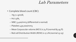
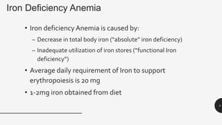
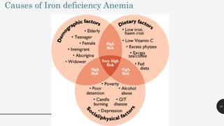
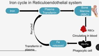
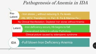
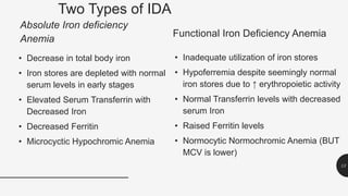
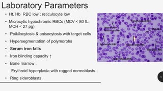
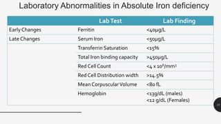
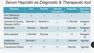
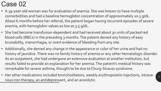
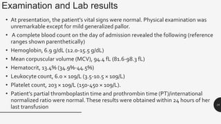
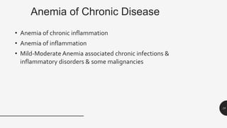
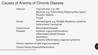
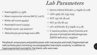
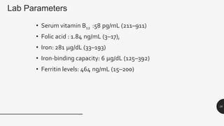
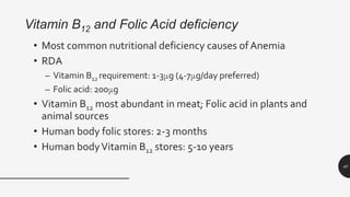
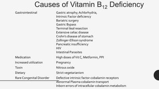
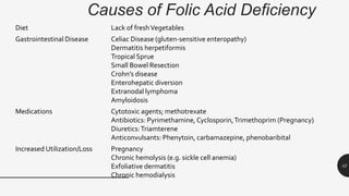
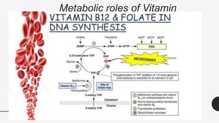
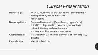
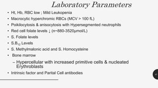
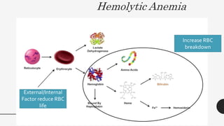
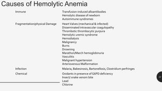
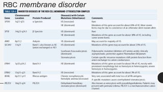
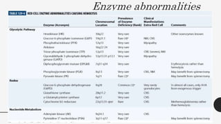
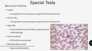
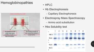
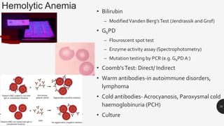
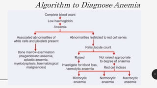
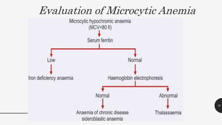
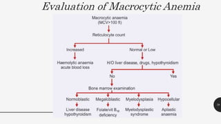
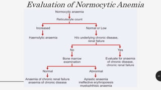
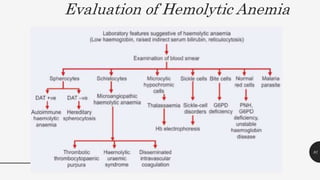
ANEMIA
PROFILE
Dr. Anisha Mathew
1
Contents • Definition of Anemia
• Types/Classification of Anemia
• Cases
• Microcytic Anemia
• Macrocytic Anemia
• Hemolytic Anemia
• Diagnosis algorithm
2
Anemia
• Condition in which number of red blood cells or their oxygen-
carrying capacity is insufficient to meet physiologic needs, which
vary by age, sex, altitude, smoking, and pregnancy status
• WHO.2019
3
O2
O2
O2
O2
Anemia from the Greek word “ναιμία”(an-haîma) meaning “without blood”
Anemia is not a disease, but
an expression of an underlying
disorder or disease
4
Classification of Anemia
5
Case 1
• A 35 year old woman is seen for easy fatigue for many months.
She is now 24 weeks pregnant with her 3rd child in 3 years. She
does not see any obstetrician and does not take any vitamins.
Lately, she has developed a taste for eating ice. She has no other
complaint. Family and past history are negative. She does not
smoke or drink. Physical examination is positive for pale
conjunctiva, mild spooning of nails, and a II/VI systolic murmur at
left lower sternal border. Stools are negative for occult blood
6
Lab Parameters
• Complete blood count (CBC)
– Hg 7.1 gm/dl,
– Hct 23%,
– WBC 5,400/mm3 (differential is normal)
– Platelets 450,000/mm3
– Mean Corpuscular volume (MCV) is 74 fl (normal 85-95 fl)
– Red cell Distribution Width (RDW) is 17.1% (normal 13-15)
7
MICROCYTIC HYPOCHROMIC ANEMIA
8
Iron Deficiency Anemia
• Iron deficiencyAnemia is caused by:
– Decrease in total body iron (“absolute” iron deficiency)
– Inadequate utilization of iron stores (“functional Iron
deficiency”)
• Average daily requirement of Iron to support
erythropoiesis is 20 mg
• 1-2mg iron obtained from diet
9
10
Causes of Iron deficiency Anemia
Iron cycle in Reticuloendothelial system
11
Iron
Plasma
Transferrin
Bone
Marrow
(RBC)
RBCs
Circulation in blood
Hb+Fe
Phagocytic cell
Fe
released
Transferrin in
plasma
Recycled
Pathogenesis of Anemia in IDA
- Iron stores ↓ without reducing S. Fe levels
-Hb, MCV, Transferrin ~n, Fe↑, S. Fe & Marrow Fe ↓
-Iron deficiency occurs; Hb begins to fall
-Generalized defect in cellular Pathology
-Full blown Iron Deficiency Anemia
12
Pre-
Latent
Latent
IDA
No Clinical Manifestation; Depleted Iron stores without Anemia
Clinical picture caused by sideropenic syndrome
Two Types of IDA
Absolute Iron deficiency
Anemia
• Decrease in total body iron
• Iron stores are depleted with normal
serum levels in early stages
• Elevated Serum Transferrin with
Decreased Iron
• Decreased Ferritin
• Microcyctic Hypochromic Anemia
Functional Iron Deficiency Anemia
• Inadequate utilization of iron stores
• Hypoferremia despite seemingly normal
iron stores due to ↑ erythropoietic activity
• Normal Transferrin levels with decreased
serum Iron
• Raised Ferritin levels
• Normocytic Normochromic Anemia (BUT
MCV is lower)
13
Laboratory Parameters
• Ht, Hb RBC low ; reticulocyte low
• Microcytic hypochromic RBCs (MCV < 80 fL,
MCH < 27 pg)
• Poikilocytosis & anisocytosis with target cells
• Hypersegmentation of polymorphs
• Serum iron falls
• Iron blinding capacity ↑
• Bone marrow :
Erythroid hyperplasia with ragged normoblasts
• Ring sideroblasts 14
Laboratory Abnormalities in Absolute Iron deficiency
LabTest Lab Finding
Early Changes Ferritin <40μg/L
Late Changes Serum Iron <50μg/L
Transferrin Saturation <15%
Total Iron binding capacity >450μg/L
Red Cell Count <4 x 10⁶/mm3
Red Cell Distribution width >14.5%
Mean CorpuscularVolume <80 fL
Hemoglobin <13g/dL (males)
<12 g/dL (Females)
15
Serum Hepcidin as Diagnostic & Therapeutic tool
Disease Iron Ferritin Soluble
Transferrin
Hepcidin Hepcidin
Therapy
Iron Deficiency
Anemia (IDA)
↓ ↓ ↓ ↓ -
Anemia of Chronic
Disease (ACD)
Normal-↑↑ Normal-↑↑ ↓ ↑↑-Normal Antagoni
st
ACD+IDA Normal Normal ↓ Normal Antagoni
st
ESA resistant ↓-Normal Normal ↓ ↑↑ Antagoni
st
Ineffective
Erythropoiesis
Normal-↑↑ ↑↑ ↑↑ ↓-Normal Agonist
Transfused Patient ↑↑ ↑↑ ↑↑ Normal Agonist
16
Case 02
• A 39-year-old woman was for evaluation of anemia. She was known to have multiple
comorbidities and had a baseline hemoglobin concentration of approximately 10.5 g/dL.
About 6 months before her referral, the patient began having recurrent episodes of severe
anemia, with hemoglobin values as low as 3.5 g/dL.
• She had become transfusion-dependent and had received about 30 units of packed red
blood cells (RBCs) in the preceding 3 months.The patient denied any history of easy
bruisability, menorrhagia, or overt evidence of bleeding from any site.
• Additionally, she denied any change in the appearance or color of her urine and had no
history of jaundice.There was no family history of anemia or any other hematologic disorder.
As an outpatient, she had undergone an extensive evaluation at another institution, but
results failed to provide an explanation for her anemia.The patient’s medical history was
remarkable for severe asthma, thought to be due to Churg-Strauss syndrome.
• Her other medications included bronchodilators, weekly erythropoietin injections, intrave-
nous iron therapy, an antidepressant, and an anxiolytic.
17
Examination and Lab results
• At presentation, the patient’s vital signs were normal. Physical examination was
unremarkable except for mild generalized pallor.
• A complete blood count on the day of admission revealed the following (reference
ranges shown parenthetically)
• Hemoglobin, 6.9 g/dL (12.0-15.5 g/dL)
• Mean corpuscular volume (MCV), 94.4 fL (81.6-98.3 fL)
• Hematocrit, 13.4% (34.9%-44.5%)
• Leukocyte count, 6.0 × 10⁹/L (3.5-10.5 × 10⁹/L)
• Platelet count, 203 × 109/L (150-450 × 109/L).
• Patient’s partial thromboplastin time and prothrombin time (PT)/international
normalized ratio were normal.These results were obtained within 24 hours of her
last transfusion 18
Anemia of Chronic Disease
• Anemia of chronic inflammation
• Anemia of inflammation
• Mild-Moderate Anemia associated chronic infections &
inflammatory disorders & some malignancies
19
Causes of Anemia of Chronic Disease
Infection Viral infection .e.g. HIV
Bacterial .e.g.Tuberculosis, Osteomyelitis, Sepsis
Parasitic: Malaria
Fungal
Cancer Hematological .e.g. Multiple Myeloma, Leukemia
Solid tumors: Carcinomas
Autoimmune
Diseases
Rheumatoid Diseases
Systemic Lupus erythromatosis
Inflammatory Bowel Disease
Sarcoidosis
Systemic Inflammatory response syndrome
Chronic rejection of solid organ transplants
Chronic Kidney Disease/Inflammation 20
Pathogenesis
21
Characteristics of ACD
Inadequate erythrocyte production
Low serum Iron
Low Iron Binding Capacity
Normocytic Normochromic Anemia- Mild Microcytic Hypochromic
Anemia
22
Laboratory Changes
• Anemia is mild to moderate (Hb 7-11gm/dL)
• Normochromic HypochromicAnemia
• Reticulocyte count is normal or slightly elevated
• Hypoferremia and decreased serumTransferrin
• Increased serum ferritin
• Marrow morphology and stainable iron are normal
23
Difference between IDA and ACD
24
Sideroblastic Anemia
Congenital X-linked inheritance
Acquired primary or idiopathic
secondary:
drugs
alcohol
lead
myeloproliferative disorders
leukaemia
secondary carcinoma
other systemic disorders (connective tissue disease)
25
Pathogenesis
26
Clinical features
• Insidious/Gradual onset
• Mild Jaundice
• Glossitis/ Angular Stomatitis
• Neuropathy
• Neural tube defects
• Increased melanin production
27
Laboratory Changes • CBC
– ↓ Hb
– ↓ MCV
– Leukopenia, Thrombocytopentia
• ↓TIBC, ↓ Ferritin
• Peripheral Smear
– Hypochromic, Microcytic
– Siderocytes with Pappenheimer bodies
– <1% Blasts
• Bone Marrow
– Similar to Myelodisplastic syndrome/IDA
– >15% Sideroblast RBCs
– >5% Blasts
28
α & β-Thalassemia
29
Pathogenesis of β-Thalassemia
30
Pathogenesis of α-Thalassemia
31
Clinical Features of Thalassemia
• failure to thrive
• intermittent infection
• severe anaemia
• extramedullary haemopoiesis → hepatosplenomegaly and
bone expansion
• Thalassaemic facies
32
Diagnosis of α-Thalassemia
• ↓Hb
• MCV <78 fL & MCH <28 pg
• HbA2 – Near borderline or ↓
• On Peripheral Smear (Hb H
disease): Cresol Blue shows
Hb H precipitates in RBCs
33
Laboratory changes of β-Thalassemia
34
• ↓Hb, ↓MCV, ↓MCH, ↓MCHC
• Peripheral Blood
• Hypochromic, Microcytic RBCs,
Target cells, Nucleated RBCs
• Anisocytosis, Poikilocytosis
• Basophilic stippling
• RBC inclusion bodies
• ↑Iron,
• ↓TIBC
• ↑Ferritin
• Thal Major: HbF : 30-98%, HbA2: 2%,
HbA: ↓↓↓
• Thal Minor: HbF: 3.5-7%, HbA2: <5%,
HbA ~n
Differential Diagnosis of
Microcytosis
35
Case 3
• A 15-year-old vegetarian boy born to non-consanguineous parents presented
with easy fatigue, breathlessness and pain in the legs on walking, noted
during the past few weeks. Paleness and icterus in the sclerae had been
noted by the parents for about 2 years, although these findings became more
obvious within the last few weeks. The vegetarian patient had not consumed
any food of animal origin for many years. Besides, the family only rarely ate
fresh fruits or vegetables.
• The patient had a history of an upper respiratory tract infection that began
about 15 days earlier and resolved 1 week later. No fever was noted. In the
• Physical examination: patient looked pale and weak, and his sclerae were
icteric.
• His heart rate was 96 bpm
36
Lab Parameters
• Haemoglobin 5.1 g/dL
• Mean corpuscular volume (MCV), 116 fL
• White cell count 2540/µl
• Neutrophil count 1230/µL
• Platelet count :107 000/mm3
• Reticulocyte percentage was 0.8%
• Serum indirect bilirubin: 4 mg/dL (0–0.8)
• LDH: 5565 U/L (135–225)
• AST: 150 U/L (8–40)
• ALT: 51 U/L (8–41)
• Uric acid levels: 8.5 mg/dL (2.4–8)
• C reactive protein, direct Coombs and
glucose-6-phosphate dehydrogenase
(G6PD) deficiency: negative
37
Peripheral: anisocytosis and poikilocytosis with macrocytes and small, fragmented red
cells (schistocytes) mimicking microangiopathic haemolytic anaemia, in addition to
hypersegmented neutrophils. No blastic cells were seen
Lab Parameters
• Serum vitamin B12 :58 pg/mL (211–911)
• Folic acid : 1.84 ng/mL (3–17),
• Iron: 281 µg/dL (33–193)
• Iron-binding capacity: 6 µg/dL (125–392)
• Ferritin levels: 464 ng/mL (15–200)
38
MACROCYTIC HYPERCHROMIC
39
Vitamin B12 and Folic Acid deficiency
• Most common nutritional deficiency causes of Anemia
• RDA
– Vitamin B12 requirement: 1-3μg (4-7μg/day preferred)
– Folic acid: 200μg
• Vitamin B12 most abundant in meat; Folic acid in plants and
animal sources
• Human body folic stores: 2-3 months
• Human bodyVitamin B12 stores: 5-10 years
40
Causes of Vitamin B12 Deficiency
Gastrointestinal Gastric atrophy;Achlorhydria,
Intrinsic Factor deficiency
Bariatric surgery
Gastric Bypass
Terminal ileal resection
Extensive celiac disease
Crohn’s disease of stomach
Zollinger-Ellison syndrome
Pancreatic insufficiency
HIV
Intestinal Parasites
Medication High doses ofVit C, Metformin, PPI
Increased utilization Pregnancy
Toxin Nitrous oxide
Dietary Strict vegetarianism
Rare Congenital Disorder Defective intrinsic factor-cobalamin receptors
Abnormal Plasma cobalamin transport
Inborn errors of intracellular cobalamin metabolism 41
Causes of Folic Acid Deficiency
Diet Lack of freshVegetables
Gastrointestinal Disease Celiac Disease (gluten-sensitive enteropathy)
Dermatitis herpetiformis
Tropical Sprue
Small Bowel Resection
Crohn’s disease
Enterohepatic diversion
Extranodal lymphoma
Amyloidosis
Medications Cytotoxic agents; methotrexate
Antibiotics: Pyrimethamine, Cyclosporin,Trimethoprim (Pregnancy)
Diuretics:Triamterene
Anticonvulsants: Phenytoin, carbamazepine, phenobaribital
Increased Utilization/Loss Pregnancy
Chronic hemolysis (e.g. sickle cell anemia)
Exfoliative dermatitis
Chronic hemodialysis
42
Metabolic roles of Vitamin
B12and Folate
43
Clinical Presentation
Hematological Anemia, usually macrocytic but normo- or microcytic if
accompanied by IDA or thalassemia
Pancytopenia
Neuropsychiatric Peripheral Neuropathy (Parasthesias, hyporeflexia)
Spinal Cord degeneration (weakness, hypereflexia,
reduced vibratory and position sense)
Memory loss, disorientation, depression
Gastrointestinal Malabsorption (weight loss, diarrhoea, abdominal pain),
glossitis
Reproductive Infertility, Fetal loss
44
Laboratory Parameters
• Ht, Hb, RBC low ; Mild Leukopenia
• Macrocytic hyperchromic RBCs (MCV > 100 fL)
• Poikilocytosis & anisocytosis with Hypersegmented neutrophils
• Red cell folate levels ↓ (n~880-3520μmol/L)
• S. Folate levels
• S.B12 Levels
• S. Methylmalonic acid and S. Homocysteine
• Bone marrow
– Hypercellular with increased primitive cells & nucleated
Erythroblasts
• Intrinsic factor and Parital Cell antibodies 45
Peripheral Blood picture Vs. Bone
Marrow
Peripheral Blood Picture Bone Marrow
46
Case 4
• A four years old girl was admitted at department of pediatrics
of Faridpur Medical College Hospital, Faridpur with
complaints of fever, cough, and sputum for three days and
gross hematuria for one day.
• She is the second girl of two sibling. Prior to this admission,
she was admitted due to hemolytic anemia with acute
tonsillitis one and half years back. On admission, she looked
actually ill, the conjunctivae were pale, the sclera were icteric
and throat was infected.The lung sound was coarse and the
liver and spleen were not palpable 47
Lab Parameters
48
Normocytic Normochromic
49
Hemolytic Anemia
50
Hemolytic Anemia
51
External/Internal
Factor reduce RBC
life
Increase RBC
breakdown
Causes of Hemolytic Anemia
52
Immune Transfusion-induced alloantibodies
Hemolytic disease of newborn
Autoimmune syndromes
Fragmentation/physical Damage HeartValves (mechanical & infected)
Disseminated intravascular coagulopathy
Thrombotic thrombocytic purpura
Hemolytic uremic syndrome
Hemodialysis
Malignancy
Burns
Drowning
Marathon/March hemoglobinuria
Vasculitis
Malignant hypertension
Arteriovenous Malformation
Infection Malaria, Babesinosis, Bartonellosis,Clostridium perfringes
Chemical Oxidants in presence of G6PD deficiency
Insect/ snake venom bite
Lead
Chlorine
RBC membrane disorder
53
Enzyme abnormalities
54
Causes of hemolysis in G6PD Deficiency
55
Intravascular causes of Hemolysis
56
Fate in Hemolytic Anemia
57
Increased red cell breakdown leads to:
• Iron ↑
• Elevated serum bilirubin (unconjugated)
– Stercobilinogen ↑
– Excess urinary urobilinogen
• Reduced plasma haptoglobin
• Abnormal red cell fragments in peripheral blood
• Increased red cell production leads to:
• Reticulocytosis
• Erythroid hyperplasia of the bone marrow
Clinical Picture of Hemolytic Anemia
58
Sickle cell disorder
59
Molecular and Cellular Changes of HbS
http://www.emedicine.com/ped/TOPIC2096.HTM
Decreased PO2
Permanent damage to RBC
Cell⟺endothelium interactions
60
Clinical Picture
61
Laboratory Changes
• CBC - ↓Hb
• Peripheral smear- sickle shaped RBCs
• Hemoglobin Electrophoresis
• HPLC: Hb S (35%-40%) (Trait)
– Hb S (80-90%)
– Hb A2 (↑Slightly)
• Trait: Sickle Solubility test
Gel Electrophoresis
62
BONE MARROW FAILURES
63
Aplastic Anemia
• Aplasia of the bone marrow with peripheral blood pancytopenia
• Bone marrow failure that arises from injury to or abnormal
expression of the stem cell
• Bone marrow becomes hypoplastic, and pancytopenia develops
• Most common cause: autoimmune suppression of hematopoiesis
64
Causes of Aplastic Anemia
Congenital Fanconi’s Anemia
Acquired Chemicals, drugs, insecticides
Ionizing radiation
Infections,Viral Hepatitis, measles
?Tuberculosis
Thymoma
Pregnancy
?Idiopathic
65
Clinical Features
• Anaemia
• Bleeding (ecchymoses, bleeding gums and
epistaxis)
• Infection (fungal infections)
66
Laboratory changes:
– Elevated serum iron
– Low haemoglobin
– White cell Count 500 / mmc
– Platelet 20,000 / mmc
– Reticulocytes virtual absent
– Hypocellular or aplastic bone
marrow
67
Approach to Patient with Anemia
68
History and Examination
History
• Fatigue and weakness
• Headache
• Tinnitus Menorrhagia
• Anorexia
• Dyspnea and palpitations
• Nausea
• Abdominal complains
Examination
• Numbness and coldness
• Pallor
• Jaundice
• Intermittent claudication
• Haemorrhages in the fundus of eyes
69
Laboratory diagnosis of Anemia
• Complete Blood Count (CBC)
– Hb (Cyanomethemoglobin)
– RBC (Impedance counting)
– Hematocrit
• Centrifuged; ratio of RBC of volume sample
• MCV:Voltage pulse impedance count/ calculated : Hct/RBC
• MCH: Hb/ RBC
• MCHC: Hb/HCT
• RDW: (Impedance/ Peripheral smear); (SD of MCV X 100)/ Mean MCV)
• WBC (Impedance)
70
Mean corpuscular volume (MCV): MCV
• <80fL : Microcytic Anemia
• 80-100 fL: Normocytic Anemia
• >100 fL: Macrocytic Anemia
Mean corpuscular hemoglobin (MCH): 27-31 pg/cell
• <26 pg/cell : Microcytosis
• >34 pg/cell: Macrocytosis
Mean corpuscular hemoglobin concentration (MCHC): 31-35 g/dL
• < 31 g/dL Hypochromic RBC
RDW -Red Cell Distribution Width: 11.5-14.5%
↑- IDA/ Hemolytic Anemia
↓- Bone marrow failures
Red blood cell indices
71
Special Tests
MicrocyticAnemia:
• Iron
• Ferritin
– Immunoradioassay
– ELISA
– Immnochemiluminesence
– Immunoflurometry
• Serum Iron
– Wong’s method using potassium
thiocynate
• Total Iron Binding Capacity
– Calculated fromTransferrin
• SerumTransferrin
– Ion Binding
• HPLC
– Hbf, HbA2,
• Capillary electrophoresis
– Hemogobinopathies
• Electrospray Mass spectrometry
– Variant identification & quantity
– Amino Acid Substitution
• DNA analysis
• HbH4 – Cresol blue in P/S
72
73
Laboratory Diagnosis of Anemia
Special Tests
Macrocytic Anemia:
• Folate
– Competent Immunoassay using Chemiluminescence
• Vitamin B12
– Competent Immunoassay using Chemiluminescence
• Hepcidin
– Immunochemical and Mass spectrometric
methodology
• Homocysteine
– ELISA &Immunoassay
• Methylmalonic Acid
– Spectrophotometry
74
Hemoglobinopathies
• HPLC
• Hb Electrophoresis
– Capillary Electrophoresis
• Electrospray Mass Spectroscopy
– Amino acid substitution
• Hbs Solubility test
75
Hemolytic Anemia • Bilirubin
– ModifiedVanden Berg’sTest (Jendrassik and Grof)
• G6PD
– Flouroscent spot test
– Enzyme activity assay (Spectrophotometry)
– Mutation testing by PCR (e.g. G6PD A-)
• Coomb’sTest: Direct/ Indirect
• Warm antibodies-in autoimmune disorders,
lymphoma
• Cold antibodies- Acrocyanosis, Paroxysmal cold
haemoglobinuria (PCH)
• Culture
76
Algorithm to Diagnose Anemia
77
Evaluation of Microcytic Anemia
78
Evaluation of Macrocytic Anemia
79
Evaluation of Normocytic Anemia
80
Evaluation of Hemolytic Anemia
81
References
• Rodgers PG,Young SN.The Bethesda Handbook of clinical hematology. 3rd edition. Wolters
Kluwers. 2015
• Greer PJ, Arber AD, Glader B, List AF, Means RT, Paraskeves F et al.Wintrobes Clinical
Hematology. 13th edition. Philadelphia. Wolters Kluwers; 2016
• HigginsT, Eckfeldt JH, Barton JC, Doumas BT.In:Teitz Fundamentals of clinical biochemistry.
7th ed. St.louis: Elsevier; p. 806–23
• Kumar,Vinay, Abul K. Abbas, and Jon C. Aster. Robbins and Cotran Pathologic Basis of Disease.
Ninth edition. Philadelphia, PA: Elsevier/Saunders, 2015.
• Turgeon ML. Clinical Hematology:Theory and Procedures. 5th edition. Philadelphia. Wolters
Kluwer; 2012
82
83

Anemia Profile- modified for study..pptx

  • 1.
  • 2.
    Contents • Definitionof Anemia • Types/Classification of Anemia • Cases • Microcytic Anemia • Macrocytic Anemia • Hemolytic Anemia • Diagnosis algorithm 2
  • 3.
    Anemia • Condition inwhich number of red blood cells or their oxygen- carrying capacity is insufficient to meet physiologic needs, which vary by age, sex, altitude, smoking, and pregnancy status • WHO.2019 3 O2 O2 O2 O2 Anemia from the Greek word “ναιμία”(an-haîma) meaning “without blood”
  • 4.
    Anemia is nota disease, but an expression of an underlying disorder or disease 4
  • 5.
  • 6.
    Case 1 • A35 year old woman is seen for easy fatigue for many months. She is now 24 weeks pregnant with her 3rd child in 3 years. She does not see any obstetrician and does not take any vitamins. Lately, she has developed a taste for eating ice. She has no other complaint. Family and past history are negative. She does not smoke or drink. Physical examination is positive for pale conjunctiva, mild spooning of nails, and a II/VI systolic murmur at left lower sternal border. Stools are negative for occult blood 6
  • 7.
    Lab Parameters • Completeblood count (CBC) – Hg 7.1 gm/dl, – Hct 23%, – WBC 5,400/mm3 (differential is normal) – Platelets 450,000/mm3 – Mean Corpuscular volume (MCV) is 74 fl (normal 85-95 fl) – Red cell Distribution Width (RDW) is 17.1% (normal 13-15) 7
  • 8.
  • 9.
    Iron Deficiency Anemia •Iron deficiencyAnemia is caused by: – Decrease in total body iron (“absolute” iron deficiency) – Inadequate utilization of iron stores (“functional Iron deficiency”) • Average daily requirement of Iron to support erythropoiesis is 20 mg • 1-2mg iron obtained from diet 9
  • 10.
    10 Causes of Irondeficiency Anemia
  • 11.
    Iron cycle inReticuloendothelial system 11 Iron Plasma Transferrin Bone Marrow (RBC) RBCs Circulation in blood Hb+Fe Phagocytic cell Fe released Transferrin in plasma Recycled
  • 12.
    Pathogenesis of Anemiain IDA - Iron stores ↓ without reducing S. Fe levels -Hb, MCV, Transferrin ~n, Fe↑, S. Fe & Marrow Fe ↓ -Iron deficiency occurs; Hb begins to fall -Generalized defect in cellular Pathology -Full blown Iron Deficiency Anemia 12 Pre- Latent Latent IDA No Clinical Manifestation; Depleted Iron stores without Anemia Clinical picture caused by sideropenic syndrome
  • 13.
    Two Types ofIDA Absolute Iron deficiency Anemia • Decrease in total body iron • Iron stores are depleted with normal serum levels in early stages • Elevated Serum Transferrin with Decreased Iron • Decreased Ferritin • Microcyctic Hypochromic Anemia Functional Iron Deficiency Anemia • Inadequate utilization of iron stores • Hypoferremia despite seemingly normal iron stores due to ↑ erythropoietic activity • Normal Transferrin levels with decreased serum Iron • Raised Ferritin levels • Normocytic Normochromic Anemia (BUT MCV is lower) 13
  • 14.
    Laboratory Parameters • Ht,Hb RBC low ; reticulocyte low • Microcytic hypochromic RBCs (MCV < 80 fL, MCH < 27 pg) • Poikilocytosis & anisocytosis with target cells • Hypersegmentation of polymorphs • Serum iron falls • Iron blinding capacity ↑ • Bone marrow : Erythroid hyperplasia with ragged normoblasts • Ring sideroblasts 14
  • 15.
    Laboratory Abnormalities inAbsolute Iron deficiency LabTest Lab Finding Early Changes Ferritin <40μg/L Late Changes Serum Iron <50μg/L Transferrin Saturation <15% Total Iron binding capacity >450μg/L Red Cell Count <4 x 10⁶/mm3 Red Cell Distribution width >14.5% Mean CorpuscularVolume <80 fL Hemoglobin <13g/dL (males) <12 g/dL (Females) 15
  • 16.
    Serum Hepcidin asDiagnostic & Therapeutic tool Disease Iron Ferritin Soluble Transferrin Hepcidin Hepcidin Therapy Iron Deficiency Anemia (IDA) ↓ ↓ ↓ ↓ - Anemia of Chronic Disease (ACD) Normal-↑↑ Normal-↑↑ ↓ ↑↑-Normal Antagoni st ACD+IDA Normal Normal ↓ Normal Antagoni st ESA resistant ↓-Normal Normal ↓ ↑↑ Antagoni st Ineffective Erythropoiesis Normal-↑↑ ↑↑ ↑↑ ↓-Normal Agonist Transfused Patient ↑↑ ↑↑ ↑↑ Normal Agonist 16
  • 17.
    Case 02 • A39-year-old woman was for evaluation of anemia. She was known to have multiple comorbidities and had a baseline hemoglobin concentration of approximately 10.5 g/dL. About 6 months before her referral, the patient began having recurrent episodes of severe anemia, with hemoglobin values as low as 3.5 g/dL. • She had become transfusion-dependent and had received about 30 units of packed red blood cells (RBCs) in the preceding 3 months.The patient denied any history of easy bruisability, menorrhagia, or overt evidence of bleeding from any site. • Additionally, she denied any change in the appearance or color of her urine and had no history of jaundice.There was no family history of anemia or any other hematologic disorder. As an outpatient, she had undergone an extensive evaluation at another institution, but results failed to provide an explanation for her anemia.The patient’s medical history was remarkable for severe asthma, thought to be due to Churg-Strauss syndrome. • Her other medications included bronchodilators, weekly erythropoietin injections, intrave- nous iron therapy, an antidepressant, and an anxiolytic. 17
  • 18.
    Examination and Labresults • At presentation, the patient’s vital signs were normal. Physical examination was unremarkable except for mild generalized pallor. • A complete blood count on the day of admission revealed the following (reference ranges shown parenthetically) • Hemoglobin, 6.9 g/dL (12.0-15.5 g/dL) • Mean corpuscular volume (MCV), 94.4 fL (81.6-98.3 fL) • Hematocrit, 13.4% (34.9%-44.5%) • Leukocyte count, 6.0 × 10⁹/L (3.5-10.5 × 10⁹/L) • Platelet count, 203 × 109/L (150-450 × 109/L). • Patient’s partial thromboplastin time and prothrombin time (PT)/international normalized ratio were normal.These results were obtained within 24 hours of her last transfusion 18
  • 19.
    Anemia of ChronicDisease • Anemia of chronic inflammation • Anemia of inflammation • Mild-Moderate Anemia associated chronic infections & inflammatory disorders & some malignancies 19
  • 20.
    Causes of Anemiaof Chronic Disease Infection Viral infection .e.g. HIV Bacterial .e.g.Tuberculosis, Osteomyelitis, Sepsis Parasitic: Malaria Fungal Cancer Hematological .e.g. Multiple Myeloma, Leukemia Solid tumors: Carcinomas Autoimmune Diseases Rheumatoid Diseases Systemic Lupus erythromatosis Inflammatory Bowel Disease Sarcoidosis Systemic Inflammatory response syndrome Chronic rejection of solid organ transplants Chronic Kidney Disease/Inflammation 20
  • 21.
  • 22.
    Characteristics of ACD Inadequateerythrocyte production Low serum Iron Low Iron Binding Capacity Normocytic Normochromic Anemia- Mild Microcytic Hypochromic Anemia 22
  • 23.
    Laboratory Changes • Anemiais mild to moderate (Hb 7-11gm/dL) • Normochromic HypochromicAnemia • Reticulocyte count is normal or slightly elevated • Hypoferremia and decreased serumTransferrin • Increased serum ferritin • Marrow morphology and stainable iron are normal 23
  • 24.
  • 25.
    Sideroblastic Anemia Congenital X-linkedinheritance Acquired primary or idiopathic secondary: drugs alcohol lead myeloproliferative disorders leukaemia secondary carcinoma other systemic disorders (connective tissue disease) 25
  • 26.
  • 27.
    Clinical features • Insidious/Gradualonset • Mild Jaundice • Glossitis/ Angular Stomatitis • Neuropathy • Neural tube defects • Increased melanin production 27
  • 28.
    Laboratory Changes •CBC – ↓ Hb – ↓ MCV – Leukopenia, Thrombocytopentia • ↓TIBC, ↓ Ferritin • Peripheral Smear – Hypochromic, Microcytic – Siderocytes with Pappenheimer bodies – <1% Blasts • Bone Marrow – Similar to Myelodisplastic syndrome/IDA – >15% Sideroblast RBCs – >5% Blasts 28
  • 29.
  • 30.
  • 31.
  • 32.
    Clinical Features ofThalassemia • failure to thrive • intermittent infection • severe anaemia • extramedullary haemopoiesis → hepatosplenomegaly and bone expansion • Thalassaemic facies 32
  • 33.
    Diagnosis of α-Thalassemia •↓Hb • MCV <78 fL & MCH <28 pg • HbA2 – Near borderline or ↓ • On Peripheral Smear (Hb H disease): Cresol Blue shows Hb H precipitates in RBCs 33
  • 34.
    Laboratory changes ofβ-Thalassemia 34 • ↓Hb, ↓MCV, ↓MCH, ↓MCHC • Peripheral Blood • Hypochromic, Microcytic RBCs, Target cells, Nucleated RBCs • Anisocytosis, Poikilocytosis • Basophilic stippling • RBC inclusion bodies • ↑Iron, • ↓TIBC • ↑Ferritin • Thal Major: HbF : 30-98%, HbA2: 2%, HbA: ↓↓↓ • Thal Minor: HbF: 3.5-7%, HbA2: <5%, HbA ~n
  • 35.
  • 36.
    Case 3 • A15-year-old vegetarian boy born to non-consanguineous parents presented with easy fatigue, breathlessness and pain in the legs on walking, noted during the past few weeks. Paleness and icterus in the sclerae had been noted by the parents for about 2 years, although these findings became more obvious within the last few weeks. The vegetarian patient had not consumed any food of animal origin for many years. Besides, the family only rarely ate fresh fruits or vegetables. • The patient had a history of an upper respiratory tract infection that began about 15 days earlier and resolved 1 week later. No fever was noted. In the • Physical examination: patient looked pale and weak, and his sclerae were icteric. • His heart rate was 96 bpm 36
  • 37.
    Lab Parameters • Haemoglobin5.1 g/dL • Mean corpuscular volume (MCV), 116 fL • White cell count 2540/µl • Neutrophil count 1230/µL • Platelet count :107 000/mm3 • Reticulocyte percentage was 0.8% • Serum indirect bilirubin: 4 mg/dL (0–0.8) • LDH: 5565 U/L (135–225) • AST: 150 U/L (8–40) • ALT: 51 U/L (8–41) • Uric acid levels: 8.5 mg/dL (2.4–8) • C reactive protein, direct Coombs and glucose-6-phosphate dehydrogenase (G6PD) deficiency: negative 37 Peripheral: anisocytosis and poikilocytosis with macrocytes and small, fragmented red cells (schistocytes) mimicking microangiopathic haemolytic anaemia, in addition to hypersegmented neutrophils. No blastic cells were seen
  • 38.
    Lab Parameters • Serumvitamin B12 :58 pg/mL (211–911) • Folic acid : 1.84 ng/mL (3–17), • Iron: 281 µg/dL (33–193) • Iron-binding capacity: 6 µg/dL (125–392) • Ferritin levels: 464 ng/mL (15–200) 38
  • 39.
  • 40.
    Vitamin B12 andFolic Acid deficiency • Most common nutritional deficiency causes of Anemia • RDA – Vitamin B12 requirement: 1-3μg (4-7μg/day preferred) – Folic acid: 200μg • Vitamin B12 most abundant in meat; Folic acid in plants and animal sources • Human body folic stores: 2-3 months • Human bodyVitamin B12 stores: 5-10 years 40
  • 41.
    Causes of VitaminB12 Deficiency Gastrointestinal Gastric atrophy;Achlorhydria, Intrinsic Factor deficiency Bariatric surgery Gastric Bypass Terminal ileal resection Extensive celiac disease Crohn’s disease of stomach Zollinger-Ellison syndrome Pancreatic insufficiency HIV Intestinal Parasites Medication High doses ofVit C, Metformin, PPI Increased utilization Pregnancy Toxin Nitrous oxide Dietary Strict vegetarianism Rare Congenital Disorder Defective intrinsic factor-cobalamin receptors Abnormal Plasma cobalamin transport Inborn errors of intracellular cobalamin metabolism 41
  • 42.
    Causes of FolicAcid Deficiency Diet Lack of freshVegetables Gastrointestinal Disease Celiac Disease (gluten-sensitive enteropathy) Dermatitis herpetiformis Tropical Sprue Small Bowel Resection Crohn’s disease Enterohepatic diversion Extranodal lymphoma Amyloidosis Medications Cytotoxic agents; methotrexate Antibiotics: Pyrimethamine, Cyclosporin,Trimethoprim (Pregnancy) Diuretics:Triamterene Anticonvulsants: Phenytoin, carbamazepine, phenobaribital Increased Utilization/Loss Pregnancy Chronic hemolysis (e.g. sickle cell anemia) Exfoliative dermatitis Chronic hemodialysis 42
  • 43.
    Metabolic roles ofVitamin B12and Folate 43
  • 44.
    Clinical Presentation Hematological Anemia,usually macrocytic but normo- or microcytic if accompanied by IDA or thalassemia Pancytopenia Neuropsychiatric Peripheral Neuropathy (Parasthesias, hyporeflexia) Spinal Cord degeneration (weakness, hypereflexia, reduced vibratory and position sense) Memory loss, disorientation, depression Gastrointestinal Malabsorption (weight loss, diarrhoea, abdominal pain), glossitis Reproductive Infertility, Fetal loss 44
  • 45.
    Laboratory Parameters • Ht,Hb, RBC low ; Mild Leukopenia • Macrocytic hyperchromic RBCs (MCV > 100 fL) • Poikilocytosis & anisocytosis with Hypersegmented neutrophils • Red cell folate levels ↓ (n~880-3520μmol/L) • S. Folate levels • S.B12 Levels • S. Methylmalonic acid and S. Homocysteine • Bone marrow – Hypercellular with increased primitive cells & nucleated Erythroblasts • Intrinsic factor and Parital Cell antibodies 45
  • 46.
    Peripheral Blood pictureVs. Bone Marrow Peripheral Blood Picture Bone Marrow 46
  • 47.
    Case 4 • Afour years old girl was admitted at department of pediatrics of Faridpur Medical College Hospital, Faridpur with complaints of fever, cough, and sputum for three days and gross hematuria for one day. • She is the second girl of two sibling. Prior to this admission, she was admitted due to hemolytic anemia with acute tonsillitis one and half years back. On admission, she looked actually ill, the conjunctivae were pale, the sclera were icteric and throat was infected.The lung sound was coarse and the liver and spleen were not palpable 47
  • 48.
  • 49.
  • 50.
  • 51.
  • 52.
    Causes of HemolyticAnemia 52 Immune Transfusion-induced alloantibodies Hemolytic disease of newborn Autoimmune syndromes Fragmentation/physical Damage HeartValves (mechanical & infected) Disseminated intravascular coagulopathy Thrombotic thrombocytic purpura Hemolytic uremic syndrome Hemodialysis Malignancy Burns Drowning Marathon/March hemoglobinuria Vasculitis Malignant hypertension Arteriovenous Malformation Infection Malaria, Babesinosis, Bartonellosis,Clostridium perfringes Chemical Oxidants in presence of G6PD deficiency Insect/ snake venom bite Lead Chlorine
  • 53.
  • 54.
  • 55.
    Causes of hemolysisin G6PD Deficiency 55
  • 56.
  • 57.
    Fate in HemolyticAnemia 57 Increased red cell breakdown leads to: • Iron ↑ • Elevated serum bilirubin (unconjugated) – Stercobilinogen ↑ – Excess urinary urobilinogen • Reduced plasma haptoglobin • Abnormal red cell fragments in peripheral blood • Increased red cell production leads to: • Reticulocytosis • Erythroid hyperplasia of the bone marrow
  • 58.
    Clinical Picture ofHemolytic Anemia 58
  • 59.
  • 60.
    Molecular and CellularChanges of HbS http://www.emedicine.com/ped/TOPIC2096.HTM Decreased PO2 Permanent damage to RBC Cell⟺endothelium interactions 60
  • 61.
  • 62.
    Laboratory Changes • CBC- ↓Hb • Peripheral smear- sickle shaped RBCs • Hemoglobin Electrophoresis • HPLC: Hb S (35%-40%) (Trait) – Hb S (80-90%) – Hb A2 (↑Slightly) • Trait: Sickle Solubility test Gel Electrophoresis 62
  • 63.
  • 64.
    Aplastic Anemia • Aplasiaof the bone marrow with peripheral blood pancytopenia • Bone marrow failure that arises from injury to or abnormal expression of the stem cell • Bone marrow becomes hypoplastic, and pancytopenia develops • Most common cause: autoimmune suppression of hematopoiesis 64
  • 65.
    Causes of AplasticAnemia Congenital Fanconi’s Anemia Acquired Chemicals, drugs, insecticides Ionizing radiation Infections,Viral Hepatitis, measles ?Tuberculosis Thymoma Pregnancy ?Idiopathic 65
  • 66.
    Clinical Features • Anaemia •Bleeding (ecchymoses, bleeding gums and epistaxis) • Infection (fungal infections) 66
  • 67.
    Laboratory changes: – Elevatedserum iron – Low haemoglobin – White cell Count 500 / mmc – Platelet 20,000 / mmc – Reticulocytes virtual absent – Hypocellular or aplastic bone marrow 67
  • 68.
    Approach to Patientwith Anemia 68
  • 69.
    History and Examination History •Fatigue and weakness • Headache • Tinnitus Menorrhagia • Anorexia • Dyspnea and palpitations • Nausea • Abdominal complains Examination • Numbness and coldness • Pallor • Jaundice • Intermittent claudication • Haemorrhages in the fundus of eyes 69
  • 70.
    Laboratory diagnosis ofAnemia • Complete Blood Count (CBC) – Hb (Cyanomethemoglobin) – RBC (Impedance counting) – Hematocrit • Centrifuged; ratio of RBC of volume sample • MCV:Voltage pulse impedance count/ calculated : Hct/RBC • MCH: Hb/ RBC • MCHC: Hb/HCT • RDW: (Impedance/ Peripheral smear); (SD of MCV X 100)/ Mean MCV) • WBC (Impedance) 70
  • 71.
    Mean corpuscular volume(MCV): MCV • <80fL : Microcytic Anemia • 80-100 fL: Normocytic Anemia • >100 fL: Macrocytic Anemia Mean corpuscular hemoglobin (MCH): 27-31 pg/cell • <26 pg/cell : Microcytosis • >34 pg/cell: Macrocytosis Mean corpuscular hemoglobin concentration (MCHC): 31-35 g/dL • < 31 g/dL Hypochromic RBC RDW -Red Cell Distribution Width: 11.5-14.5% ↑- IDA/ Hemolytic Anemia ↓- Bone marrow failures Red blood cell indices 71
  • 72.
    Special Tests MicrocyticAnemia: • Iron •Ferritin – Immunoradioassay – ELISA – Immnochemiluminesence – Immunoflurometry • Serum Iron – Wong’s method using potassium thiocynate • Total Iron Binding Capacity – Calculated fromTransferrin • SerumTransferrin – Ion Binding • HPLC – Hbf, HbA2, • Capillary electrophoresis – Hemogobinopathies • Electrospray Mass spectrometry – Variant identification & quantity – Amino Acid Substitution • DNA analysis • HbH4 – Cresol blue in P/S 72
  • 73.
  • 74.
    Special Tests Macrocytic Anemia: •Folate – Competent Immunoassay using Chemiluminescence • Vitamin B12 – Competent Immunoassay using Chemiluminescence • Hepcidin – Immunochemical and Mass spectrometric methodology • Homocysteine – ELISA &Immunoassay • Methylmalonic Acid – Spectrophotometry 74
  • 75.
    Hemoglobinopathies • HPLC • HbElectrophoresis – Capillary Electrophoresis • Electrospray Mass Spectroscopy – Amino acid substitution • Hbs Solubility test 75
  • 76.
    Hemolytic Anemia •Bilirubin – ModifiedVanden Berg’sTest (Jendrassik and Grof) • G6PD – Flouroscent spot test – Enzyme activity assay (Spectrophotometry) – Mutation testing by PCR (e.g. G6PD A-) • Coomb’sTest: Direct/ Indirect • Warm antibodies-in autoimmune disorders, lymphoma • Cold antibodies- Acrocyanosis, Paroxysmal cold haemoglobinuria (PCH) • Culture 76
  • 77.
  • 78.
  • 79.
  • 80.
  • 81.
  • 82.
    References • Rodgers PG,YoungSN.The Bethesda Handbook of clinical hematology. 3rd edition. Wolters Kluwers. 2015 • Greer PJ, Arber AD, Glader B, List AF, Means RT, Paraskeves F et al.Wintrobes Clinical Hematology. 13th edition. Philadelphia. Wolters Kluwers; 2016 • HigginsT, Eckfeldt JH, Barton JC, Doumas BT.In:Teitz Fundamentals of clinical biochemistry. 7th ed. St.louis: Elsevier; p. 806–23 • Kumar,Vinay, Abul K. Abbas, and Jon C. Aster. Robbins and Cotran Pathologic Basis of Disease. Ninth edition. Philadelphia, PA: Elsevier/Saunders, 2015. • Turgeon ML. Clinical Hematology:Theory and Procedures. 5th edition. Philadelphia. Wolters Kluwer; 2012 82
  • 83.

Editor's Notes

  • #61 SCD is a state of inflammation characterized by vascular endothelium activation and increased blood cell⇔endothelium interactions. Adhesion of sickle RBCs involves contribution from both sickle RBC abnormalities (induced by repeated sickling, expression of adhesion molecules, and dense RBC formation) and up-regulation of endothelial adhesion molecules. Ischemic and reperfusion events in the microcirculation may lead to endothelial oxidant generation, endothelial activation, and up-regulation of adhesion molecules. There is evidence that HbS participates in reactions (peroxidation) that damage cells.