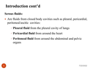
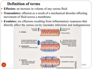
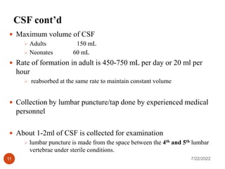
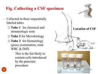
1. Cell counts on CSF and other
body fluids
CHAPTER 14
Aschalew K. (MSc.)
7/22/2022
1
2. Objectives
At the end of this chapter, students will be able to:
Define terms related to body fluids
Identify different types of body fluids
Define CSF
Define Serous fluids: pleural, pericardial, peritoneal (Ascitic),
and Synovial fluid
Explain the analysis of CSF
Discuss how to perform cell count on other body fluids
Apply QC measures in body fluid examination
Determine sources of error during CSF analysis
7/22/2022
2
3. Brainstorming questions
1. What are the common body fluids that used in the laboratory for
diagnosis of the patients?
2. What are serous fluids?
3. What is the difference between transudate and exudate effusions?
4. Would you explain about hydrostatic and colloid osmotic
pressures?
5. Where anatomical place of CSF in the body?
6. How to collect the specimen of CSF?
7. Would you remember method of CSF analysis?
8. What is the clinical significance of CSF analysis?
4. 14.1. Introduction to Body Fluids
Body fluids
Are ultra-filtrates of plasma
Fluids serve as lubricants as membranes move against each other
Body fluids commonly analyzed in hematology lab include:
CSF
Serous fluid
Synovial fluid (fluid from the joints)
Semen
7/22/2022
4
5. Introduction cont’d
Serous fluids:
Are fluids from closed body cavities such as pleural, pericardial,
peritoneal/ascitic cavities:
Pleural fluid from the pleural cavity of lungs
Pericardial fluid from around the heart
Peritoneal fluid from around the abdominal and pelvic
organs
7/22/2022
5
6. Definition of terms
Effusion: an increase in volume of any serous fluid
Transudates: effusion as a result of a mechanical disorder affecting
movement of fluid across a membrane
Exudates: are effusions resulting from inflammatory responses that
directly affect the serous cavity (includes infections and malignancies)
7/22/2022
6
8. Characteristics of Serous Effusions: Transudate
versus Exudate
Observation / Test Transudes Exudates
Appearance Watery, clear, pale
yellow, does not clot
Cloudy, turbid, purulent, or bloody;
may clot (fibrinogen present)
WBC count Low, < 1,000/µL with
> 50% mononuclear
cells
>1,000 cells/µL, with increased PMNs,
increased lymphocytes with TB or
rheumatoid arthritis
Red cell count Low, unless from a
traumatic tap
> 100,000/µL, especially with a
malignancy, trauma, or pulmonary
infarction
Total protein Low >3g/dl (or > than half the serum level)
Lactate
dehydrogenase
Varies with serum
level
Increased (>60% of serum level
because of cellular debris)
Glucose Not applicable Lower than serum level with some
infections and high cell counts
7/22/2022
8
9. 14.2. Cerebrospinal fluid (CSF) analysis
I. About CSF
Fluid in the space called sub-arachnoid space between the
arachnoid mater and pia mater
Protects the underlying tissues of the central nervous system
(CNS)
Serve as mechanical interface to:
prevent trauma
regulate the volume of intracranial pressure
circulate nutrients
remove metabolic waste products from the CNS
Act as lubricant
Has composition similar to plasma except that it has less
protein, less glucose and more chloride ion
7/22/2022
9
11. CSF cont’d
Maximum volume of CSF
Adults 150 mL
Neonates 60 mL
Rate of formation in adult is 450-750 mL per day or 20 ml per
hour
reabsorbed at the same rate to maintain constant volume
Collection by lumbar puncture/tap done by experienced medical
personnel
About 1-2ml of CSF is collected for examination
lumbar puncture is made from the space between the 4th and 5th lumbar
vertebrae under sterile conditions.
7/22/2022
11
12. Fig. Collecting a CSF specimen
Location of CSF
Collected in three sequentially
labeled tubes
Tube 1 for chemical and
immunologic tests
Tube 2 for Microbiology
Tube 3 for Hematology
(gross examination, total
WBC & Diff)
This is the list likely to
contain cells introduced
by the puncture
procedure
7/22/2022
12
14. CSF ont’d
II. Clinical Significance
Diagnosis of meningitis of bacterial, fungal, mycobacterial and
amoebic origin or differential diagnosis of other infectious
diseases
Subarachnoid hemorrhage or intracerebral hemorrhage
III. Principle of CSF analysis
CSF specimen examined visually and microscopically and total
number of cells can be counted and identified
i.e. Routine:- Gross examination, Cell counts and diff count,
Glucose and Protein values. Further more:- Cultures, Stains
(Gram, Acid Fast), Cytology, electrophoresis…..etc.
7/22/2022
14
15. CSF ont’d
IV. Specimen: the third tube in the sequentially collected tubes*
must be counted within 1 hour of collection (cells disintegrate
rapidly). If delay, store at 2-8oC.
All specimens should be handled as biologically hazardous
7/22/2022
15
Composition of normal CSF
Protein 15-45mg/dl
Glucose 50-80mg/dl
Urea 6.0-16mg/dl
Uric acid 0.5-3.0mg/dl
Creatinine 0.6-1.2mg/dl
Cholesterol 0.2-0.6mg/dl
Ammonia 10-35mg/dl
Sodium 135-150mEq/L
Potassium 2.6-3.0mEq/L
Chloride 115-130mEq/L
Magnesium 2.4-3.0mEq/L
16. Lab analysis
V. Equipment and Reagents: same as for WBC counting on whole
blood
VI. Method
Gross appearance
Is visual assessment of CSF for turbidity, color and viscosity
Normal CSF is clear in appearance with viscosity comparable to
water
Abnormal CSF may appear
cloudy, smoky, hazy, turbid or grossly bloody
7/22/2022
16
17. Method: cont’d
1. Turbidity
Turbidity may be graded from 0 to 4+ as follows:
0 = crystal clear fluid
1+ = faintly cloudy, smoky or hazy with slight turbidity
2+ = turbidity clearly present but news print easily read through tube.
3+ = news print not easily read through tube
4+ = grossly turbid, news print cannot be seen through tube.
Note:
Slight haziness indicates WBC count of 200-500/uL
Turbidity indicates WBC count of > 500/uL
Turbidity in general could result from large number of leukocytes
or bacteria, or increase in proteins or lipids
7/22/2022
17
18. Method: cont’d
2. Bloody specimens
Can result from a traumatic spinal tap (often occur in children)
Grossly blood specimen: may indicate subarachnoid hemorrhage or
intra-cerebral hemorrhage
If the specimen is bloody:
There is a need to differentiate between a traumatic tap and a
patient’s clinical condition
If the specimen in the 1st tube is bloody and is clear in the last tube, it
indicates traumatic tap
If the specimen has the same bloody color in all the three tubes, it
indicates clinical condition
7/22/2022
18
19. Method: cont’d
3. Color
Any color should be reported (N.B. normal CSF is clear)
Xanthochromia: is yellow coloration of CSF
Yellow color could be due to:
Result of release of hemoglobin from lysed red blood cells
increase in bile pigments
Specimen collected 2 hours post arachnoid hemorrhage
Xanthochromia of the CSF refers to a pink, orange, or yellow
color of the supernatant after the CSF has been centrifuged.
7/22/2022
19
20. Method: cont’d
4. Viscosity
Normal CSF has viscosity comparable to that of water
Viscosity may be associated with:
clotting
increased fibrinogen
a traumatic tap
or rarely may be associated with meningitis or subarachnoid
block
7/22/2022
20
21. Method: cont`d
Microscopic cellular enumeration
Cell count is performed by manual method
Electronic methods should be used with care
RBC counts have limited value
WBC counts are useful in developing differential diagnosis
NR:
0-5 WBC/µL or 0-5 x 10⁶/L
Neonates have higher value of 0-30 cells/µL
Low WBC with turbidity could indicate high concentration of bacteria
WBC between 100-10,000 x 106/L could indicate acute untreated bacterial
meningitis
7/22/2022
21
22. Method: cont’d
Differential count is performed when WBC >30 cells/ µL
Smear is prepared from centrifuged CSF settlement
Total Leucocyte Count on CSF
If CSF is clear
Mix well the undiluted CSF and properly charge the improved
Neubauer counting chamber
Count the cells in all 9mm2 area
Multiply the number by 10/9 to get the number of WBC/mm3
WBC/mm3 = Number of WBCs X 10
9
7/22/2022
22
23. Method: cont’d
If CSF is slightly turbid
prepare a 1:10 dilution with 10% acetic acid (1 drop CSF and 9
drops 10% acetic acid)
count the cells in all 9mm2 area in the improved Neubauer
counting chamber
Multiply counted cells by 100/9
If CSF is purulent, proceed with the dilution and counting as for a
blood sample
7/22/2022
23
24. Method: cont`d
Technique for Counting Mixture of WBC and RBC
This is done to find the true WBC count when the CSF is bloody
due to
Traumatic tap
Perform the WBC and RBC counts on the patient’s blood and CSF.
Multiply the ratio of RBC count on CSF to RBC count on blood
by the blood leucocyte count and subtract this product from the
WBC count of CSF.
RBCCSF x WBCblood= WBCADDED
RBCBLOOD
Corrected WBCCSF = WBCCSF – WBCADDED
7/22/2022
24
25. Excercise
Example:
RBCBLOOD = 5 x 10⁶/mm3
RBCCSF = 2,500/mm3
WBCBLOOD = 12 x 103/mm3
WBCCSF = 70/mm3
WBCADDED = 2,500 x 12,000
5 x 106
= 60/mm3
There fore:- Corrected WBCCSF = 70 – 60 = 10/mm3
7/22/2022
25
26. Method: cont`d
Differential Leukocyte Count on CSF
Centrifuge the CSF at 2500 rpm for 10 min
Remove supernatant (can be saved for other analysis)
Re-suspend the sediment
Prepare a smear from the re-suspended sediment
Stain using Wright stain
Wash off stain with water and air-dry
Identify the types of leucocytes (PMNs or mononuclear cells) and
their number may be expressed as percentage of the total count
Count at least 100 cells using the oil immersion objective
Artifacts due to distortion of cells can lead to misidentification
7/22/2022
26
27. Method: cont`d
Differential Leukocyte Cont`d
Cells in the spinal fluid may include:
granulocytes
mature or reactive lymphocytes,
momonuclear phagocytes,
plasma cells, blast cells and
malignant cells (indicating primary tumors of brain and
spinal cord)
Others like nucleated red cells, and intracellular bacteria.
Other rare cells unique to spinal fluid (ependymal cells,
choroidal cells) may be found
7/22/2022
27
28. VII. Quality control
Count both sides of hemocytometer (18 mm2 area) for the total
WBC
Increasing the number of cells to be counted for differential count
(instead of 100 cells count 200 cells)
Check staining quality (e.g. staining time, pH, filtering)
Proper centrifugation (Speed and time)
Properly follow SOP
7/22/2022
28
29. VIII. Sources of errors
General sources of error in dilution, charging, counting ,
calculating ….etc that were discussed for WBC count also apply
here
Delay in analysis
Centrifugation time and speed during sediment preparation for
Diff count
Staining time
Improper handling of sample
7/22/2022
29
30. IX. Interpretation of CSF diff count
Normal range <5 cells/mm3
Increased Neutrophil indicate bacterial infection
Meningitis
Sub arachnoid hemorrhage
metastasis
Increased Eosinophils indicate:
Systemic parasitic and fungal infection
systemic drug reaction
Idiopathic eosinophilic meningitis
Increased basophils indicate
Chronic basophilic leukemia
Chronic granulocytic leukemia
Purulent meningitis
Inflammatory processes
Parasitic infections 7/22/2022
30
31. Interpretation of CSF diff cont’d
Increased lymphocytes indicate:
viral infections
viral meningeal encephalitis
Aseptic meningitis syndrome (majority
of the cases)
Fungal meningitis
Partially treated bacterial meningitis
Syphilitic meningeal encephalitis
Non-infectious cases of increased
lymphocytes may indicate multiple
sclerosis.
7/22/2022
31
32. Increased monocytes (>2%)
indicate:
Tuberculosis meningitis
Syphilis and viral encephalitis
Meningeal irritation
Subarachnoid hemorrhage
Leukemic infiltration of the
meninges and infectious state
Increased macrophages
Infectious diseases
CNS leukemia
Lymphoma
Malignant myeloma, and other
metastatic tumors spreading to
the brain 7/22/2022
32
Interpretation of CSF diff cont’d
33. Plasma cells increased in:
Viral disorders such as herpes
simplex infection
Meningeal encephalitis
Syphilitic involvement of the CNS
Post subarachnoid hemorrhage
Erythrocytes increased in:
Traumatic tap specimens
Patients with a bleeding
subarachnoid hemorrhage or
intracerebral hemorrhage
Chronic myelogenous leukemia or
erythroleukoblastic conditions
7/22/2022
33
Interpretation of CSF diff cont’d
34. Associated findings
Glucose and protein values should be correlated with macro and
microscopic hematological findings
Generally decreased glucose in CSF with normal blood
glucose indicates bacterial utilization correlates with
increased PMNs in the Diff
Elevated protein suggestive of inflammatory reaction or
bacterial infection
Viral infections will not have dramatic effect on either CSF
glucose or protein levels
7/22/2022
34
35. Other body fluids
Pleural fluid, pericardial fluid, peritoneal/ascitic fluid, synovial
fluid, semen
Same protocol followed as with CSF
Macroscopic examination for:
Turbidity
Color
Viscosity
Microscopic
Total white cell count
Differential count using Wright`s stain
Gram stain and culture is done in bacteriology laboratory
7/22/2022
35
36. Review Questions
1. What is the function of body fluids?
2. What is the role of CSF in our body?
3. Mention at least three different types of body fluids
4. What is the difference between transudates and exudates
5. Define serous fluids
6. Define CSF
7. How do you perform the total leukocyte count on a:
a) clear CSF b) slightly turbid CSF c) purulent CSF
8) How do you correct the total leukocyte count to a true value
when the count is performed on a sample of CSF that is slightly
turbid due to traumatic tap?
9) How is the differential leukocyte count performed on a sample
of CSF?
7/22/2022
36