Gutell 027.faseb.j.1993.07.0223
•
1 like•304 views

This document presents the complete small subunit ribosomal RNA (SSU rRNA) sequences of Giardia ardeae, G. muris, G. duodenalis, and the diplomonad Hexamita. The sequences are compared to analyze phylogenetic relationships. The Giardia sequences range from 1432 to 1453 nucleotides in length, while Hexamita's is 1550 nucleotides. Secondary structure analysis reveals both typically eukaryotic and variable regions. A phylogenetic tree groups the Giardia species separately from Hexamita and Vairimorpha, suggesting they represent a separate kingdom within the domain Eucarya.

Recommended
More Related Content
What's hot
What's hot (20)
Viewers also liked
Viewers also liked (20)
Similar to Gutell 027.faseb.j.1993.07.0223
Similar to Gutell 027.faseb.j.1993.07.0223 (20)
More from Robin Gutell
More from Robin Gutell (20)
Recently uploaded
Recently uploaded (20)
Gutell 027.faseb.j.1993.07.0223
- 1. Unique phylogenetic position of Diplomonadida based on the complete small subunit ribosomal RNA sequence of Giardia ardeac, G. muris, G. duodenalis and Hexamita sp. HARRY VAN KEULEN,5’ ROBIN K. GUTELL,t MICHAEL A. GATES SCOTT K. CAMPBELL, SThNLEY L. ERLANDSEN,1 EDWARD L. JARROLL’ JAROSLAV KULDA,S AND ERNEST A. MEYER11 Departinent of Biolog Cleveland State Universit Cleveland, Ohio 44115, USA; tMolecu!ar, Cellular and Developmental Biology University of Colorado, Boulder, Colorado 80309, USA; tDepartment of Cdl Biology and Neuroanatomy, University of Minnesota Medical School, Minneapolis, Minnesota 55455, USA; SDepai.tnein of Parasitolog Charles University, Vinicna, 12844 Prague, Czechoslovakia; and ‘Department of Microbiology and ImmunoIog Oregon Health Sciences University, Portland, Oregon 97201, USA 0892-6638/9310007-02231$O1.50. © FASEB 223 ABSTRACT Complete small-subunit rRNA (SSU-rRNA) coding region sequences were determined for two species of the intestinal parasite Giardia: G. ardeae and G. muris, both belonging to the order Diplomonadida, and a free- living member of this order, Hexamita sp. These se- quences were compared to published SSU-rDNA se- quences from a third member of the genus Giardia, G. duodenalis (often called G. intestinalis or G. lamblia) and various representative organisms from other taxa. Of the three Giardia sequences analyzed, the SSU-rRNA from G. muris is the smallest (1432 bases as compared to 1435 and 1453 for G. ardeae and G. duodenalis, respectively) and has the lowest G + C content (58.9%). The Hexamita SSU-rRNA is the largest in this group, containing 1550 bases. Because the sizes of the SSU-rRNA are prokaryotic rather than typically eukaryotic, the secondary structures of the SSU-rRNAs were constructed. These structures show a number of typically eukaryotic signature se- quences. Sequence alignments based on constraints im- posed by secondary structure were used for construction of a phylogenetic tree for these four taxa. The results show that of the four diplomonads represented, the Giardia species form a distinct group. The other diplomonad Hexamita and the microsporidium Vairimorpha necatrix appear to be distinct from Giardia.-van Keulen, H., Gutell, R. R., Gates, M. A., Campbell, S. K., Erlandsen, S. L., Jarroll, E. L., Kulda, J., Meyer, E. A. Unique phylogenetic position of Diplomonadida based on the complete small subunit ribosomal RNA sequence of Giardia ardeae, G. mw-is, G. duodenalis, and Hexamita sp. FASEBJ. 7: 223-231; 1993. Key Words: ribosomal RNA Diplomonadida Giardia Hexamita p/zylogenetic relationship ALTHOUGH MORE THAN 40 DIFFERENT SPECIES of the genus Giardia have been described (for review, see ref 1), presently only five species can be well defined, based on morphological and electrokaryotype characteristics. These are Giardia duo- denalis, G. ardeae, G. muris, G. agilis, and G. psittaci (2-4). All are cosmopolitan intestinal parasitic protozoa of several ver- tebrate classes (1). In humans, Giardia duodenalis (syn. G. lam- blia or C. intestinalis) can cause a chronic enteritis (1); the name G. duodenalis will be used here. This same morphologi- cal type of Giardia has been reported from nonhuman sources also,resulting in the establishment of several axenic isolates from different host species that were previously designated as unique Ciardia species (1-6). These various isolates of C. duo- denalis, although similar morphologically, display variations in their karyotypes (7), and this has engendered some con- troversy about their taxonomic status (1). Clearly, both sys- tematic and epidemiological studies of the genus Giardia would benefit greatly if reliable identification criteria were available. Adequate ta.xonomic criteria could include electrophoretic karyotype analysis, DNA-level comparisons by restriction site analysis, and sequence analysis of selected genes. The ribosomal RNA (rRNA)2 genes are conserved in function, organization, and sequence in all organisms, yet their intrin- sic variability permits detailed analysis of sequence similar- ity. Therefore, these genes, especially the highly conserved small subunit (SSU) rRNA genes, have been used exten- sively in analyzing phylogenetic relationships (8). G. duodenalis, G. math, and G. ardeae differ in the organiza- tion of their rRNA genes (9). This study presents the SSU- rDNA sequences of C. muris and G. ardeae to provide a broader database for discussing phylogeny among Giardia species and to provide useful information for designing molecular probes to identify individual Giardia species. These SSU-rDNA sequences are compared with each other, with those of C. duodenalis (syn. C. lamblia (10)) and the free- living diplomonad, Hexamita sp., and with several represen- tative pro- and eukaryotic SSU-rRNAs. Clustering of the Giardia species in a phylogenetic tree based on SSU-rRNA sequences appears to place them in a group separate from Hexamita and Vairimorpha. We propose that these organisms represent a separate kingdom in the domain Eucarya (11). MATERIALS AND METHODS DNA sources The Giardia ardeae and G. daodenalis isolatesused here were described previously (7). The source of C. muris DNA is from cysts isolated from CFI mice (7); the original host was a ‘To whom correspondence should be addressed, at: Department of Biology, Cleveland State University, 1983 E. 24th St., Cleveland, OH 44115, USA. 2Abbreviations: rRNA, ribosomal RNA; SSU, small subunit; kbp, kilobase pair; nt, nucleotides.
- 2. 224 Vol. 7 January 1993 The FASEBJournal VAN KEULEN ET AL. naturally infected golden hamster (12). Hexamila sp. was iso- lated from a lake in Czechoslovakia. DNA techniques Plasmid DNA containing various cloned rDNA from Giardia (9) was isolated by the alkaline minipreparation procedure with the modifications described previously (9). Genomic DNA from Hexamita was isolated in the same manner as Ciardia genomic DNA (7). All cloned DNA used for subclon- ing in M13 vectors was digested with the appropriate restric- tion enzymes and separated from vector DNA by agarose gel electrophoresis. The DNA fragments were purified by the freeze-phenol procedure (13). DNA sequence analysis Sequence analysis was by the chain termination procedure (14). The vectors M13mp18 and mpl9 were used with host strain JM 109. Isolation of single-stranded DNA and the se- quencing reactions were as described in the protocols sup- plied with the Sequenase and TAQuence kits (United States Biochemicals Corp., Cleveland, Ohio). The sequences for both strands were obtained by using the universal sequenc- ing primers for the appropriate clones and were sup- plemented with internally positioned oligonucleotide primers. The position of the primers are given relative to the sequence of C. duodenalis SSU-rDNA (10). For C. ardeae, primer A, reverse, these were positions 301-285, 5’GCTCTCCGGAGTCGAAC3’, for Ha, reverse, positions 1214-1200, 5’GCCGGCTTGGCGGGTCG3, and for C. muris, primer 1, forward, positions 10-22, 5’GAThCTGCCGGAC3. Primer I is a universal primer and was also used to complete the C. ardeae sequence. The se- quence around the 3’ end of the G. duodenalis SSU-rDNA was determined for five different isolates (MR4, Dl, P1(9), AB, and CM). In the case of Hexamita rDNA, fragments were generated with Saa3AI and RsaI to provide fragments small enough to sequence both strands. Sequence analysis was by electrophoresis on 8% polyacrylamide sequencing gels with 8 M urea. Areas with extreme band compressions were ana- lyzed by using dITP with Sequenase or 7-deaza-dGTP with TAQuence in the reaction labeling mixture. When necessary, the standard 8% polyacrylamide, 8 M urea sequencing gels were substituted by 8% polyacrylamide, 8 M urea, 20% for- mamide gels. The resulting nucleotide sequences were aligned and ana- lyzed with the DNASTAR programs (DNASTAR Inc., Madison, Wis.). Values for total sequence similarity calcula- tions were obtained from global alignments over 3411 nucleo- tides according to Woese et al. (15). The sequence similarity values were calculated as similarity = matches/(matches + mismatches + gaps/2). A gap larger than five nucleotides was taken as five (16). The similarity values were converted to distance values (the number of evolutionary changes per 100 positions) (16). These distances were used to construct a phylogenetic tree by using the neighbor-joining method of Saitou and Nei (17). The algorithms of Nei and Miller (18) were used for tree construction. Secondary structure The secondary structure model is based on comparative se- quence analysis (15) and drawn in a format similar to that of the Escherichia coli 16S rRNA model, which may be regarded as the standard or prototype structure (19). The figures were prepared with the assistance of a new RNA graphics program, XRNA, developed by B. Weiser (unpub- lished results). Sequences were manually aligned with the alignment editor AE2, developed by T. Macke (unpublished results). Identification of the variable regions in V1-V9 and numbering of some of the stems (E8-1) are as described by Neefs et al.(20). RESULTS The rDNAs of Ciardia duodenalis, G. ardeae, and C. muris were cloned, and physical and genetic maps were constructed (9). The SSU-rDNA from Hexamita was identified by Southern blot analysis of genomic DNA digested with various restric- tion enzymes and probed with specific Giardia rDNA probes (data not shown). A 1.7-kilobase pair (kbp) PstI fragment ap- peared to contain the entire SSU-rDNA and was cloned in pUCI8. Subclones that covered relevant regions of the SSU- rDNA were prepared in M13 DNA by using the available restriction enzyme recognition sites. The entire SSU-rRNA genes of C. ardeae, G. muris, and Hexamita were sequenced from both strands. Although the entire sequence of the C. duodenalis SSU-rRNA gene has been published (10, 21), some sections were resequenced due to minor differences ob- served, especially at the 3’ end of the SSU-rRNA gene; the differences are included in the presented sequence and are explained later. The sequences of the Ciardia and Hexamita SSU-rRNAs are shown in Fig. 1, where they are aligned with each other and with that of E. coli. The 5’ and 3’ boundaries of the ma- ture SSU-rRNA genes were defined by comparison with previously reported sequences (10, 21). The sequences of C. duodenalis were taken from 50gm et al. (10) (C. lamblia) and Healey et al.(21) (C. inlestinalis). Three alterations are included in the C. duodenalis sequence, based on sequence analysis of the SSU-rDNA of five different C. duotfrnalis isolates: at position 1241, an extra G was found- around position 1275, three Cs instead of two were found; and the last four bases were CTCG in five different isolates of C. duodenalis, instead of TCTA. These alterations were in- cluded in the alignments and in the construction of the secondary structure. This sequence analysis shows that C. muris has the smallest sized SSU-rRNA of the three Ciardia species, namely, 1432 nucleotides (nt), followed by 1435 for C. ard.eae and 1453 for C. duodenalis. The Hexamita rRNA is the largest, with 1550 nt. The G + C contents of C. duodenalis and C. ardeae are similar, 74.8 and 71.2%. However, the G + C content of G. math SSU-rRNA is lower, namely, 58.9%; that of Hexamita is 51.4%. Secondary structures of the three Ciardia and Hexamita SSU-rRNAs were generated; the one for C. muris is shown as a representative structure in Fig. 2. The regions that show the highest degree of variation are identified as V1-V9 (20). Because these regions are variable in both primary and secondary structure, the secondary structures of all four diplomonad rRNA are shown separately in Fig. 3 for com- parison of similarities and differences in some of the variable regions. An inventory of specific nucleotides of all Ciardia species and Hexamita was made by using the domain-specific bases and base pairs originally identified by Winker and Woese (22). Based on the E. coli 16S rRNA positions, these were scored for the presence of bases that represent each of the three domains Bacteria, Archaea, Eucarya, or combinations of these. Because SSU-rRNA of the microsporidium Vairimorpha necatrix resembles that of diplomonads (see be- low), Vairimorpha was included in this analysis. The results
- 3. 104 79 77 GG 76 GG 76 GA $2 213 -- 161 139 )- 13$ I 13$ )- 16$ 273 233 251 236 236 233 391 371 370 374 374 404 -- 497 99 390 . 389 9c 390 9c. 390 434 376 507 506 303 503 556 Lcd GCACGCAGGCGIGUUUGUUAAC GkAAkUCCCGG( ( CCGGMCCMUGMJACUGC ACC<- - 651 Oa*e cGGcGucG I ------- .-------------. ( . <#{231} q#{231} igg I N99C5 99C9 09UU99 567 G.iu,.s G5CcGGA;cCI---- ------------------ 4....... g#{231} ugg#{231} iguqagua 566 GJ.rmair..lls GccCGUAcUZI-------- .------------. (--. l---------------- <c .c ca iqg - uc 564 GJ.b1a GCCCGUAGUl ------- .-- I ---j 2 56 He.ita GcUCGUAGcCGI--------- -------------. (----. I ---------------------------ttati 616 Lcd! - - ---------- ----------------------,u G GG #{149}90CC 4 1?. 713 o ccaccqcoc 9051 5qog------- --. i.qq------------- 64$ Oi, gu a sac- ----.uqt.qq------->--- 651 GJ.aIils cc i qcqc ccc 99------->--- 0.1bHg cc i scgca#{231} ccci rs- --- N ai iaocgcu#{231} ,a j iacaaa iaauacauuqauccocac C aauoaaaauauD- 3Q 00 G Z 90 GQG G _ .A1 “IGG531 XCSCCAM 739 #{163}soU WCGGC GMG)GCGGCCccCG&CGA1 SAct .- GGGACW ACCCAMaC GGA03q 33$ Ga*..e J m a_I, :j 773 0 IJ 799 GJ.b3a Ii KisUa G6 VUUCAACAGC u I6 cc I--. 39 IC USAC) Al _ - .CIC GGGIGU&CGGC 3GGCC #{149} OC IGW 2C5U 952 9ucq- -GGCI IGMA) -20C cG t G ( MCRMG0CP 2GGI 1UUGC $84 - 014g,... . - .GUcAUCG?J -- (GMA) XCUCI 900 4 GC $90 - o9c9o9c ) -. cGccGGccGw ci c 917 - -. W2GCUCi c 91$ U ---- 3UU- 90C 973 - - A 5W3G 1034 G 1 #{163}GCG SC?. XUCCl 9 X c GG n CACC .C OGGU.C)Gc 701 ‘ - 970 Gc k xcc s a wcc cci : -cacs - -- 976 s I ccacc-- - - 1033 s ccG -GG > 1034 25 I GC9 - ------- - .---- 1063 icc na GcCA I Ul IAAlCDUOCC osG 034 C- 1139 -- 1 1 --cGGGcAlz I C AGI------ .- Cc 1043 -- I - I - ----GG0QCAIX I IGC I 1Al-- -. C - Cc 1049 1’ 4- ----CGGWGUGCAUG GCCGCCklGCGU GGC (GMCCCGUnC (UcCAUU) GcGACAAcCCGCkGGI --- ( <c 1016 -- .- I - ------CGGlGWCA I ;ccc IGCI W) ICGACAACGSCCG12ACCGGI - - 4 - Ca 1077 - .-- - I GCC 21 I 2GUUUAA ACUCUGC CW UU) ICGUUJ 10 XG1 23CMCCAI -- 4.. ( 113$ ----->co ;GAAcw.scGl G5CGCkCCC1 1244 cacac (-ccqc) ququgg>- . I 1141 qg)qg). .-------. 7 1147 cgcqg(-gcq-) oogcgg>-. . 7 1179 o9o9g(-qcq-)coqc- . 7 1190 .-- 6AGcGcc I GcccwAGAcAcxuooGcAcG03w.cw.cA 1243 cAuAcMAG?AAocG&cclx 0 < - -i - )GA #{163}334 03C .- <qc.qcgo#{231}CGCGCG0t GCIS4GICCGG9cg 9co aq) -0i9uwJwawG5u..C -C)OC #{163}224 Gos ----- --. uqwac1 XUa .#{248} -- 2G 1220 OGG------------. .- <cc.q-. )2AC goq aqc> -Cc’-w??-1-w2MN-wCvwC -C)OC 1245 GG- -- . - ----. ;a)CACGC .109..- VC> ----------------- .-c)cc 1246 ‘ ----- -- I <gccaqoc .c --------- U)UU 1322 : ai GGAMS GG& 9CVu*C #{163}441 :4 Al GGMU a IqC- #{163}346 a ai GGAA z - #{163}342 ,aA.zn.,tMAd,.cGMa=nwunn 1363 ccccGcGcAccAGGakcGcGccos (cCcCCAc) #{163}366 (UW.WAC) 1450 C- (uucg-) (GGAA)CCUGcGGUUGGkUCAcU I 1542 -- (ccccq-) ----gu9a*cGGcGkcGAGccc;GcGccuGGAGGAAGGsG.AGUcGuMcsAGGuA-JAGG WGAA)CCUGcGG1OGGMC-CW.G I 1433 - (gauga.) ----ccqaaCACCGACGMCCGGAGGCUUGGACGAAGGAAAGUcGUMCAkGGW-1Cn75CG (M)CCUGCGGMGGMC--AG I 1432 - (ccgc-) -------q--GGGccGcc’7ccccc0cGccuGGAGGAAcGAGAAUAAcAAGGuM1uccGUAGG (AP.) CCUGcGGMJGGAUCC-----C I 1452 - (ogcqc-) ------q*aGGGAocccccGccuGG&GGAAGGAcM.GucGUA&cAAGGU&-4cGUAGG (AA)CCUGCGG&UGGAUCC-----UCW.G I 1454 9’, (cqcq-)qcaugcacqaaquuCtcGAAcCCCAAtUGGAGGAACCAG?.AGUCkUMCAAGGCU-GCUGUAGG (GGAA) CCUGCAGcCGGAUC?.----UU&GCCI 1550 Figure 1. Alignment of the nucleotide sequences of Giardia and Hexamita with E. coli SSU-rRNA. The entire nucleotide sequences are given for Giardia ardeae, C. muris, and G. duodenalis, divided here into C. lamblia, based on Sogin et al. (10), and for G. intestinalis, based on Healey et al. (21). The names are as they appear in GenBank. The sequence of E. coli l6S rRNA is GenBank number V00348. Dashes (-) indicate adjustments of the sequence to allow for optimal alignment. PHYLOGENETIC POSITION OF DIPLOMONADIDA 225
- 4. uc U U AUOA S50 -0-00-0 1050U . AA0 0C*A00AAAeA%0 e U A.0AS U OOUAA #{216}#{216}U0 #{149}1 II 11111.111 #{149}11 a iii ii ji.i ii oVi ;UOOoCA.o4OoUOOUUAU0.OA 00 0A 0-a U00000 aaeouo aQuA U U I c-a IA CAQ Qi 0-0 #{149}Oa A j ( Vs AiIuu iibA A0 UUA #{149}U-A AA:A_ U A 0-0 U S A C a o C A A A$00-a #{149}A ,cL AA0-C a / 0-0 U,0, U #{149}#{149}141U OU 0AQ’JAC0 U-A A #{149} a ,C0 U #{149} AU-A #{149} 0l A-U A U A A I A....UA 0 A. GA-U 0A0/ A COU00 A O 0 0 0 CCCOO 0-0 0#{149} A A0 c#{193}O #{176}0....* 000.U U o V4 11111 I. U a-C / U UA O CU IAACC io / V5 ,A0#{149} A0.AO: #{149} 0 A A ( GAO #{176}CAaAO’ 50_A U SU A A/00O00U 4 U0. #{149}a-C uu0OA SC0 a O”A .0-0 A a 00A %“UA 0 0U#{176}%A - A 000A000 O a Au 000 AA 0 caUuUCAC A l555110_ #{149} 0 - 450_A U 500 A-U 0 A 0 -0-0 A0-0 0 0A A00A #{149} 0AU0 Co A I - AA 0 U 150 Vs U .. #{149} U UA A 50 U#{149} 000AOUAU00000 UA I 0a-C-U C 0-C C A-UI C A AC UAa00’,#{176}0, : S50-t Asa iUUOA U000A 00#{149}A0A #{149} A#{149}a I #{149}% A A 0-0 Iull.lulSI.I % U a,,A0 C CIeos::1 AUUOAOOA0000UA A #{149}U 0 CA o A #{149} U400A u.s #{149} ACUQUUOOCCOU 0 AAUOQQA Al C 0 U * sum A ts#{176},%*u 0 C oJ_0.12oo ,OUACOOU A .a U0 ,. . I 0 AA C o U 0 A #{149}a0,% %_a AGaCa050.eAa A 1300-c 0 A aI U _oA #{149}1400#{149}a,cCaUCH 11111 C A U’AUC00UAa0U uA,iI. A 0-a .SIIul#{149}IulAC a A 350 #{149} A U-A aUA0000UOOA” #{149}A c-a 0 #{149} A*O - - 0:a O a-CA 5I -4 _ S0-0 - *0 A-U 0S A a I c-a I 00d’,AO0CAOC S #{149}A IA#{149}0 I ‘AC’ A I Aa.u u_O #{149}C CA -U.S o AaUOOa hi II II 0 C-a- #{149} i 0CA ca #{149}UAUO U-A A 00 0U0* o o Aa,,O0A.A ... / C 0 C A #{149} a A Vg U-. _Aa 0 o O a0 0-0A #{149}u_C_S c-s0-0 U.a 250 Q:oA _,‘.ioo a-CAa #{149}A p - a-c0 0 #{149}A 0 A A#{149}0 O A..U0 a-o1350 a-c 0-0 U U a A #{149}SU%t1 - Es -1 AUaA U0 a-C / 5:: *00 200 0_GA #{176}I CO COCUGOC IS 111111 A o*#{176} 000A000 0 U- 0AAC V2 150 C U A C O A o A A A0 0 Figure 2. Secondary structure of SSU-rRNA of Giardia muris. Every 10th base is marked and every 50th is numbered in the sequence beginning at the 5’ end. The numbering of variable regions as V1-V9 and the eukaryotic specific stem E 8-1 are as described by Neefs et a!. (20). Region V6 is absent in eukaryotes but is shown here to indicate where its prokaryotic equivalent appears. The stem structure is eukaryotic stem 35 (20). Regions V2 and V4 are left unstructured. 226 Vol. 7 January 1993 The FASEB Journal VAN KEULEN ET AL.
- 5. Vi vs r vs Giardia duodenalis Ci ardi a ardeae vi Giardia muris V. Hexami ta sp PHYLOGENETIC POSITION OF DIPLOMONADIDA 227 Figure 3. Secondary structure model of diplomonad rRNAs. Line drawings of SSU-rRNA of all four diplomonad rRNAs are given. The names of the organisms are shown in the figures. The regions VI to V9 are indicated as in Fig. 2.
- 6. E. coli Position’ C. muris G. duodenatis G. ardeae Hexainita Type Vairimorpha Base pair TypeBase pair Type Base pair 8 Ub ar(eu)b C eu C eu 9:25 C:G eu+ar C:G eu+ar C:G eu+ar 10:24 C:G ar U:A eu A:U ba # S 33:551 A:U eu+ba A:U eu+ba A:U eu#{247}ba 52:359 G:C eu+ar G:C eu+ar C:G (ba) # S 53:358 C:G eu+ar C:G eu+ar C:G eu+ar 113:314 C:G eu+ar C:G eu+ar C:G eu+ar !21 A eu A eu U (ba) # S 292:308 A:U eu A:U eu U:U u # S 307 C eu (ba) C eu (ba) U (eu) # S 335 C ba+ar C ba+ar A eu #S 338 A eu+ba A eu+ba A eu+ba 339:350 C:G eu + ba G:G u A:U u # 5 341:348 U:A eu U:G u U:A eu 5 361 C eu+ar C eu+ar C eu+ar 365 A eu + ar A eu + ar A eu + 367 U eu+ba U eu+ba U eu+ba 377:386 C:G eu+ar U:A (eu) #{149} G:C (ba) #$ 393 A eu+ba A eu+ba A eu+ba 500:545 G:C ba+ar U:A eu U:A eu 514:537 G:C eu+ar G:C eu+ar A:U u#5 549 C eu+ba C eu+ba U ar #S 558 G ba A eu* U ar#5 569:881 G:C eu G:C eu G:C eu 585:756 U:A eu C:G ar* U:A eu+ar$ 675 U eu+ar U eu+ar U eu+ar 684:706 G:C eu+ar G:C eu+ar G:C eu+ar 716 C eu+ar C eu+ar C eu+ar 867 C eu + ar C eu + ar U eu + ar# S 880 U eu U eu U eu 884 G eu G eu G eu 923:1393 A:U eu+ba A:U eu+ba A:U eu+ba 928:1389 G:C ba+ar A:U eu A:U eu 930:1387 G:C eu G:C eu A:U ar # 5 931:1386 G:C eu+ar A:U u #{149} A:U u 933:1384 A:U eu+ar A:U eu+ar A:U eu+ar 962:973 U:G eu U:G eu U:G eu 966 U eu + ar U eu + ar U eu + ar 974 A ba C ba A ba C ba A ba (5) 1098 G eu + ar G eu + ar A u # 5 1109 A eu+ar A eu+ar A eu#{247}ar 1110 G eu+ar G eu+ar G eu+ar 1194 A eu G ar A eu G ar A eu (5) 1201 U eu U eu C ba+ar# 5 1211 U eu+ba C eu C eu U eu+ba C eu (5) 1212 A eu+ar A eu+ar A eu+ar 1381 C eu+ar C eu+ar C eu+ar 1487 A eu A eu G ba+ar# 5 1516 U eu G ar (ba) A ba # 5 ‘The positions are numbered according to Escherichia co/i 16S rRNA; the signatures are indicated as being like the signature sequence of Bacteria (ba), Archaea (ar), or Eucarya (eu) or a combination of two of these, based on the data from Winker and Woese (22). Unique sequences are indicated as U. The signatures in parentheses indicate minor forms of a signature feature. .6The signatures that are shared among all Giardia species are shown in the columns marked with superscript b; the ones that differ are indicated slightly to the left and right of these two columns. Indicates signatures that differ between all 46 shared Giardia signatures and Hexamita; # indicates where the Vairimorpha signatures differ from the 46 Giardia signatures, and S indicates where Vairimorp/za differs from Hexamita in the same 46 signatures. The (5) indicates the difference between Vairimorpha and Hexamita in the three signatures that vary in the Giardia group. 228 Vol.7 January1993 The FASEBJournal VAN KEULEN FT AL. are presented in Table 1 and further analyzed in Table 2. In Giardia SSU-rRNA, two positions are typical for the Archaea (three in the case of C. duodenalis), two for the Bacteria, and three for both Archaea and Bacteria. In contrast, 20 posi- tions are like Archaea plus Eucarya, and 14 to 15 are unique for Eucarya. For Hexamita these numbers are 3, 1, 18, and 15, respectively. The sequence of Hexamita shows three unique TABLE 1. Comparison of 16S-lik.e rRNA sequence signatures signature features. Note that of the signature sequences described by Winker and Woese (22), 22 are shared between Archaea and Eucarya, 6 between Bacteria and Eucarya, 12 between Archaea and Bacteria, and 9 are unique among the three groups. Of the 49 positions, all Giardia species have 46 in common. The signatures ofHexamita differ in 11 positions from the ones that all Giardia species have in common. The
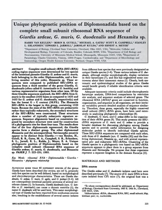
- 7. Oxytricha nova - Tetrahymena thensophiia 0.01 TABLE 2. Distribution of signature sequences PHYLOGENETIC POSITION OF DIPLOMONADIDA 229 Eu Eu + Ar Eu + Ba Ar + Ba Ar Ba unique Giardiamuris 14(15) 20 8 3 2(1) 2(3) 0 Giardiaduodenalis 14(15) 20 7 3 3(2) 2(3) 0 Giardiaardea 15(16) 20 7 3 2(1) 2(3) 0 Hexamita 15(16) 18 7 1 3(2) 1(3) 3 Vairimorpha 12(13) 15 5 2 3 3(6) 5 The number of domain-specific signatures is indicated. The abbreviations are as in Table 1. The number in parentheses indicates minor forms of a signaturefeature. total number of signature differencesbetween Hexamita and C. muris is 13,whereas these totalsare 14 with C. ard#{128}aeand 12 with C. duodenalis, corresponding to 26.5, 28.5, and 24.4%, respectively.The differencein signature sequences among the three Giardiaspecies is between 2 and 6% (see Ta- ble 1 and Table 2). Comparison with Vairimorpha shows a larger number of different signatures, a total of 20 of the shared 46 of Ciardia (43%). Structural similarity values and distance data were gener- ated for the SSU-rRNA of the three Ciardia species, Hexam- ita, and a number of representative organisms from eubac- terial (Bacteria), archaebacteria! (Archaea), and eukaryotic (Eucarya) origin. The entire SSU-rRNAs sequences were aligned on the basis of primary and secondary structure con- servation of the 16S-like rRNA (19; and R. R. Gutell, un- published analysis). A phylogenetic tree was constructed us- ing the neighbor-joining method (Fig. 4). The Ciardia cluster together, leaving the other diplomonad Hexamita and the microsporidium Vairimorpha to group with other eukaryotes. DISCUSSION Sequence similarities of the SSU-rRNA genes within a genus can be as close as 98.6 to 100% (23). However, within the ge- nus Giardia, the sequence similarity is much lower. The similarities of Ciardia muris with C. ardeae and C. duodenalis are only 80 and 76%, respectively. Within the suborder Diplomonadida a comparable result is obtained when Hex- amita sp., a free-livingrepresentative of this group, is in- cluded (approximately 63% similarity with all Ciardia). All four organisms have a SSU-rRNA that shows a relatively low degree of sequence similarity, which results in long branch lines in the estimated phylogenetic tree (see below). However, the diplomonads also share a number of charac- teristic features. First of all, they have the smallest rDNAs se- quenced so far, with the exception of the SSU-rRNA gene of Vairimorpha necatriy, which is only 1244 nt in size. The SSU- rRNA of Ciardiamuris is the next shortest, with 1432 nt; all these rRNAs are much smaller, for instance, than 16S rRNA Ie.nOpus laevlB Zea maya I__I chlamydoinonaa reinharcftii I SaCCharOmYCCS ce.revislae Cracllariopsls op. Plasmodium malarlae LlictyOBtelium discoideum rrypan050ma brucel - crith1da fascicuiata - Euglena grac.iLia Valrlmorpba neca trix Hexamlta op. Glardia ardeae Giardia duodezza1.j Giardla murio II Halobacteriimt vol canil - Suit olobua soltataricus Thezmotcga maritima Hacberlch.ia coil Figure 4. Phylogenetic dendrogram constructed for SSU-rRNA. The tree was constructed using the neighbor-joining method. The horizontal component of separation represents the evolutionary distance between organisms, measured in units of the average number of base changes per sequence position.Sequences are quoted by accession number in Genbank and EMBL nucleotide sequence libraries: Xenopus laevis X02995; Zea mays K02202; Chiamydomonas reinhardtii M32703; Saccharomyces cerevisiaeM27607; Gracilariopsissp. M33639; Ox,- tricha nova X03948; Tetrahyinena therinophila M10932; Plasmodium malariae M54897; Dictyostelium discoideum K02641; Trypanosoma brucei M12676; Critlzidiafasciculata X03450; Euglena gracilis M12677; Vairimorpha necatrixY00266; Giardia duodenalis M19500; C. mum X65063; Halobacterium K00421; Sulfolobus solfataricus X03235; Therinotoga rnaritima M21774; Escheric/zia coli V00348; Giardia ardeag Z17210; Hexamita sp. Z17224.
- 8. 230 Vol. 7 January 1993 The FASEB Journal VAN KEULEN FT AL. from E. coli (1542 nt). With this small size in mind, it is tempting to attribute prokaryotic features to the Hexamita and Giardia rRNA. However, close inspection of the secon- dary structure reveals the presence of typical eukaryotic sig- natures in these rRNAs, as shown in Table 1. The presence of these signatures is independent of the G + C content, as 46 of the 49 are shared among the three Ciardia. Other typi- cal eukaryotic features are that the eukaryote-specific loop E 8-1 around position 120, though small, is present in all diplomonads. Also, variable region 2 is not split into two stems as in all eubacteria, and the more problematic V4 region is unlike the typical eubacterial stem. However, it is also unlike the V4 region of many eukaryotes, irrespective of which folding model is used (20, 24). Region V6 has the typi- cal stem structure found in archaebacterial and eukaryotic SSU-rRNA (22). However, a number of “difficult” regions are present in all the diplomonad rRNAs, namely, V2 and V4, for which no phylogenetically conserved structure can be presented. Note that the V4 region of G. duodenalis differs from all other diplomonad rRNA structures (Fig. 3). The se- quence that makes up region V7 in all other SSU-rRNA structures, with the exception of Vairimorpha, is the most truncated in the diplomonads (20). Other individual nt or nt pairs show a strong resemblance with the domain-specific signatures of Eucarya (Table 1). Finally, the bulge at position 810 (ACG; Fig. 2) is a typical eukaryotic feature (R. R. Gutell, unpublished results). For phylogenetic tree construction, a number of options are possible. The entire sequence can be used or a selection can be made disregarding “difficult” regions. Second, several tree construction procedures are available, given an estimate of the sequence distance matrix. To get a simple overall pic- ture, the entire SSU-rRNA sequences of all taxa were used for alignment and the resulting distance matrix was analyzed only by the neighbor-joining method (Fig. 4). The resulting dendrogram shows that, of the diplomonads, all those representing the genus Giardia form a distinct group. As observed by others (10), the Ciardia spe- cies form the first branching in the eukaryotic line when the neighbor-joining procedure is used. After this come Hexamita and Vairimorpha, in that order. However, no definitive state- ment on grouping can or should be made here, due to the lack of additional diplomonad species and microsporidian representatives. A more selective procedure that restricts the number of nucleotide positions might give different results, as might other methods of phylogenetic tree analysis. These variations are not reported in this study in view of the overall robust- ness of the tree procedure selected (25-27). However, the positioning of the Ciardia species as presented here agrees with other information: all Giardia and the single Hexainita appear to have the smallest SSU-rRNAs known, with only Vairimorpha as an exception. All have a smaller than usual, but free, 5.8S rRNA (9; unpublished data). The positioning of Hexamita with respect to Ciardiais supported by ultrastruc- tural characteristics, which place Giardia the furthest away from a hypothetical common diplomonad ancestor (28). Analysis of the sequence signatures shows that Hexamita differs from Giardiain 11 of the 46 common Ciardiapositions. The Hexamita signatures include three positions that are unique. Vairimorpha,which also has a unique position in the tree, shows a large difference in signatures with both Ciardia and Hexamita. It differs in 20 of the 46 positions with Ciardia and in 19 of the same 46 positions with Hexamita (a total of 22 ifall 49 positions are compared). With respect to the three Ciardia species, the unique posi- tion of Ciardia muris and the closer relationship between C. ardeae and C. duodenalis are also not unexpected. As observed previously (9), the rDNA operon of C. muris differs from the other two Ciardia rDNAs in a number of ways: the distance between the SSU-rDNA and LSU-rDNA is shorter in C. muris; the spacer is longer than in C. duodenalis and is heter- ogeneous. The SSU-rRNA is the shortest of the three Ciardia, and it has the lowest G + C content. These sug- gested affinities are sustained by morphological evidence. The morphological features of C. muris are unique when compared with those of the other two Ciardia; it is rounder in shape, and has a different median body morphology and a different ultrastructure for the ventrolateral flange (29). Also, the host specificity of C. muris is different and more res- tricted (rodents) than that of C. duodenalis (many different mammals and birds). However, C. ardeae is also, as far as is known, restricted to a few avian hosts (wading birds, e.g., great blue heron, egret, and green heron). Although the SSU-rRNA genes of G. duodenalis and C. ardeae show a greater degree of similarity when compared with each other than when either one is compared with C. muris, it is clear from these data that C. ardeae is sufficiently different in its ribosomal genes to be recognized as a distinct species. Mor- phological differences between C. duodenalis and C. ard.eae are subtle, so that the present analysis, together with the previ- ously described karyotypic evidence (7), supports the propo- sition that C. ardeae is a distinct species (4). Taken together, the position of the parasites Vairimorpha and Giardia and of the free-living diplomonad Hexamita in the phylogenetic trees and their overall eukaryotic, but also unique signatures (the unresolved V2 and V4 regions), ap- pear to place these organisms in a unique systematic realm. These organisms could be arranged in a separate kingdom within the domain of Eucarya, as suggested by Woese et al. (11). Cavalier-Smith (30) has suggested the name Archezoa for the kingdom that includes phyla with organisms that lack mitochondria and peroxisomes. The size and organization of the rRNA genes substantiates this division. However, the new data presented here have complicated this view; now two new Ciardiaspecies and another diplomonad are added. The tree shows that the three species of Ciardiaconsistently group together, leaving both Hexamita and Vairimorpha out- side this group, which is in agreement with the differences in signature sequences. A superkingdom of Archezoa, which can then be divided into individual kingdoms, appears to be a more appropriate classification. One of these kingdoms could comprise the three species of the genus Ciardia. To es- tablish several kingdoms as indicated and to solve the problems of species identification within the genus Ciardia, more information is needed from a greater number of Diplomonadida, both parasitic and free living. This work was supported by grants from the Ohio Boards of Re- gents Academic and Research Challenge Programs (H.V.K., E.L.J.), the U.S. Environmental Protection Agency cooperative agreement CR-816637-01-0 to the University of Minnesota (S.L.E.) and Cleveland State University (E.L.J.), and National Institutes of Health grant GM 48207 (R.R.G.). We acknowledge the technical assistance of Mrs. J. M. Robles-Resto, and we are grateful to Dr. M. Nei for providingcomputer programs for the neighbor-joining algorithm. We would also like to thank the W. M. Keck foundation for its generous support of RNA science on the Boulder campus. R.R.G. is an associate in the Evolutionary Biology Program of the Canadian InstituteforAdvanced Research.
- 9. REFERENCES PHYLOGENETIC POSITION OF DIPLOMONADIDA 231 1. Thompson, R. C. A., Lymbery, A. J., and Meloni, B. P. (1990) Genetic variation in Giardia Kunstler, 1982: taxonomic and epidemiological significance. Protozool. Abstr. 14, 1-28 2. Filice, F. P. (1952) Studies on the cytology and life history of a giardiafrom the laboratory rat. Univ. Ca4f Pub!. Zoo!. 57, 53-146 3. Erlandsen, S. L., and Bemrick, W. J. (1987) SEM evidence for a new species, Giardiapsittaci.j Parasito!.73, 623-629 4. Erlandsen, S. L., Bemrick, W. J., Wells, C. L., Feely, D. E., Knudson, L., Campbell, S. R., van Keulen, H., andJarroll, E. L. (1990) Axenic culture and characterization of Giardia ardeae from the great blue heron (Ardea hewdias). j Parasitol.76, 717-724 5. Meyer, E. A. (1976) Giardia lamblia: isolation and axenic cultiva- tion. Exp. Parasitol.39, 101-105 6. Wenman, W. M., Meuser, R. U., and Wallis, P. M. (1986) Anti- genie analysis of Giardia duodenalis strains isolated in Alberta. Can. J. Microbiol. 32, 926-929 7. Campbell, S. R., van Keulen, H., Erlandsen, S. L., Senturia, J.B., and Jarroll, E. L. (1990) Giardia sp.: comparison of dee- trophoretic karyotypes. Exp. Parasitol.71, 470-482 8. Woese, C. R. (1987) Bacterial evolution. MicrobioL Rev. 51, 221-2 71 9. van Keulen, H., Campbell, S. R., Erlandsen, S. L., andJarroll, E. J. (1991) Cloning and restriction enzyme mapping of ribosomal DNA of Giardia duodenalis, Giardia ardeae and Giardia muris. Mo!. Biochem. Parositol.46, 275-284 10. Sogin, M. L., Gunderson, J.H., Elwood, H. J.,Alonso, R. A., and Peattie, D. A. (1989) Phylogenetic meaning of the kingdom concept: an unusual ribosomal RNA from Giardia !amb!ia. Science 243, 75-77 11. Woese, C. R., Kandler, 0., and Wheelis, M. L. (1990) Towards a natural system of organisms: proposal for the domains Ar- chaea, Bacteria,and Eucarya. Proc. Nat!. Acad. &i. USA 87, 4576-4579 12. Roberts-Thomson, I. C., Stevens, D. P., Mahmoud, A. A. F., and Warren, K. S. (1976) Giardiasis in the mouse: an animal model. Gostroenterology 71, 57-61 13. Benson, S. A. (1984)A rapid procedure forisolationof DNA fragments from agarosegels. BioTechniques2, 66-68 14. Maniatis, T., Fritsch, E. F., and Sambrook, J.(1982) Molecular CloningA LaboratoryManual. Cold Spring Harbor Laboratory, Cold Spring Harbor, New York 15. Woese, C. R., Gutell, R., Gupta, R., and Noller, H. F. (1983) Detailed analysis of the higher order structure of 16S-like ribosomal ribonucleic acids. Microbiol. Rev. 47, 621-669 16. Olsen, G. J.(1988) Phylogenetic analysis using ribosomal RNA. Meth. Enzymo!. 164, 793-812 17. Saitou, N., and Nei, M. (1987) The neighbor-joining method: a new method for reconstructing phylogenetic trees. MoL BioL EvoL 4, 406-425 18. Nei, M., and Miller, J. C. (1990) A simple method for estimat- ing average number of nucleotide substitutions within and be- tween populations from restriction data. Genetics 125, 873-879 19. Gutell, R. R., Larsen, N., and Woese, C. R. (1993) Lessons from an evolving ribosomal RNA: 16S and 235 rRNA structure from a comparative perspective. In Ribosomal RNA: Structure, Evolution, Gene expression and Function in Protein Synthesis (Zimmer- mann, R. A., and Dahlberg, A. E., eds) CRC press, Boca Ra- ton, Florida, In press 20. Neefs, J-M., Van de Peer, Y., Hendriks, L., and Dc Wachter, R. (1991) Compilation of small ribosomal subunit RNA se- quences. Nucleic Acids Res. 18 (Suppl.), 2237-2317 21. Healey, A., Mitchell, R., Upcroft, J.A., Boreham, P. E L., and Upcroft, P. (1990) Complete nucleotide sequence of the ribosomal RNA tandem repeat unit from Giardiaintestinalis. Nucleic AcidsRes.18, 4006 22. Winker, S., and Woese, C. R. (1991) A definition of the domains Archaea, Bacteria and Eucarya in terms of small subunit ribosomal RNA characteristics. Syst. AppL MicrobioL 14, 305-310 23. Schlegel, M., Elwood, H. J., and Sogin, M. L. (1991) Molecular evolution in hypotrichous diiates: sequence of the small subunit ribosomal RNA genes from Onychodromus quadricorn Ut us and Ox,- tricha granu4fera(Oxytrichidae,Hypotrichidae,Ciiophora).J. MoL Evol.32, 64-69 24. Nickrent, D. L., and Sargent, M. L. (1991) An overview of the secondary structureof the V4 region of eukaryotic small - subunit ribosomal RNA. Nucleic Acids Res. 19, 227-235 25. Nei, M. (1991) Relative efficiencies of different tree-making methods for molecular data. In Phylogenetic Analysis ofDNA Se- quences(Miyamoto, M. M., and Cracraft,J.,eds) pp. 90-128, Oxford UniversityPress,New York 26. Rzhetsky, A., and Nei, M. (1992) A simple method for estimat- ing and testing minimum-evolution trees. MoL BioL EvoL 9, 945-967 27. DeBry, R. W. (1992) The consistency of several phylogeny- inference methods under varying evolutionary rates.MoL BioL EvoL 9, 537-555 28. Siddell, M. E., Hong, H., and Desser, S. S. (1992) Phylogenetic analysis of the Diplomonadida (Wenyon, 1926) Brugerolle, 1975: evidence for heterochrony in protozoa and against Ciardia lamblia as a “missing link”. j ProtozooL 39, 361-367 29. Sogayar, M. I., and Gregorio,E. A. (1989) Giardia muris and Giardia duodenalis groups: ultrastructuraldifferences between the trophozoites.Rev.Inst. Med. Trop. Sao Paulo31, 242-247 30. Cavalier-Smith, T (1989) Archaebacteria and Archezoa. Nature (London) 339, 100-101 Received for publicationSeptember30, 1992. Acce/fled forpublicationNovember3, 1992.