1. International Journal of Biomedical Research
ISSN: 0976-9633 (Online)
Journal DOI:10.7439/ijbr
CODEN:IJBRFA
IJBR (2014) 05 (10) www.ssjournals.com
Case Report
Gorlin-Goltz syndrome: Case report
Mayank Mahajan, Sagar Gupta,Yash shah, Honeypalsinh H Maharaul*
and Sanjay Vaghani
Sumandeep Vidyapeeth University, India
*Correspondence Info:
Dr. Honeypalsinh H Maharaul
Sumandeep Vidyapeeth University, India
E-mail: mailhanipal_19@yahoo.com
1.Introduction
Gorlin-Goltz syndrome, also known as basal cell nevus syndrome, is an uncommon, autosomal dominant inherited disorder, which is
characterized by numerous basal cell carcinomas (seen in 50–97% of people with the syndrome), maxillary keratocysts (present in about 75% of patients)
and musculoskeletal malformations. It was first reported by Jarisch and White in 1894. Binkley and Johnson in 1951, and Howell and Caro in 1959
suggested a relationship between basal cell epitheliomas and developmental malformations. Robert J.Gorlin and Robert W. Goltz described the distinct
syndrome, consisting of the presence of multiple nevoid basal cell epitheliomas, jaw cysts, and bifid ribs1
. The incidence of this disorder is estimated to be 1
in 50,000 to 150,000 in the general population, varying by region2
. It appears in all ethnic groups, but most often in whites; males and females are equally
affected3
. Along with multiple basal cell carcinomas (BCC), jaw cysts and musculoskeletal anomalies are lesser known manifestations of this disorder
involving the skin, central nervous system, ophthalmic, endocrine, urogenital system, and so on4-7
.
2. Case report
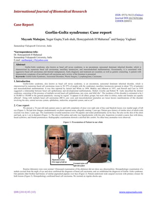
We present a 74-year-old male patient came to opd with complaints of ulcer over right side of face and blackish lesion over medial angle of left
eye (Figure.1). He had skin changes, predominantly on photo exposed areas, allegedly starting 1 year ago. Patient gave history of similar ulcer of which wide
excision was done 2 years ago. The examination revealed numerous (over 50) papules and nodes predominantly on his face, but also on the skin of the chest
and back, up to 1 cm in diameter (Figure. 1). The skin of his palms and soles was hyperkeratotic with tiny pits. Inspection revealed a coarse face with dense,
fused eyebrows, and frontal prominences. Radiographic examination showed a calcified falx cerebri. No other bony anomalies were observed.
Figure 1: Presentation of Patient in our clinic
Routine laboratory tests were normal. Ultrasound examination of the abdomen did not show any abnormalities. Histopathologic examination of a
nodule excised from the angle of eye and ulcer confirmed the diagnosis of basal cell carcinoma, and we established the diagnosis of Gorlin- Goltz syndrome.
Our patients elder brother had history of similar pigmented papules over face (Figure 2). Patient underwent wide surgical excision with primary closure for
both lesions (Figure 3). Histopathology confirmed basal cell carcinoma. We insisted on regular checks-ups.
Abstract
Gorlin-Goltz syndrome, also known as basal cell nevus syndrome, is an uncommon, autosomal dominant inherited disorder, which is
characterized by numerous basal cell carcinomas, maxillary keratocysts, and musculoskeletal malformations. Occasionally, it is associated with
aggressive basal cell carcinomas and internal malignancies. Early diagnosis and treatment are essential, as well as genetic counseling. A patient with
characteristic symptoms of nevoid basal cell carcinoma and a review of the literature is presented.
Keywords: Gorlin-Goltz Syndrome, Autosomal Disorders, Plastic Surgery, Lymphangioma, Carcinoma
2. Mahajan et al 660
IJBR (2014) 05 (10) www.ssjournals.com
Figure 2: Patient’s Brother having similar Lesions
Figure 3: Post-operative image of Patient
3. Discussion
Gorlin-Goltz syndrome is autosomal dominant with a high penetrance and variable expressivity. It is causedby mutations in the patched tumor
suppressor gene (PTCH), a human homologue of the Drosophila genemapped to chromosome 9q21-231,4
. Chromosomal mapping and genetic studies suggest
that the underlying basis for this disease is an abnormality in the Hedgehog (Hh) signaling pathway. The role of this pathway in embryogenesis is well
known. The PTCH gene product is part of a receptor for the protein called Sonic Hedgehog (SHH), which is involved in embryonic development8
. More
recent investigations reveal the role of the Hh pathway in cell cycle regulation in adults. In the Drosphila model, the primary receptor for the Hh signaling
pathway has two transmembrane protein components: Patched (Ptc) and Smoothened (Smo). In the absence of Hh protein, the Ptc protein inhibits the Smo.
Under normal conditions, Hh, when present, binds Ptc, releasing Smo to affect downstream events such as cellgrowth and differentiation. Based on this
model, inactivation of Ptc or constituitive activity of Smo or Hh could lead to overactivity of Smo, resulting in neoplasm formation1
.
The diagnostic criteria for nevoid basal cell carcinoma were established by Evans et al., and modified by Kimonis et al. in 19973
. According to
them, diagnosis of Gorlin-Goltz syndrome can be established when two major or one major and two minor criteria are present as described below2,3
:
I. Major criteria:
– More than two basal cell carcinomas or one basal cell carcinoma at younger than 30 years of age or more than 10 basal cell nevi.
– Any odontogenic keratocyst (proven on histology) or polyostotic bone cyst.
– Three or more palmar or plantar pits (present in about 65% of patients).
– Ectopic calcification: Lamellar or early at younger than 20 years of age.
– Falx cerebri calcification.
– Positive family history of nevoid basal cell carcinoma.
Some authors take plurilamellar appearance of the falx cerebri calcification as a pathognomonic symptom of Gorlin-Goltz syndrome9
).
II. Minor criteria:
– Congenital skeletal anomalies; fused, splayed, missing, or bifid ribs, wedged or fused vertebrae.
– Occipital-frontal circumference more than 97%.
– Cardiac or ovarian fibroma.
– Medulloblastoma.
– Lymphomesenteric cysts.
– Congenital malformations such as cleft lip or palate, polydactylism or eye anomalies (cataract, coloboma, microphthalmus).
Other diagnostic findings in adults with Gorlin-Goltz syndrome are:
I. Skeletal anomalies:
Hemivertebrae, scoliosis, syndactyly, polydactyly, shortened 4th metacarpal.
3. Mahajan et al 661
IJBR (2014) 05 (10) www.ssjournals.com
II. Craniofacial anomalies:
Frontal bossing: increased size of calvaria (occipitofrontal circumference 60 cm or more in adults); brachycephaly; macrocephaly, coarse face,
heavy fused eyebrows; broadened nasal root; calcification of the falxes; tentorium cerebelli calcification; bridged sella turcica; low positioning of occiput;
congenital blindness due to corneal opacity; congenital or precocious cataract or glaucoma; coloboma of iris, choroids, or optic nerve; convergent or
divergent strabismus; and nystagmus
III. Neurological anomalies:
Agenesis/dysgenesis of corpus callosum; congenital hydrocephalus; meningioma; mental retardation;schizoid personality.
IV. Oropharyngeal anomalies:
Cleft lip/palate; high arched palate or prominentridges.
V. Anomalies of the reproductive system
VI. Cardiac anomalies
Tumors accompanying this syndrome include parathyroid adenoma, adrenal cystic lymphangioma, ovarian fibroma, and other neoplasms. This
syndrome is followed by multiple complications, predominantly aggressive basal cell tumors invading surrounding structures, or distant metastases, causing
death. Medulloblastoma causes death during infancy. Recurrence of odontogenic keratocysts causes varying degrees of jaw deformity.
Diagnosis and therapy of this syndrome require a multi disciplinary approach (dermatologists, surgeons, dentists, maxillary surgeons, and
neurologists). It consists of removal of tumors (surgical excision, topical chemotherapies, and laser ablation) and adequate treatment of maxillary cysts. A
new treatment strategy, based on the understanding of the Hh signaling pathway and the premise that tumors arise due to it’s over activity, supposes that
inhibition of this pathway with specific pharmacological treatment might suppress tumor growth1
.
Patients with Gorlin-Goltz syndrome require consistent sun protection. Genetic counseling that considers the genetic risks is advisable for all
patients with this syndrome, both familial and sporadic.
Reference
1. Kimonis VF, Goldstein AM, Pastakia B, Yang ML, Kase R, Di Giovanna JJ, Bale AF, Bale SJ. Clinical manifestations in 105 persons with nevoid basal
cell carcinoma syndrome. Am J Med Genet. 1997; 69:299–308.
2. Ly JQ, Beall DP. Gorlin’s syndrome: diffuse appendicular skeletal involvement with scintigraphic correlation. Australas Radiol. 2003; 47:318–21.
3. Mortele KJ, Hoier MR, Mergo PJ, Ros PR. Bilateral cystic lymphangiomas in nevoid basal cell carcinoma (Gorlin-Goltz) syndrome: US, CT and MR
findings. J Comput Assist Tomogr. 1999; 23:562–4.
4. Lambrecht JT, Stubinger S, Siewert B, Harle F. Calcification of the falx cerebri. A pathognomonic symptom of Gorlin-Goltz syndrome. HNO. 2005;
53:701–6.
5. Diaz-Fernandez JM, Infante-Cossio P, Belmonte-Caro R, Ruiz-Laza L, Garcia-Perla-Garcia A, Gutierrez-Perez JL. Basal cell nevus syndrome.
Presentation of six cases and literature review. Med Oral Patol OralCir Bucal. 2005; 10 Suppl 1:E57–66.
6. Patil K, Mahima VG, Gupta B. Gorlin syndrome: A case report. J Indian Soc Pedod Prev Dent. 2005;23:198–203
7. Tilli CMLJ, Van Steensel MAM, Krekels GAM, Neumann HAM, Ramaekers FCS. Molecular Aetiology and Pathogenesis of Basal Cell Carcinoma. Br
J Dermatol. 2005; 152:1108–24.
8. Boutimzine N, Laghmari A, Karib H, Karmane M, Bencherif M, Albouzidi A, Cherkaoui O, Mohcine Z.Gorlin-Goltz phacomatosis: ophthalmological
aspects in one case. J Fr Ophtalmol. 2000; 23:180–6.