
Recommended
More Related Content
Similar to NEPHROBLASTOMA.pptx
Similar to NEPHROBLASTOMA.pptx (20)
Recently uploaded
Recently uploaded (20)
NEPHROBLASTOMA.pptx
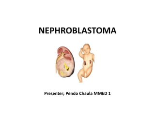
- 1. NEPHROBLASTOMA Presenter; Pendo Chaula MMED 1
- 2. INTRODUCTION Nephroblastoma also known as Wilms tumor, is the most common renal malignancy affecting one in 10,000 children <15 years old it accounts for about 95% of all paediatric tumors of the kidney with long term survival above 90% for localised disease and 75% for metastatic disease In patients with unilateral involvement, the median age at diagnosis is 43 months in girls and 37 months in boys Bernstein L, Linet M, Smith MA, et al. Cancer incidence and survival among children and adolescents: United States SEER Program 1975-1995 SEER Program. Bethesda, MD, National Cancer Institute 1999. p.79 Breslow N, Olshan A, Beckwith JB, Green DM. Epidemiology of Wilms tumor. Med Pediatr Oncol 1993; 21:172
- 3. The peak age is between 24 and 36 months and 75% of patients will be less than 60 months of age at diagnosis and 95% of patients will be less than 120 months of age at diagnosis Children with bilateral disease are diagnosed at an earlier age (median age, girls at 31 months and boys at 24 months): Patients with associated congenital anomalies are also diagnosed at an earlier age • The overall survival rate approaches 90% in the developed world but in developing countries the survival rates are much less and in some sub-Saharan countries it is only 40% at 8 months after diagnosis Bernstein L, Linet M, Smith MA, et al. Cancer incidence a SEER Program. Bethesda, MD, National Cancer Institute 1999. p.79 Breslow N, Olshan A, Beckwith JB, Green DM. Epidemiology of Wilms tumor. Med nd survival among children and adolescents: United States SEER Program 1975-1995 Pediatr Oncol 1993;
- 4. ASSOCIATED CONGENITAL SYNDROMES • Wagr syndrome- refers to the presence of Wilms tumor, aniridia, genitourinary anomalies, and mental retardation • Denys-Drash syndrome- This includes male pseudo- hermaphroditism and progressive renal failure starting in infancy. 90% developes WT. • Beckwith-wiedemann syndrome- hemihypertrophy, pancreatic enlargement, hypertrophic kidneys, omphalocele, ear creases, macrosomia, and macroglossia. 5% to 10% developes WT. • Other congenital anomalies like Perlman syndrome, Setos syndrome, Simpson-Golabi-Behmel syndrome, Trisomy 18 (Edward's syndrome), Frasier syndrome, Bloom syndrome, Li- Fraumeni syndrome, etc.
- 5. EPIDEMIOLOGY The risk of developing Wilms tumor varies among ethnic groups, with a greater risk in African-Americans and a lower risk in the Asian population, Black – African children have an increased prevalence of WT (11 cases per million children Accounts for 6-7% of cases of childhood cancer in the developed world and 12% in South Africa In children less than 15 years of age, the annual prevalence rate is about 7 to 10 cases per million USA. Breslow N, Olshan A, Beckwith JB and Green DM. ‘Epidmiology of Wilms' tumor’, Med PediatrOncol. 1993; vol.21, pp.172-181. Bernstein L, Linet M, Smith MA, Olshan AF. 1999, ‘Renal tumors’, In: Ries LAG, Smith MA, Gurney JG, et al (eds), Cancer Incidence and Survival among Children and Adolescents: United States SEER Program 1975-1995, National Cancer Institute, SEER Program (Publication No. 99-4649), National Institutes of Health, Bethesda.
- 6. In Tanzania the prevalence is 6.7% ( Mgaya E et al., 2000), “third from leukemia and lymphoma” (Shakilu J, 2017) The overall survival rate of nephroblastoma approaches 90% in the developed world but in developing countries the survival rates are much less and in some sub-Saharan countries it is only 40% at 8 months after diagnosis Wilms tumor is primarily a sporadic disease and only 1-2% of individuals with Wilms tumor have a relative with the disease Stiller CA, Parkin DM. Geographic and ethnic variations in the incidence of childhood cancer. Br Med Bull 1996; 52:682 Huff V. Wilms' tumours: about tumour suppressor genes, an oncogene and a chameleon gene. Nat Rev Cancer 2011; 11:111
- 7. Etiology • The cause is not precisely known, but it is believed to be due to genetic alterations that deal with the normal embryological development of the genitourinary tract • It is thought to develop from persistent metanephric tissue or nephrogenic rests • These may occur in 1% of infantile kidneys but typically regress during childhood. • These abnormal metanephric cells are found in up to 100% of cases of bilateral Wilms but only 35% of unilateral tumors
- 8. PATHOGENESIS Caused by abnormal renal development, resulting in proliferation of the metanephric blastema without normal tubular and glomerular differentiation Is thought to arise from foci of persistent metanephric cells referred to as nephrogenic rests or nephroblastomatosis Nephrogenic rests normally occur in 1% of newborn kidneys and regress early in childhood: in contrast, they are present in 35% of kidneys with unilateral Wilms tumor and almost 100% of kidneys with bilateral disease Beckwith JB, Kiviat NB, Bonadio JF. Nephrogenic rests, nephroblastomatosis, and the pathogenesis of Wilms' tumor. Pediatr Pathol 1990; 10:1. Beckwith JB. Precursor lesions of Wilms tumor: clinical and biological implications. Med Pediatr Oncol 1993; 21:158.
- 9. Has been associated with loss of function mutations of a number of tumor suppressor and transcription genes These include mutations of the WT1, p53, FWT1, and FWT2 genes and at the 11p15.5 locus The role of these gene mutations in the pathogenesis of Wilms tumor remains unknown Coppes MJ, Haber DA, Grundy PE. Genetic events in the development of Wilms' tumor. N Engl J Med 1994; 331:586
- 10. Most Wilms tumors are solitary lesions however 5-7% of patients have bilateral renal involvement and 10% have multifocal loci within a single kidney May include cysts, hemorrhage or necrosis The tumor is typically surrounded by a pseudocapsule, which may help distinguish it from other renal tumors, which have an infiltrative border Fernandez C, Geller JI, Ehrlich PF, et al. Renal tumors. In: Principles and Practice of Pediatric Oncology, 6th ed, Pizzo P, Poplack D (Eds), Lippincott Williams & Wilkins, St. Louis 2011. p.861
- 11. Histologically, the classic favorable histology Wilms tumor is comprised of three cell types: Blastemal cells – Undifferentiated cells Stromal cells – Immature spindle cells and heterologous skeletal muscle, cartilage, osteoid or fat Epithelial cells – Glomeruli and tubules Some contain only one or two cell types: tumors with only one cell type, it is often difficult to make the diagnosis of Wilms tumor
- 12. Histopathology • Grossly, Wilms tumors are usually well-circumscribed and have a pseudo-capsule • Histologically, Wilms is divided into "Favorable" and "Unfavorable" histologies • "Favorable" Histology: 90% of Wilms tumors will demonstrate "favorable" histology which generally has a better prognosis
- 13. • Classical histological features of a "favorable" Wilms tumor include a triphasic pattern of blastema, epithelial, and stromal tissues • The blastema is the most undifferentiated
- 14. • malignant component. It consists of collections of small, round blue cells with very active mitotic activity and overlapping nuclei • The epithelial component can demonstrate wide variations in differentiation from an early tubular formation with primitive epithelial rosette-like structures to differentiating tubules or glomeruli-like structures, which represent nephrogenesis at different developmental stages
- 15. • The stromal component may include densely packed undifferentiated mesenchymal cells or loose cellular myxoid areas • The latter areas may be difficult to distinguish from non- tumorous stroma associated with chemotherapy- induced change • Heterologous differentiation of neoplastic stroma in the form of well-differentiated smooth or skeletal muscle cells, fat tissue, cartilage, bone, and even glial tissue is present in some cases, especially in tumors that have undergone preoperative chemotherapy
- 16. CLINICAL PRESENTATION Most children with Wilms tumor present with an abdominal mass or swelling without other signs or symptoms • Abdominal pain 30-40%, hematuria 12-25%, fever and hypertension 25% (HTN will normalize after nephrectomy). Subcapsular hemorrhage can present with rapid abdominal enlargement and anemia. Wilms tumor in boys may also present with cryptorchidism, varicocele or hypospadias Fernandez C, Geller JI, Ehrlich PF, et al. Renal tumors. In: Principles and Practice of Pediatric Oncology, 6th ed, Pizzo P, Poplack D (Eds), Lippincott Williams & Wilkins, St. Louis 2011. p.861
- 17. Although the lung is the most common metastatic site, children rarely present with respiratory symptoms (dyspnea or tachypnea). 10% of affected girls will have congenital uterine anomalies and other renal congenital abnormalities such as duplication and renal ectopia. Fernandez C, Geller JI, Ehrlich PF, et al. Renal tumors. In: Principles and Practice of Pediatric Oncology, 6th ed, Pizzo P, Poplack D (Eds), Lippincott Williams & Wilkins, St. Louis 2011. p.861
- 18. DIAGNOSIS The definitive diagnosis of Wilms tumor is made by histologic confirmation at the time of either surgical excision or biopsy Laboratory testing: renal function, urinalysis, liver function, serum calcium, complete blood count, coagulation studies and serum electrolyte. Cytogenetics studies to look for 1p and 16q deletion. Abdominal imaging: USS, CT/MRI Chest imaging: CXR or chest CT scan for metastasis https://www.ncbi.nlm.nih.gov/books/NBK442004/?report=printable 4/10
- 19. Tanzania treatment guideline 2021
- 20. DIFFERENTIAL DIAGNOSIS Imaging studies and tissue histology differentiate Wilms tumor from other disorders: Neuroblastoma-patients who are post-radiation and post-chemotherapy are at increased risk. Clear cell sarcoma of the kidney-high mortality and relapse rate. Metastasize to bone. Histologically similar to WT. Rhabdoid tumor of the kidney-seen before age of 2yrs almost never in children older than 5yrs. Metastatic at initial presentation. 80% mortality rate. Congenital mesoblastic nephroma-typically found in the first year of life, most often by USS. Hypertension and elevated renin levels usually accompany it. Renal cell carcinoma- is rare in the pediatric age group. However, when present, it is often at a more advanced stage than in adults. Renal medullary carcinoma-very aggressive and dangerous cancer that is found almost exclusively in individuals with sickle cell disease, usually trait. It tends to be highly locally-invasive and metastasizes early.
- 21. STAGES Staging criteria for Wilms tumor are based upon the anatomic extent of the tumor without consideration for genetic, histologic or biological markers: National Wilms Tumor Study (NWTS): The NWTS system is based upon surgical evaluation prior to the administration of chemotherapy International Society of Pediatric Oncology (SIOP): The SIOP system is based upon post-chemotherapy surgical evaluation Metzger ML, Dome JS. Current therapy for Wilms' tumor. Oncologist 2005; 10:815
- 22. Staging Stage I indicates the tumor was completely contained within the kidney without any breaks or spillage outside the renal capsule and no vascular invasion. This stage accounts for 40% to 45% of all Wilms tumors. Stage II would be a tumor that has grown outside the kidney to some degree, such as into surrounding fatty tissue. Usually, the tumor would be completely removable by surgery, and regional lymph nodes are negative, About 20% of all Wilms tumors are at this stage.
- 23. Stage III comprises about 20% to 25% of all Wilms tumors and indicates a tumor which could not be completely removed surgically such as the following: Cancer has spread to the regional lymph nodes but not to more distant nodes, such as in the chest Cancer has grown into nearby vital structures so it could not be surgically removed completely Deposits of the tumor (tumor implants) are found in the peritoneum, or there are positive surgical margins
- 24. Cancer cells were accidentally “spilled” into the abdominal cavity during surgery The tumor was removed in separate pieces surgically; such as one piece from the kidney and another from the adrenal gland A renal biopsy of the tumor was done before it was surgically removed
- 25. Stage IV tumors are those that have spread through the vascular system to distant organs such as the lungs, liver, brain, or bones, or to distant lymph nodes. These account for about 10% of all Wilms tumors. Stage V are those cases where both kidneys are involved with tumor at the time of initial diagnosis. About 5% of all Wilms tumors are at this stage. Individual staging of each renal unit is needed as well.
- 26. TREATMENT Surgery is the main treatment for Wilms tumor Radical nephrectomy for unilateral: cancer along with the entire kidney, surrounding lymphnodes and tissues are removed and partial nephrectomy on the contralateral side Lymphnodes sampling is performed to assess degree of spread within the abdomen Neoadjuvant treatment shrinks the tumor and adjuvant therapy prevents recurrence
- 27. Chemotherapy • Give vincristine IV, • actinomycin D IV, • doxorubicine IV, • carboplatin IV, • etoposide IV, • cyclophosphamide IV, • doxorubicin IV 6/23/2021 Wilms Tumor - StatPearls - NCBI Bookshelf https://www.ncbi.nlm.nih.gov/books/NBK442004
- 28. Treatment / Management chemotherapy first and do the nephrectomy later. The opposite kidney may be explored to ensure that cancer has not spread although this is not necessary for low stage tumors with favorable histology if imaging is negative. Lymph nodes around the aorta are sampled for staging and to improve survival. Open surgery, however, typically provides more lymph nodes in the surgical specimen. Routine biopsies are not recommended except in unusual circumstances as a biopsy automatically increases tumor staging to Stage III. This stage requires radiation and chemotherapy.
- 29. Postoperative radiation may or may not be administered depending on tumor histology and extent of spread. For patients without metastases who will be receiving radiation, initiation of therapy within 14 days of surgery appears to improve overall survival Chemotherapy is usually administered for more aggressive disease. In children with bilateral disease, immediate nephrectomy is not performed. Some experts attempt high- dose chemotherapy to kill the tumor cells and hopefully salvage the kidney.
- 30. • Repeat biopsies are required to determine if the tumor is responding to therapy. • Patients who relapse after initial combined therapy tend to have a worse prognosis than newly discovered Wilms.
- 31. PROGNOSIS Several prognostic factors at the time of initial diagnosis are associated with an increased risk of tumor recurrence or death: The most important determinants of outcome in children with WT are the histopathology and tumor stage. Tumor histology: anaplastic histology, blastemal type histology Tumor stage Molecular and genetic markers: e.g. loss of heterozygosity at chromosome 16q, 1p, 11p15; and 1q gain Age >2 years Dome JS, Graf N, Geller JI, et al. Advances in Wilms Tumor Treatment and Biology: Progress Through International Collaboration. J Clin Oncol 2015; 33:2999
- 32. The prognosis varies by tumor stage and histology. Favorable histology has survival rates of 99% to 86% while unfavorable histology survival ranges from 84% to 38% depending on the stage. A poorer prognosis is associated with the following characteristics: Anaplastic histology in stage II to IV tumors Diffuse anaplasia is worse than focal 6/23/2021 Wilms Tumor - StatPearls - NCBI Bookshelf https://www.ncbi.nlm.nih.gov/books/NBK442004/?report=printable 6/10
- 33. A poorer prognosis has been linked to TP53 and with the loss of heterozygosity at chromosomes 1p, 1q, 11p15 and 16q. Loss of heterozygosity at chromosomes 1p, 1q, 11p15 and 16q or presence of TP53 Higher stage (most epithelial predominant tumors are stage I; most blastema predominant tumors are stage III and IV)
- 34. Age older than two years Higher positive lymph node density Large tumor size Even small tumor foci can be associated with a poorer prognosis due to resistance to chemotherapy
- 35. complications Increased risk of secondary malignancies years later in life due to Radiation and chemotherapy It is well established that radiation therapy will increase the risk for bone, breast, colon and thyroid cancers later on in life It will also increase the risk of osteoporosis Islam M, Saltzman AF, Amini A, Carrasco A, Cost NG. Factors Influencing Overall Survival of Children, Adolescents, and Young Adults With High-risk Renal Tumors. Urology. 2018 Oct;120:222-230. [PubMed: 30076944] Islam M, Saltzman AF, Amini A, Carrasco A, Cost NG. Factors Influencing Overall Survival of Children, Adolescents, and Young Adults With High-risk Renal Tumors. Urology. 2018 Oct;120:222-230. [PubMed: 30076944] ,
- 36. Chemotherapy with dactinomycin, doxorubicin and vincristine contributes to a higher risk of secondary malignancies as well as specific toxicities such as hearing (carboplatin), cardiac function (adriamycin) and peripheral neuropathy (vincristine) About 5% to 10% of Wilms patients will present with Von Willebrand's disease which can complicate treatment Baxter PA, Nuchtern JG, Guillerman RP, Mahoney DH, Teruya J, Chintagumpala M, Yee DL. Acquired von Oostveen RM, Pritchard-Jones K. Pharmacotherapeutic Management of Wilms Tumor: An Update. Paediatr Drugs. 2019 Feb;21(1):1- 13. [PubMed: 30604241]
- 37. RECOMMENDATION Advocate early health care seeking behavior in the communities for early diagnosis and management Networking with pediatric cancer charity organizations to assist parents with financial constraints for timely investigations Therapeutic foods program establishment at different hospitals eg Benjamin Mkapa hospital
- 38. TAKE HOME MESSAGE Early and thorough diagnosis thus treatment helps reduce morbidity and mortality
Editor's Notes
- [17] [17]