
Recommended
More Related Content
Similar to Genome Shiffling
Similar to Genome Shiffling (20)
More from berciyalgolda1
More from berciyalgolda1 (20)
Recently uploaded
Recently uploaded (20)
Genome Shiffling
- 1. By Assist.Prof Dr. Berciyal Golda. P VICAS Genome Shiffling
- 2. Genetic Recombination Exchange of genes between two DNA molecules to form new combinations of genes on achromosome contributes to apopulation’s genetic diversity (source of variation in evolution) Recombination is more likely than mutation to be beneficial Less likely destroy agene's function May bring together combinations of genes
- 10. Double-strand breaks in DNA initiate recombination (part I)
- 11. Double-strand breaks in DNA initiate recombination (part II)
- 15. 2. Site specific recombination Viruses and transposable elements often integrate their genomes into the host chromosome Site specific recombination is used by both eukaryotes and prokaryotes to regulate gene expression and to increase the organisms genetic range
- 16. Site specific recombination FIGURE 25–38 A site-specific recombination reaction. (a) The reaction is carried out within a tetramer of identical subunits. Recombinase subunits bind to a specific sequence, often called simply the recombination site. 1 One strand in each DNA is cleaved at particular points within the sequence. The nucleophile is the OH group of an active-site Tyr residue, and the product is a covalent phosphotyrosine link between protein and DNA. 2 The cleaved strands join to new partners, producing a Holliday intermediate. Steps 3 and 4 complete the reaction by a process similar to the first two steps. The original sequence of the recombination site is regenerated after recombining the DNA flanking the site. These steps occur within a complex of multiple recombinase subunits that sometimes includes other proteins.
- 17. WHY SSR??? • Transformation through Agrobacterium & direct DNA transfer leads to complex integration of GOI. • Complex integration is classified into, Single copy – multi locus integration Multi copy – single locus integration • Complex integration leads to gene silencing.
- 18. • Ideal integration is single copy – single locus and integration of GOI without intervening with functional gene. • The best way out of this issue is promoting the development of system/technique that facilitates site specific recombination.
- 19. • Two systems have been developed for facilitating site specific recombination, namely, Cre/lox system FLP/FRT system
- 20. Cre/lox system • Derived from P1 bacteriophage. • Cre recombinase catalyses the recombination between two loxP(site for recombination) sites. • loxP site consists of an 8-bp core sequence, where recombination takes place, and two flanking 13-bp inverted repeats.(In total 34-bp)
- 21. loxP site
- 23. FIGURE 25–39 Effects of site-specific recombination. The outcome of site-specific recombination depends on the location and orientation of the recombination sites (red and green) in a double-stranded DNA molecule. Orientation here (shown by arrowheads) refers to the order of nucleotides in the recombination site, not the 5n3 direction. (a) Recombination sites with opposite orientation in the same DNA molecule. The result is an inversion. (b) Recombination sites with the same orientation, either on one DNAmolecule, producing a deletion, or on two DNAmolecules, producing an insertion.
- 24. FLP/FRT system • Derived from 2 micrometer yeast plasmid • FLP recombinase enhances recombination of sequences between two short Flippase Recognition Target (FRT). • FRT consists of an 8-bp spacer and two flanking 13-bp inverted repeats, where recombination takes place (In total 34-bp).
- 25. FRT site
- 26. DNA Transposition recombination that allows the movement of transposable elements, or transposons. These segments of DNA, found in virtually all cells, move, or “jump,” from one place on a chromosome (the donor site) to another on the same or a different chromosome (the target site). DNA sequence homology is not usually required for this movement, called transposition; the new location is determined more or less randomly. Insertion of a transposon in an essential gene could kill the cell, so transposition is tightly regulated and usually very infrequent. Transposons are perhaps the simplest of molecular parasites, adapted to replicate passively within the chromosomes of host cells. In some cases they carry genes that are useful to the host cell, and thus exist in a kind of symbiosis with the host
- 27. Bacteria have two classes of transposons. 1. simple transposons Insertion sequences contain only the sequences required for transposition and the genes for proteins (transposases) that promote the process. 2.Complex transposons contain one or more genes in addition to those needed for transposition. These extra genes might, for example, confer resistance to antibiotics and thus enhance the survival chances of the host cell. The spread of antibiotic-resistance elements among disease-causing bacterial populations that is rendering some antibiotics ineffectual is mediated in part by transposition. Bacterial transposons vary in structure, but most have short repeated sequences at each end that serve as binding sites for the transposase. When transposition occurs, a short sequence at the target site (5 to 10 bp) is duplicated to form an additional short repeated sequence that flanks each end of the inserted transposon (Fig. 25–42). These duplicated segments result from the cutting mechanism used to insert a transposon into the DNA at a new location. Classes of Transposons
- 28. FIGURE 25–42 Duplication of the DNA sequence at a target site when a transposon is inserted. The duplicated sequences are shown in red. These sequences are generally only a few base pairs long, so their size (compared with that of a typical transposon) is greatly exaggerated in this drawing.
- 29. There are two general pathways for transposition in bacteria. In direct or simple transposition (Fig. 25–43, left), cuts on each side of the transposon excise it, and the transposon moves to a new location. This leaves a double-strand break in the donor DNA that must be repaired. At the target site, a staggered cut is made (as in Fig. 25–42), the transposon is inserted into the break, and DNAreplication fills in the gaps to duplicate the target site sequence. In replicative transposition (Fig. 25–43, right), the entire transposon is replicated, leaving a copy behind at the donor location. A cointegrate is an intermediate in this process, consisting of the donor region covalently linked to DNAat the target site. Two complete copies of the transposon are present in the cointegrate, both having the same relative orientation in the DNA. In some well-characterized transposons, the cointegrate intermediate is converted to products by site-specific recombination, in which specialized recombinases promote the required deletion reaction.
- 30. FIGURE 25–43 Two general pathways for transposition: direct (simple) and replicative. 1 The DNA is first cleaved on each side of the transposon, at the sites indicated by arrows. 2 The liberated 3- hydroxyl groups at the ends of the transposon act as nucleophiles in a direct attack on phosphodiester bonds in the target DNA. The target phosphodiester bonds are staggered (not directly across from each other) in the two DNA strands. 3 The transposon is now linked to the target DNA. In direct transposition, replication fills in gaps at each end. In replicative transposition, the entire transposon is replicated to create a cointegrate intermediate. 4 The cointegrate is often resolved later, with the aid of a separate site-specific recombination system. The cleaved host DNA left behind after direct transposition is either repaired by DNA end-joining or degraded (not shown). The latter outcome can be lethal to an organism.
- 31. Genetic Transfer Vertical genetransfer From parents to offspring Horizontal genetransfer From one microbe to another Between different strains and species of bacteria and viruses Leads to recombination
- 32. Horizontal gene transfer Part of total DNA from Donor cell integrated into Recipient cell. Remaining amount of DNA from donor cell degraded. Recipient cell with DNA from donor is called Recombinant. 1% of population might undergo recombination
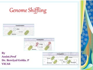
- 33. Recombination in prokaryotes occurs through three mechanisms 1. Transformation 2. Transduction 3. Conjugation Horizontal gene transfer - Mechanisms
- 34. Transformation Transfer of naked DNA from donor to recipient cell Discovered by Frederick Griffith in 1928 in Streptococcus pneumoniae Showed that DNA is the genetic material Can be transferred between a donor and a recipient cell Griffith’s expt: Avirulent S. pneumonia became virulent when exposed to heat killed virulent cell
- 36. Bacterial transformation without mice Broth containing non-encapsulated living bacteria and dead encapsulated bacteria incubated After incubation, encapsulated living virulent bacteria were found Non-encapsulated bacteria received genes from dead encapsulated for forming a capsule The material responsible for transmission of this character was not known
- 37. Experiment: DNA is the genetic material In 1944, Oswald T Avery, Colin M Macleod, Maclyn Mccarty proved that DNA is the genetic material
- 38. Competent cells & competence Competence: ability of a recipient bacterium to take up DNA from the environment Competent cells: cells which can be transformed E.coli cannot undergo transformation naturally It is made competent by artificial transformation procedures (Calcium chloride or Electroporation)
- 39. Mechanism of transformation After death, cell lysis leads to release of DNA from bacteria Other bacteria take up DNA and integrate into their chromosomes by recombination Recipient cell with this combination of genes will now become a hybrid or recombinant Works best between closely related species Transformation in nature: Bacillus,Haemophilus, Streptococcus,Staphylococcus,Neisseria etc.
- 40. Mechanism of transformation Step1: The DNA binding receptor on a competent bacterium binds double stranded DNA As the DNA enters the cell, one strand is degraded, & the other strand is coated with single- strand DNA-binding protein. Step2: The single strand of donor DNA is integrated into the chromosome of the recipient cell producing a recombinant DNA
- 42. Conjugation Transfer of genes between cells that are in physical contact with another First demonstration of recombination in bacteria: Jhosua Lederberg & Edward Tatum in 1946 Found that, genetic traits could be transferred among two different strains of E. coli, if they are in physical contact
- 43. F+ and F- FACTORS William Hayes, Francois Jocob and Eli H Wolman (1950) Conjugating bacteria are of two mating types:- Male types which donates their DNA, these are called F+ cells Female types which are recipient of DNA donated by F+ cells and are called F- cells These F+ and F- are called fertility factor or F- factor or sex factor
- 44. Process of Conjugation The F Pili of the F+ donor cell make contact with the F- recipient cell & pull the cell together. Rolling circle replication transfer one strand of the F factor into the recipient cell. Transfer of F factor is completed, yielding two F+ factor bacteria.
- 46. Process of Conjugation In donor F+ cells, F factor may integrate into the host chromosome becoming Hfr (High Frequency of Recombination). Thus F+ cells become Hfr cells Conjugation between Hfr and F- cells results in replication of the chromosome with F factor. Asingle parental strand is transferred from Hfr cell to the F- cells.
- 48. Process of Conjugation Complete transfer of the chromosome does not take place Only a small piece of F factor leads the chromosomal genes into F- cells Small strand containing chromosomal genes recombines with the DNA of F- cells Thus F- cells receive only a part of chromosomal genes and hence do not get converted to F+ cells
- 49. Transduction Transduction occurs when a phage (virus) carries bacterial genes from one host cell to another Discovered by Norton Zinder and Joshua Lederberg in 1952 Bacteriophage Twotypes: 1. Bacteriophage T4 2. Bacteriophage λ Lifecycle 1. Lytic cycle 2. Lysogenic cycle
- 50. Lytic & Lysogenic Cycle
- 51. Lytic & Lysogenic Cycle Bacteriophage attaches to donor bacteria Inject their nucleic acid (DNA) into bacterium DNA replicates rapidly, and also directs the synthesis of new phage protein Then, the new DNA combines with new proteins, to make whole phage particles These are then released by destruction of cell wall and lysis of the cell
- 52. Process of Transduction These phages may composed of DNA of the host This phage attacks the another host and infect it Recipient DNA integrates with this DNA Results in the transfer of DNA Recipient cell is now called transducedcell
- 53. Types of Transduction Mainly there are two types Generalised or Non-specialised Transduction Restricted or Specialized Transduction
- 54. Generalized Transduction All fragments of bacterial DNA have a chance to enter a transducing phage
- 55. Specialized Transduction Certain phages can transfer only a few restricted genes of the bacterial chromosomes Only those bacterial genes adjacent to prophage in bacterial chromosomes Mediates the exchange of only limited numbers of specific genes Mediated by Bacteriophage λ
- 56. Specialized Transduction λ phage can only incorporate into a specific site (attλ) gal gene is on one side of attλ and bio gene (biotin synthesis) is on the other side Wrong cross over of λ phageat the end of the lysogenic phase Piece of the E. coli chromosome incorporated into λ phage chromosome gal gene or bio gene can be transferred