1. The document describes the development of the male and female genital systems from early embryonic stages through formation of the internal and external genitalia.
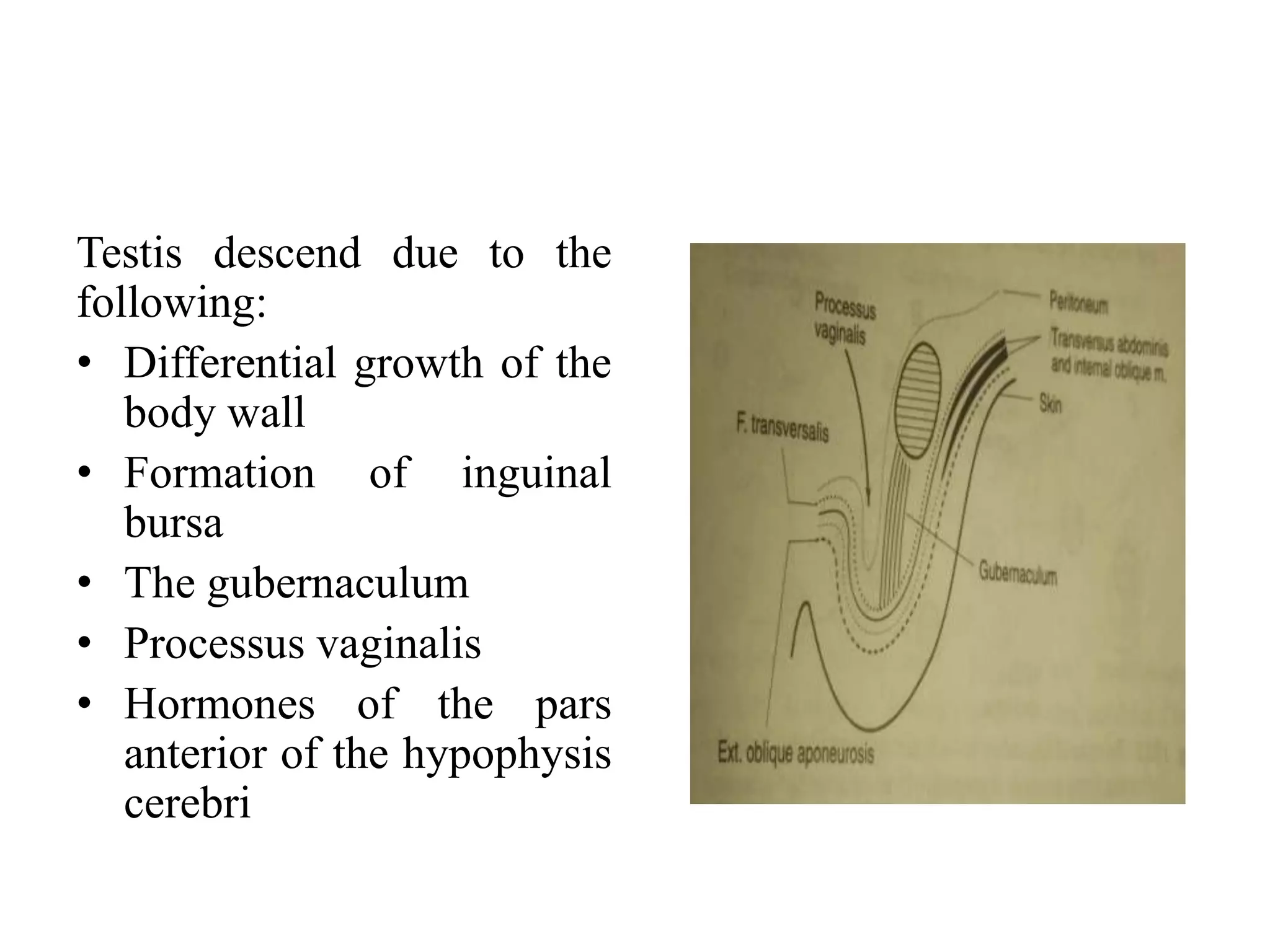
2. Key events include formation of the genital ridges which develop into testes in males and ovaries in females, descent of the testes into the scrotum, development of the duct systems including the vas deferens and epididymis in males and Mullerian duct regression leading to formation of the uterus and vagina in females.
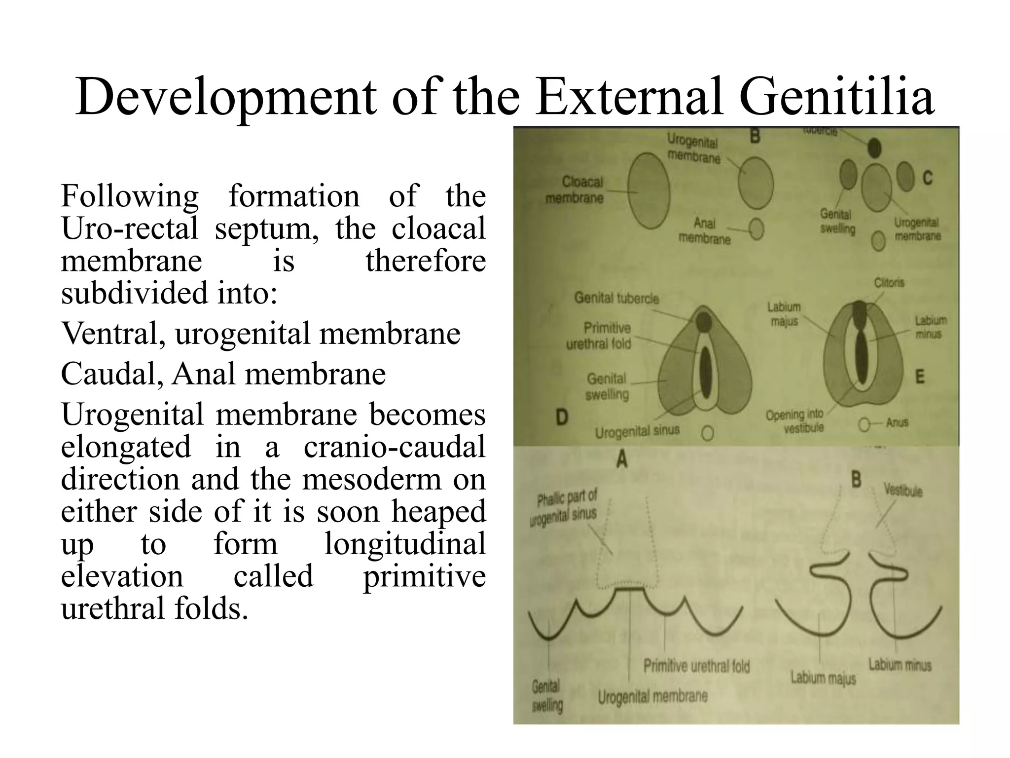
3. External genitalia develop from the genital tubercle and swellings, with the penis and scrotum forming in males and clitoris, labia, and vagina developing in females.
The development of the female genital system is determined at fertilization by the presence of two X chromosomes. In female embryos, the primitive sex cords dissociate and are replaced by the ovarian medulla and cortex. The paramesonephric ducts develop into the uterus, fallopian tubes, and upper vagina, while the sinovaginal bulbs form the lower vagina. Defects can occur if the paramesonephric ducts fail to fuse properly, resulting in conditions like a septate, bicornuate, or didelphys uterus. The genital tubercle forms the clitoris and genital swellings become the labia, with the urethral folds
The document summarizes the development of the genitourinary system during intrauterine life. It discusses the development of the pronephros, mesonephros and metanephros. It describes how the ureteric bud forms the collecting system of the definitive kidney. It also discusses the development of the gonads, testis, male external genitalia and prostate. Congenital anomalies that can affect the kidney, ureter and bladder are also summarized.
This document describes the development of the male genital system from the indifferent stage through differentiation and formation of the testes and external genitalia. It begins with the formation of the genital ridges and primordial germ cells. In males, under the influence of SRY gene, the ridges develop into testes containing seminiferous tubules and Leydig cells. The testes descend into the scrotum and the mesonephric ducts form the epididymis, vas deferens and seminal vesicles. Initially the genital ducts and external genitalia are indifferent, later differentiating into male structures.
The document summarizes the development of the male and female reproductive systems from the early undifferentiated gonads through sex determination and the formation of internal and external genitalia. It describes how in males, the testes secrete testosterone and MIF to develop the testes, penis, and scrotum. In females, the absence of these factors leads to ovarian development and the formation of the clitoris, labia, and uterus/vagina. It also discusses several congenital abnormalities that can occur during this development.
1. The genital system develops from the intermediate mesoderm, celomic epithelium, and part of the cloaca. Genetic sex is established at fertilization as XY or XX.
2. Between 4-12 weeks, the genital ridges develop into testes or ovaries depending on the presence of SRY gene on Y chromosome. This gene encodes TDF which leads to male development.
3. The genital ducts initially consist of the mesonephric and paramesonephric ducts. In males, the mesonephric ducts form the epididymis, vas deferens, and ejaculatory duct while the paramesonephric ducts regress. In females
The urogenital system develops from two embryonic structures - the intermediate mesoderm and cloaca. The intermediate mesoderm forms the urogenital ridge which gives rise to the urinary and genital systems. The cloaca divides into the primitive rectum and urogenital sinus. Kidneys develop from the nephrogenic cord and ureteric buds. The mesonephric ducts give rise to parts of the urinary tract in males and regress in females. External genitalia are initially bipotential and differentiate into male or female forms based on sex hormones.
The document summarizes the development of the female reproductive system. It begins with the formation of the genital ridge in the intermediate mesoderm at 3 weeks. At 5-6 weeks, primordial germ cells form the indifferent gonad. In the absence of the Y chromosome, the gonad develops into an ovary with cortical cords and primordial follicles. The ovaries descend into the pelvis guided by the gubernaculum. Meanwhile, the paramesonephric ducts form the fallopian tubes, uterus and upper vagina. The vagina develops from the sinovaginal bulbs and vaginal plate. Remnants of the mesonephric ducts include the epoophoron and
The document summarizes the anatomy and development of the female reproductive system. It describes the ovaries, oviducts, uterus, cervix, vagina, vulva, and their structure and function. It also discusses the embryonic development of the gonads, genital ducts, and external genitalia in both males and females. Key hormones produced by the hypothalamus and pituitary glands that regulate the reproductive system are also outlined.
The genital system develops from three main sources: the primordial germ cells, the indifferent gonads that develop into either ovaries or testes, and the genital ducts including the Müllerian and Wolffian ducts. In males, testosterone causes the Wolffian ducts to form the epididymis, vas deferens and seminal vesicles while regressing the Müllerian ducts. In females, the lack of testosterone causes the Müllerian ducts to form the fallopian tubes, uterus and upper vagina while regressing the Wolffian ducts. The external genitalia initially develop in an indifferent state before differentiating into either male or female forms based on hormone levels.
Development of the female genital systemPukar Sapkota
Gonadal development in females occurs slowly, with the ovaries not being identifiable until 10 weeks. Between 16-18 weeks, cortical cords in the developing ovaries break up and form primordial follicles, each containing an oogonium surrounded by follicular cells. Approximately 2 million primordial follicles are present at birth, though many degenerate during fetal development. The paramesonephric ducts play a leading role in female development, fusing to form the uterovaginal primordium which develops into the uterus and upper vagina. Estrogen produced by the placenta and fetal ovaries leads to the feminization of the external genitalia.
Development of the female reproductive systemAhmad Atere
The female reproductive system develops from indifferent gonads that are initially the same in males and females. In the absence of the SRY gene, the gonads develop into ovaries containing primordial follicles. The paramesonephric ducts fuse to form the uterus and fallopian tubes, while the mesonephric ducts regress. The vagina develops from sinovaginal bulbs, and external genitalia are feminized under the influence of estrogen to form the clitoris and labia.
Embryology Course IX - Urogenital SystemRawa Muhsin
This session discusses the development of the urogenital system and includes:
1. Development of the kidneys and ureters
2. Development of the bladder and urethra
3. Development of the gonads and genital ducts
4. Development of the external genitalia
1. The urinary and genital systems develop from a common intermediate mesoderm and initially share a common cavity called the cloaca.
2. The kidneys develop through three successive stages - the pronephros, mesonephros, and metanephros - with the metanephros forming the permanent kidneys.
3. The ureters develop from the mesonephric ducts and later join the bladder, which develops from the urogenital sinus. The bladder remains connected to the umbilicus by the urachus in early development.
USMLE REPRODUCTIVE 06 Development of female genital system.pdfAHMED ASHOUR
The development of the female genital system is a complex process involving the differentiation of structures that eventually form the reproductive and associated organs.
Understanding the embryonic development of the female genital system is crucial for surgeons, obstetricians, and gynecologists, especially in the context of congenital anomalies or surgical interventions.
The reproductive systems of male and female Ascaris lubricoides worms are described. The male system includes a single coiled testis, vas deferens, seminal vesicle, ejaculatory duct, and two penial spicules. The female system includes two long coiled ovaries, oviducts, uteri which store fertilized eggs, and a vagina. Gametes are formed through a process called telogony, where gametogonia bud off from the gonads and differentiate into gametocytes as they move through zones of proliferation, growth, and maturation.
This document provides an overview of urinary system development. It discusses how the urinary system develops from the intermediate mesoderm and includes three sets of nephric structures - the pronephros, mesonephros, and metanephros. The metanephros becomes the adult kidney. It also describes the development of other urinary structures like the ureters, bladder, and urethra in both males and females. Finally, it notes some common congenital anomalies that can result from abnormalities during urinary system development.
The female reproductive system consists of two ovaries, two uterine tubes, a uterus, a vagina, and external genitalia. The ovaries contain primordial follicles which mature into graafian follicles that release eggs. The uterine tubes transport eggs and allow for fertilization to occur. The uterus allows for implantation and fetal development. The vagina is the birth canal. The broad ligament supports the uterus and contains vessels and ligaments connecting the ovaries and uterus.
The male and female reproductive systems develop initially embryonically "indifferent", it is the product of the Y chromosome SRY gene that makes the "difference".
♂ - Male ♀ - Female
The reproductive organs are developed from the intermediate mesoderm.
The permanent organs of the adult are preceded by a set of structures which are purely embryonic, and which with the exception of the ducts disappear almost entirely before the end of fetal life.
These embryonic structures are the mesonephric ducts (also known as Wolffian ducts) and the paramesonephric ducts, (also known as Müllerian ducts). The mesonephric duct remains as the duct in males which gives rise to seminal vesical, epididymes and vas deferens, and the paramesonephric duct as that of the female.
Importantly its sex chromosome dependence, late embryonic/fetal differential development, complex morphogenic changes, long time-course, hormonal sensitivity and hormonal influences make it a system prone to many different abnormalities.
Gonads:
Gonads Produce eggs and sperm cells, transport and sustain egg and sperm cells, nurture developing offspring, and produce hormones.
The gonads, ovary or testis, also develop in the intermediate mesoderm.
They originally form as swellings that lie just ventral to the anterior mesonephric kidney.
A mullarian duct also develops in the intermediate mesoderm near the mesonephric duct.
Due to fusion or failure of 1st ridge to differentiate, some vertebrates (agnathans, some female lizards & crocodilians, & most female birds) have a single testis or ovary.
Hormones cause differentiation of early gonads into either testes or ovaries.
As males develop the mesonephric duct makes connection with the testis as the primary sperm conducting duct, and the mullerian duct is lost.
The document summarizes the development of the female genital system from indifferent gonads to internal and external structures. It describes how in the absence of the SRY gene, the gonads develop into ovaries through proliferation of surface epithelium into cortical cords containing primordial follicles. The paramesonephric ducts fuse to form the uterovaginal canal and give rise to the uterus and upper vagina. The lower vagina develops from sinovaginal bulbs. External structures like the labia, clitoris and vestibule form from the genital tubercle and urogenital folds. Anomalies can occur if fusion of ducts is incomplete.
Development of the male& female genital system.pptxJwan AlSofi
The document summarizes the development of the male and female genital systems from an indifferent stage. It describes how in males, the presence of SRY leads testes to develop from indifferent gonads, while in females without SRY ovaries develop. It outlines the development of testes, ovaries, male ducts including epididymis and vas deferens, and female ducts including uterus and vagina from indifferent ducts. External genitalia also develop differently in males under testosterone versus females.
The document summarizes the anatomy and functions of the male and female reproductive systems. It describes the organs involved in both systems, including the testes, ovaries, ducts, and external genitalia. It explains sperm and egg production, hormone regulation, and fertilization. Key concepts covered are the structures of the testes, ovaries, uterus, and vagina, as well as spermatogenesis, oogenesis, and the menstrual cycle.
1) The document describes the development of the genital system including the formation of gonadal ridges and primitive sex cords in early development of the gonads.
2) It explains the development of testes and ovaries including the roles of genes like SRY and the migration of primordial germ cells.
3) The descent of testes and ovaries is summarized along with the development of duct systems and external genitalia in males and females.
4) Common anomalies affecting genital development like Klinefelter's syndrome and hermaphroditism are also mentioned.
This document discusses abnormalities of the female urogenital tract, including their embryological development and classification. It notes that the urinary and genital systems develop from a common intermediate mesoderm and cloaca. Abnormalities can include defects in the development of the uterus, vagina, or urogenital sinus. Evaluation involves history, physical exam, imaging like ultrasound and MRI, and hormonal/genetic testing. Classification systems relate to the level of confluence between the urethra and vagina. Treatment is multidisciplinary and involves gender assignment and surgical reconstruction if needed.
How to Setup Default Value for a Field in Odoo 17Celine George
In Odoo, we can set a default value for a field during the creation of a record for a model. We have many methods in odoo for setting a default value to the field.
A Free 200-Page eBook ~ Brain and Mind Exercise.pptxOH TEIK BIN
(A Free eBook comprising 3 Sets of Presentation of a selection of Puzzles, Brain Teasers and Thinking Problems to exercise both the mind and the Right and Left Brain. To help keep the mind and brain fit and healthy. Good for both the young and old alike.
Answers are given for all the puzzles and problems.)
With Metta,
Bro. Oh Teik Bin 🙏🤓🤔🥰
How to Download & Install Module From the Odoo App Store in Odoo 17Celine George
Custom modules offer the flexibility to extend Odoo's capabilities, address unique requirements, and optimize workflows to align seamlessly with your organization's processes. By leveraging custom modules, businesses can unlock greater efficiency, productivity, and innovation, empowering them to stay competitive in today's dynamic market landscape. In this tutorial, we'll guide you step by step on how to easily download and install modules from the Odoo App Store.
THE SACRIFICE HOW PRO-PALESTINE PROTESTS STUDENTS ARE SACRIFICING TO CHANGE T...indexPub
The recent surge in pro-Palestine student activism has prompted significant responses from universities, ranging from negotiations and divestment commitments to increased transparency about investments in companies supporting the war on Gaza. This activism has led to the cessation of student encampments but also highlighted the substantial sacrifices made by students, including academic disruptions and personal risks. The primary drivers of these protests are poor university administration, lack of transparency, and inadequate communication between officials and students. This study examines the profound emotional, psychological, and professional impacts on students engaged in pro-Palestine protests, focusing on Generation Z's (Gen-Z) activism dynamics. This paper explores the significant sacrifices made by these students and even the professors supporting the pro-Palestine movement, with a focus on recent global movements. Through an in-depth analysis of printed and electronic media, the study examines the impacts of these sacrifices on the academic and personal lives of those involved. The paper highlights examples from various universities, demonstrating student activism's long-term and short-term effects, including disciplinary actions, social backlash, and career implications. The researchers also explore the broader implications of student sacrifices. The findings reveal that these sacrifices are driven by a profound commitment to justice and human rights, and are influenced by the increasing availability of information, peer interactions, and personal convictions. The study also discusses the broader implications of this activism, comparing it to historical precedents and assessing its potential to influence policy and public opinion. The emotional and psychological toll on student activists is significant, but their sense of purpose and community support mitigates some of these challenges. However, the researchers call for acknowledging the broader Impact of these sacrifices on the future global movement of FreePalestine.
CapTechTalks Webinar Slides June 2024 Donovan Wright.pptxCapitolTechU
Slides from a Capitol Technology University webinar held June 20, 2024. The webinar featured Dr. Donovan Wright, presenting on the Department of Defense Digital Transformation.
Philippine Edukasyong Pantahanan at Pangkabuhayan (EPP) CurriculumMJDuyan
(𝐓𝐋𝐄 𝟏𝟎𝟎) (𝐋𝐞𝐬𝐬𝐨𝐧 𝟏)-𝐏𝐫𝐞𝐥𝐢𝐦𝐬
𝐃𝐢𝐬𝐜𝐮𝐬𝐬 𝐭𝐡𝐞 𝐄𝐏𝐏 𝐂𝐮𝐫𝐫𝐢𝐜𝐮𝐥𝐮𝐦 𝐢𝐧 𝐭𝐡𝐞 𝐏𝐡𝐢𝐥𝐢𝐩𝐩𝐢𝐧𝐞𝐬:
- Understand the goals and objectives of the Edukasyong Pantahanan at Pangkabuhayan (EPP) curriculum, recognizing its importance in fostering practical life skills and values among students. Students will also be able to identify the key components and subjects covered, such as agriculture, home economics, industrial arts, and information and communication technology.
𝐄𝐱𝐩𝐥𝐚𝐢𝐧 𝐭𝐡𝐞 𝐍𝐚𝐭𝐮𝐫𝐞 𝐚𝐧𝐝 𝐒𝐜𝐨𝐩𝐞 𝐨𝐟 𝐚𝐧 𝐄𝐧𝐭𝐫𝐞𝐩𝐫𝐞𝐧𝐞𝐮𝐫:
-Define entrepreneurship, distinguishing it from general business activities by emphasizing its focus on innovation, risk-taking, and value creation. Students will describe the characteristics and traits of successful entrepreneurs, including their roles and responsibilities, and discuss the broader economic and social impacts of entrepreneurial activities on both local and global scales.
BPSC-105 important questions for june term end exam
Genital system 2206.pptx
1. Development of the Genital
System
Mohammed Alhaji Buba B.Sc., M.Sc., MCIPDM
2. Development of the ovaries
Early developmental stages of the ovary are exactly the same
as the testis
1. The coelomic epithelium on the medial side of the
mesonephros becomes thickened to form the genital ridges
2. Cords of cells proliferate to germinal epithelium and
grows into the underlying mesoderm
3. Primordial germ cells that are formed in relation to the
yolk sac migrate to the region of the developing ovary and
give rise to the oocytes
4. The sex cords become broken into small masses. The cells
of each mass surrounds one primordial germ cell or oocyte
to form aprimordial follicle.
3. Descends of the ovary
Ovary descend from the lumber region where it is
formed to the pelvis. There is formation of
gubernaculum as in male and extends from the
ovary to the labia majus.
It becomes attached to the uterus at its junction with
the uterine tube the part of the gubernaculum that
persist between the ovary and uterus becomes the
round ligament of ovary. The between the uterus
and the labia majus becomes the round ligament of
the uterus.
4. Anomalies of the ovary
1.Absence of the ovary on one or both sides
2.Duplication of the ovary
3.Descend of the ovary into the inguinal canal or
labium majus
4.adrenal or thyroid tissue may be present in the
ovary. the sometimes contain cells that are capable
of differenciating into various tissues like bone,
cartilage, hair e.t.c and growth of the rest of the cells
can give rise to perculiar tumour called teratoma,
5. Paramesonephric duct
This structure remains
rudimentary in males, the greater
part of it disappears however, the
cranial end of it persist as a small
rounded body attached to the
testis (appendix of the testis).
It has generally been considered
that the prostate utricle represents
the utero-vaginal canal in male,
therefore a homologue of the
uterus however, it is now believed
that it corresponds mainly to the
vagina.
6. DEVELOPMENT OF THE UTERUS
The uterus develops from
two major sources;
a. Uterovaginal canal:
develops the epithelium of
the body of the uterus.
b. Mesoderm sorrounding
the uterovaginal canal:
develops muscles of the
uterus (myometrium).
c. Unfused part of the
paramesonephric duct:
develops the fundus of the
uterus.
7. Development of the External Genitilia
Following formation of the
Uro-rectal septum, the cloacal
membrane is therefore
subdivided into:
Ventral, urogenital membrane
Caudal, Anal membrane
Urogenital membrane becomes
elongated in a cranio-caudal
direction and the mesoderm on
either side of it is soon heaped
up to form longitudinal
elevation called primitive
urethral folds.
8. External genitilia in Females
• The genital tubercle becomes cylindrical and
forms the clitoris
• The genital swellings enlarge to form the
Labia majus their posterior end fuse across the
midline to form the posterior commissure.
• The urogenital membrane breaks down so that
continuity is established between the
urogenital sinus and the exterior.
9. External genitilia in Females
• The primitive urethral folds forms the labia
minora.
• Note: labia minora is lined on the outside by
ectoderm and on the inside by the endoderm.
11. Development of the testis
• It develops from coelomic
epithelium that covers the
medial side of the
mesonephros of the
corresponding side.
• Genital ridge is formed on
the region where the testis is
to develop to thickening of
the germinal epithelium.
Proliferation of the germinal
epithelium results in in the
formation of number of slid
sex cords which grow into
the under lying
messenchyme and reach
deep into the gonad and are
called medullary cords
12. Development of the testis cont.
• The sex cords soon
canalised to form
seminiferous tubules.
• The primary primordial
germ cells migrate to the
region of the underlying
testis and get incorporated
into the seminiferous
tubules.
• Interstitial cells are derived
from
1. Sex cords that are not
canalised
2. Sorounding messenchyme
13. Development of the testis
Duct system of the testis
• Testis develops in close proximity with the
mesonephros and mesonephric duct
• Mesonehric duct degenerates, however some
persist. The persist ones along with the
mesonephric duct forms the duct system of the
testis
14. Development of the testis cont.
1. Rete testis is formed as a result of
anastomoses between the ends of the
seminiferous tubules.
2. Vasa defferencia is formed when the rete
testis establishes contact with the existing
mesonephric tubules.
3. Epiddedymis is formed when the cranial part
of the mesonephric duct becomes highly
coiled on itself.
15. • Note : seminal vesicle arises on either side as a
diverticulum from the lower end of the
mesonephric duct.
• The part of the mesonephric duct between its
opening into the prostatic urethra and the
origin of this diverticulum forms the
ejaculatory duct.
16. Descend of the testis
• In relation to the posterior
abdominal wall
• Descend to the scrotum and
reach the iliac fossa during
the 3rd month and lie at the
side of the deep inguinal
ring at the 7th month of intra
uterine life.
• They pass through the
inguinal canal in the 7th
month and reach the
scrotum by the eight month.
17. Testis descend due to the
following:
• Differential growth of the
body wall
• Formation of inguinal
bursa
• The gubernaculum
• Processus vaginalis
• Hormones of the pars
anterior of the hypophysis
cerebri
18. Anomalies of the testis
• Absence of the testis on one or both sides
• Testis may be reduplicated
• The two testis may be fused together
• cryptochidisms
19. Anomalies of descend
The testis may failed to descend and may be in
the following position
Lumber region
Inguinal canal
Illiac fossa
Upper part of the scrotum
20. Anomalies of the duct system
1.The seminiferous tubules may fail to establish
connection with the vasa deferencia
2.The ductus deference may absent , in whole or
in part on one or both sides
3.The ductus deference may have no connection
with the epididymis
21. Anomalies of processus vaginalis
Part of the processus vaginalis that extends from
the deep inguinal ring up to the tunica vaginalis
normally disappears, this may persist in whole or
in part. Abdominal content may enter it to
produce various forms of inguinal hernia.
Alternatively fluid may accumulate in it
producing the condition called hydrocoele.
23. • The genital tubercle is now cylindrical called
the Phallus. It undergoes great enlargement to
form the penis. As Phallus grows, the glans
becomes distinguishable by the appearance of
coronary sulcus.
• The prepuce is formed by reduplication of the
ectoderm covering the distal part of the
phallus.
24. We have seen that the urogenital membrane lies
in a linear groove flanked on either side by the
primitive urethral folds.
As phallus grows the groove elongates and
extends on the undersurface. The groove is lined
by ectoderm and is termed as primitive
urethral groove.
25. • It is clear that the phallus is closely related to
the endodermal lining of the phallic part of the
UGS.
• The endodermal cells proliferate and grow into
the phallus in the form of solid plate of cells
called the urethral plate.
26. • The urogenital membrane soon breaks down so
that UGS (Phallic part) opens to the outside in
the caudal part of the primitive urethral
groove.
At the same time the cells forming the core of
the urethral plate degenerate along with the
ectodermal cells lining the primitive urethral
groove.
27. In this way a deeper groove called the definitive
urethral groove lined by endodermal cells is now
formed on the undersurface of the phallus.
At the base of the phallus, this groove is
continuous with the cavity of the UGS. The
margins of this groove is called the primitive
urethral folds.
This folds now approach and fuse with each
other.
28. The fusion begins posteriorly in the regions of
the UGS and extends forward onto the Phallus.
The penile urethra is formed as a result of this
fusion. It will now be apparent that the wall of
the:
1. Original endodermal lining of the phallic part
of the UGS
2. Endodermal cells of the urethral plate.
29. The penile urethra formed in this way extends
only up to the glans penis.
The distal most part of the urethra is ectodermal
in origin and is formed by the canalization of the
solid mass of ectodermal cells
The genital swellings fuse with each other, in the
midline to form the scrotal sac into which testis
later descend.