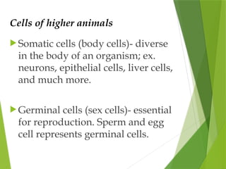
Cells of higheranimals
Somatic cells (body cells)- diverse
in the body of an organism; ex.
neurons, epithelial cells, liver cells,
and much more.
Germinal cells (sex cells)- essential
for reproduction. Sperm and egg
cell represents germinal cells.
3.
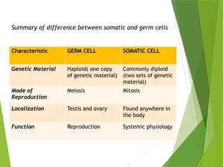
Summary of differencebetween somatic and germ cells
Characteristic GERM CELL SOMATIC CELL
Genetic Material Haploid( one copy
of genetic material)
Commonly diploid
(two sets of genetic
material)
Mode of
Reproduction
Meiosis Mitosis
Localization Testis and ovary Found anywhere in
the body
Function Reproduction Systemic physiology
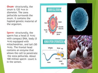
Ovum- structurally, the
ovumis 120 m in
diameter. The zona
pellucida surrounds the
ovum. It contains the
haploid genetic material of
the organism.
Sperm- structurally, the
sperm has a head (5 m)
with compact DNA, body (5
m) equipped with
mitochondrion, and tail (50
m). The frontal head
contains an enzyme that
allows the cell to penetrate
the zona pellucida. About
100 million sperm count is
in the semen.
6.
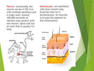
Neuron- structurally, the
neuroncan be 4-150 m
with multiple dendrites and
a single axon. Around
100,000 networks of
neurons may connect with
one neuron. Spine cell has
an axon that is usually 1m
long.
Melanocytes- are epithelial
cells that contain long
branches that lie in
keratinocytes. Its function
is to pass the pigment to
the melanosome.
7.
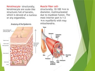
Keratinocyte- structurally,
keratinocyte arescale-like
structures full of keratin,
which is devoid of a nucleus
or any organelles.
Muscle fiber cell-
structurally, 10-100 m in
diameter, multinucleated
due to myoblast fusion. The
most interior part is 1-2
m myofibrils with may
mitochondria.
8.
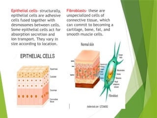
Epithelial cells- structurally,
epithelialcells are adhesive
cells fused together with
desmosomes between cells.
Some epithelial cells act for
absorption secretion and
ion transport. They vary in
size according to location.
Fibroblasts- these are
unspecialized cells of
connective tissue, which
can commit to becoming a
cartilage, bone, fat, and
smooth muscle cells.
9.
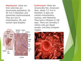
Hepatocytes- these are
livercells that are
structurally polyhedral, 20-
30 m in diameter, and
sometimes multinucleated.
They are rich in
mitochondria, ER, and
contain lipid droplets
Erythrocytes- these are
structurally flat, biconcave
discs, about 7.2 m in
diameter. It does not
contain mitochondria,
nucleus, and ribosomes.
They have a lifespan of 120
days. There are normally 5
million cells per blood
sample, and they transport
oxygen.
10.
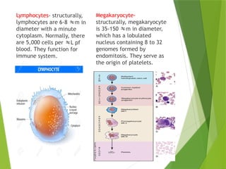
Lymphocytes- structurally,
lymphocytes are6-8 m in
diameter with a minute
cytoplasm. Normally, there
are 5,000 cells per L pf
blood. They function for
immune system.
Megakaryocyte-
structurally, megakaryocyte
is 35-150 m in diameter,
which has a lobulated
nucleus containing 8 to 32
genomes formed by
endomitosis. They serve as
the origin of platelets.
11.
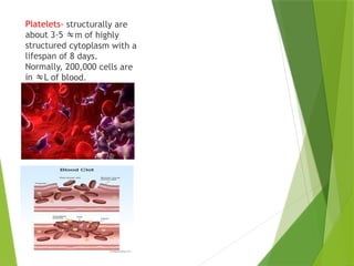
Platelets- structurally are
about3-5 m of highly
structured cytoplasm with a
lifespan of 8 days.
Normally, 200,000 cells are
in L of blood.
Tissues result fromthe differentiation of
several groups of cells that will form a group
of cells having the same function.
Histology- the study of different tissues.
Different tissues arise from a particular germ
layer during embryonic development.
In animals, the tissues are either epithelial,
connective,muscular, or nervous tissue.
14.
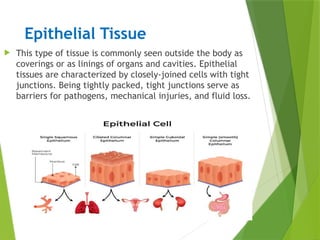
Epithelial Tissue
Thistype of tissue is commonly seen outside the body as
coverings or as linings of organs and cavities. Epithelial
tissues are characterized by closely-joined cells with tight
junctions. Being tightly packed, tight junctions serve as
barriers for pathogens, mechanical injuries, and fluid loss.
15.
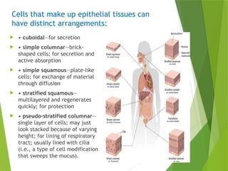
Cells that makeup epithelial tissues can
have distinct arrangements:
• cuboidal—for secretion
• simple columnar—brick-
shaped cells; for secretion and
active absorption
• simple squamous—plate-like
cells; for exchange of material
through diffusion
• stratified squamous—
multilayered and regenerates
quickly; for protection
• pseudo-stratified columnar—
single layer of cells; may just
look stacked because of varying
height; for lining of respiratory
tract; usually lined with cilia
(i.e., a type of cell modification
that sweeps the mucus).
16.
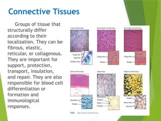
Connective Tissues
Groups oftissue that
structurally differ
according to their
localization. They can be
fibrous, elastic,
reticular, or collagenous.
They are important for
support, protection,
transport, insulation,
and repair. They are also
responsible for blood cell
differentiation or
formation and
immunological
responses.
17.
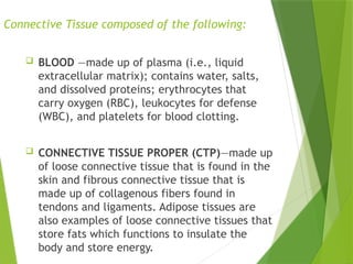
Connective Tissue composedof the following:
BLOOD —made up of plasma (i.e., liquid
extracellular matrix); contains water, salts,
and dissolved proteins; erythrocytes that
carry oxygen (RBC), leukocytes for defense
(WBC), and platelets for blood clotting.
CONNECTIVE TISSUE PROPER (CTP)—made up
of loose connective tissue that is found in the
skin and fibrous connective tissue that is
made up of collagenous fibers found in
tendons and ligaments. Adipose tissues are
also examples of loose connective tissues that
store fats which functions to insulate the
body and store energy.
18.
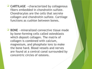
CARTILAGE —characterizedby collagenous
fibers embedded in chondroitin sulfate.
Chondrocytes are the cells that secrete
collagen and chondroitin sulfate. Cartilage
functions as cushion between bones.
BONE —mineralized connective tissue made
by bone-forming cells called osteoblasts
which deposit collagen. The matrix of
collagen is combined with calcium,
magnesium, and phosphate ions to make
the bone hard. Blood vessels and nerves
are found at a central canal surrounded by
concentric circles of osteons.
19.
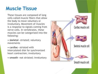
Muscle Tissue
These tissuesare composed of long
cells called muscle fibers that allow
the body to move voluntary or
involuntary. Movement of muscles
is a response to signals coming from
nerve cells. In vertebrates, these
muscles can be categorized into the
following:
• skeletal—striated; voluntary
movements
• cardiac—striated with
intercalated disk for synchronized
heart contraction; involuntary
• smooth—not striated; involuntary
20.
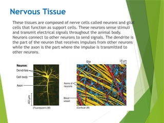
Nervous Tissue
These tissuesare composed of nerve cells called neurons and glial
cells that function as support cells. These neurons sense stimuli
and transmit electrical signals throughout the animal body.
Neurons connect to other neurons to send signals. The dendrite is
the part of the neuron that receives impulses from other neurons
while the axon is the part where the impulse is transmitted to
other neurons.
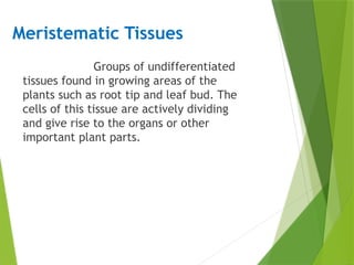
Meristematic Tissues
Groups ofundifferentiated
tissues found in growing areas of the
plants such as root tip and leaf bud. The
cells of this tissue are actively dividing
and give rise to the organs or other
important plant parts.
23.
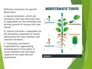
Different meristems forspecific
plant parts:
A. Apical meristems- which are
located on root tips and shoot tips,
is responsible for the formation and
primary growth of various cells and
tissues.
B. Lateral meristem- responsible for
the horizontal expansion of tissues
particularly the cork cambium and
vascular meristem.
C. Intercalary meristems-
responsible for regenerating
removed parts of the plant. It
occurs between the old tissue
regions of the node and leaf
attachment.
24.
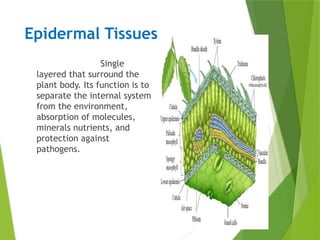
Epidermal Tissues
Single
layered thatsurround the
plant body. Its function is to
separate the internal system
from the environment,
absorption of molecules,
minerals nutrients, and
protection against
pathogens.
25.
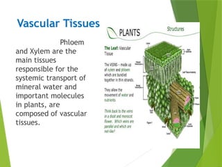
Vascular Tissues
Phloem
and Xylemare the
main tissues
responsible for the
systemic transport of
mineral water and
important molecules
in plants, are
composed of vascular
tissues.
26.
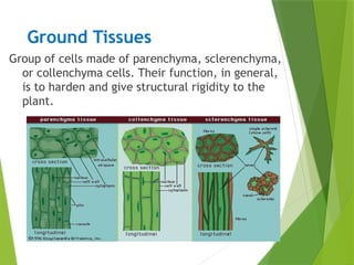
Ground Tissues
Group ofcells made of parenchyma, sclerenchyma,
or collenchyma cells. Their function, in general,
is to harden and give structural rigidity to the
plant.


![animal_tissues_by_Deepak_b[presentation].pptx](https://cdn.slidesharecdn.com/ss_thumbnails/animaltissuesbydeepakb1-250228175730-fe92da00-thumbnail.jpg?width=640&height=640&fit=bounds)


