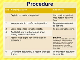
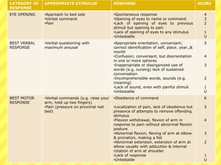
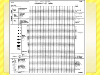
2. Introduction
GCS is a quick, practical, & standardized system for
assessing the degree of impaired consciousness.
Three indicators of response are evaluated:
1. Opening of eyes
2. The best verbal response
3. & the best motor response
Specific behaviours observed as responses to the
testing stimulus in each of these areas are given a
numeric value & can be plotted on a graph.
The subscale scores are particularly important if
a patient is untestable in one area. for example severe
periorbital edema may makes eye opening impossible.
the total GCS score is the sum of the numeric values
assigned to each of the three areas are evaluated. the
highest GCS score is 15 for a fully alert person, & the
lowest possible score is 3. a GCS score of 8 or less is
generally indicative of coma.
3. INDICATIONS FOR USE:
Conscious/semi-conscious patient due to any reason
Obvious or suspected HI
To assess the arousal aspect of consciousness.
Post neurosurgery or vascular surgery to carotid vertebral
artery
Severe unexplained headache
Neuro impairment
Prolonged epileptic seizure
Drug overdose
The nurses responsibility is to elicit the best response on each of the scales: the
higher the scores, the higher the level of brain functioning. a graph can be used
to determine whether the patient is stable, improving, or deteriorating.
Articles
GCS proforma (in some situations a neurological assessment
chart).
A pen torch (PENLIGHT)
A tray for the assessment of vital signs
4. Eye opening:
Score 4 if patient’s eyes are open upon approaching the bed, or remain
open when not spoken to.
Score 3 if patient’s eyes open when spoken to or shouted at.
Score 2 if patient’s eyes open after applying a central pain stimulus.
Score 1 if patient’s eyes don’t open at all.
Put “C” if eye closed due to swelling. (1 point)
Verbal response:
Score 5 if patient knows who they are, the day, and where they are.
Score 4 if patient’s responses to questions are incorrect, but appropriate.
Score 3 if patient’s response to questions are inappropriate or irrelevant
to question.
Score 2 if patient’s response incomprehensible (eg moans).
Score 1 if no response.
Put “T” if patient scores 1 because they’re intubated.
Motor response:
Use the best response at time of assessment, normally arms, but may be legs.
Score 6 if patient obeys simple commends. Ensure pateint’s hand squeeze is not
grasp reflex by asking them to release their grip also.
Score 5 if patient localizes with hands/arms to sternal rub.
Score 4 if patient withdraws hand, briskly, to nail bed pressure.
Score 3 if patient has abnormal flexion to central pain stimulus (ie Decorticate
posturing).
Score 2 if patient extends to central pain stimulus (ie Decerebratye posturing or
abnormal extension).
Score 1 for no movement.
5. Procedure
s.n
.
Nursing action Rationale
1 Explain procedure to patient Unconscious patient
may retain ability to
hear.
2 Keep patient in comfortable position To promote comfort
to patient.
3 Score responses in GCS sheets. To assess GCS score.
4 Add total score at bottom of sheet
during each assessment.
5 Assess vital signs for completion of
procedure
Vital Signs
It is important that the patient's
temperature, blood pressure,
pulse and respiration are also
included because changes in
vital signs may indicate a
compression or damage in the
brain stem.
6 Document accurately & report changes
if any.
To maintain accuracy
in records.
6. CATEGORY OF
RESPONSE
APPROPRIATE STIMULUS RESPONSE SCORE
EYE OPENING •Approach to bed side
•Verbal command
•Pain
•Spontaneous response
•Opening of eyes to name or command
•Lack of opening of eyes to previous
stimuli but opening to pain
•Lack of opening of eyes to any stimulus
•Untestable
4
3
2
1
U
BEST VERBAL
RESPONSE
•Verbal questioning with
maximum arousal
•Appropriate orientation, conversant;
correct identification of self, place. year.,&
month
•Confusion; conversant, but disorientation
in one or more spheres
•Inappropriate or disorganized use of
words (e.g. cursing) lack of sustained
conversation
•Incomprehensible words, sounds (e.g.
moaning)
•Lack of sound, even with painful stimuli
•Untestable
5
4
3
2
1
U
BEST MOTOR
RESPONSE
•Verbal commands (e.g. raise your
arm, hold up two fingers)
•Pain (pressure on proximal nail
bed)
•Obedience of command
•Localization of pain, lack of obedience but
presence of attempts to remove offending
stimulus
•Flexion withdrawal, flexion of arm in
response to pain without abnormal flexion
posture
•Abnormal flexion, flexing of arm at elbow
& pronation, making a fist
•Abnormal extension, extension of arm at
elbow usually with adduction & internal
rotation of arm at shoulder
•Lack of response
•Untestable
6
5
4
3
2
1
U
7.
8.
9. Note:
۞Document if patient has GCS of 3 due to paralysis or
sedation.
۞If eye closed due to swelling document “C” under eye
opening.
۞Docuement “T” for verbal response if patient
intubated or has a tracheostomy.
۞If motor response differs from R to L document “R” or
“L” in appropriate boxes.
۞Make note if patient is a quadriplegic/paraplegic.
۞Responses to peripheral pain stimuli (nail bed
pressure) may be possible even with a complete cord
transection. Therefore peripheral stimulus is an
inappropriate stimulus for assessing the GCS.
10. Pupillary assessment
Pupillary assessment is an important part of neurological
assessment because changes in the size, equality and reactivity of
the pupils can provide vital diagnostic information in the critically
ill patient .
Both pupils should be the same shape, size and react equally to
light. Although not part of the Glasgow Coma Scale ,examination
of the pupils is an essential adjunct to it, especially when the
patient’s level of consciousness is impaired
Anatomy and physiology
The pupil is the ‘black hole’ in the centre of the iris, a flattened
muscular diaphragm which is attached to the ciliary
body.Relaxation and contraction of the muscles of the iris causes
it to dilate (in darkness) or constrict (in bright light).
Evaluation of pupillary reaction is effectively an assessment of the
third cranial nerve (oculomotor nerve), which controls constriction
of the pupil. Compression of this nerve will result in fixed dilated
pupils.
11. Pupil size and shape
Pupil size should be measured, ideally with reference to a neurological
observation chart or similar. The average size is 2-5mm (Bersten et al,
2003). The pupils should be equal in size.
Pupil shape should be ascertained. It should be round; abnormal shapes
may indicate cerebral damage; oval shape could indicate intracranial
hypertension.The pupils should be identical in shape.
If the 3rd cranial nerve (occulomotor) is compressed, the pupil on the
affected side becomes larger until it fully dilates.
If ICP continues to increase, both pupil dilates.
Reaction to a bright light
Pupil reaction to light should be brisk and after removal of the light source,
the pupil should return to its original size. There should also be a
consensual reaction to the light source, that is the opposite pupil also
constricts when the light source is applied to one eye. Pupil reaction should
be documented as per local policy, for example B (brisk), S (sluggish) or N
(no reaction). Both pupils should react equally to light.
Sluggish reaction can indicate early pressure on 3rd CN.
A fixed pupil unresponsive to light stimulus usually indicates increased ICP.
Unreactive pupils can be caused by an expanding mass, for example a
blood clot exerting pressure on the third cranial nerve; a fixed and dilated
pupil may be due to herniation of the medial temporal lobe.
12. Pre Procedure assessment
Prior to undertaking pupillary assessment:
Note if the patient has any pre-existing irregularity with the
pupils, for example cataracts, false eye or previous eye injury;
Check if there are any pre-existing factors that can cause
pupillary dilation, for example medications including
tricyclics, atropine and sympathomimetics and traumatic
mydriasis.
Check if there are any pre-existing factors that can cause
pupillary constriction, for example medications including
narcotics and topical beta-blockers.
Assemble equipment: a pen torch and the patient’s observation
chart (in some situations a neurological assessment chart).
If possible, dim the overhead light source (a darkened room is
ideal but practically this will rarely be possible); dim light will
facilitate a better view of the pupils and their reaction to light.
13. Procedure
s.n
.
Nursing action/ step of procedure Rationale
1 Wash and dry hands. To prevent cross –
infection.
2 Explain procedure to patient, even if she or
he is unconscious.
To gain patient’s
cooperation.
Unconscious patient
may retain ability to
hear.
3 Adopt a position in front of the patient. To perform pupillary
assessment.
4 Look into each of the patient’s eyes,
examining the size of the pupils. Note
the size (mm) of each pupil; if
available, use the scale printed on the
neurological assessment chart as a
comparison. Compare the sizes of the
pupils.
To assess pupils size.
5 Note the shape of each pupil.
Compare the shapes of the pupils. to assess pupils shape
14. s.n
.
Nursing action/ step of procedure Rationale
6 6.1. After providing prior warning to
the patient, move the torchlight
from the side of the head towards
the pupil and note any change in
pupil size and the speed of the
reaction (brisk or sluggish). Taking
care to avoid shining the light in
the other eye, observe whether the
opposite pupil also reacts
(consensual reflex).
6.2. Repeat the above procedure in
the opposite eye.
To assess pupillary
reaction to bright
light.
7 Document the findings of the pupillary
assessment as per local policy and
guidelines. If necessary, inform the
nurse in-charge of any changes or
abnormalities.
To maintain accuracy
in records.