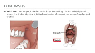
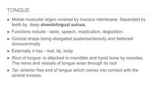
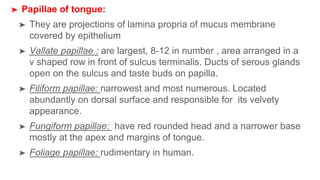
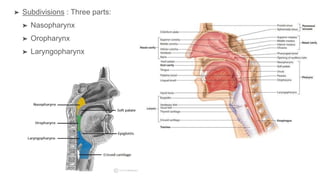
SlideShare a Scribd company logo
1 of 95
ORAL CAVITY
➤ Vestibule: narrow space that lies outside the teeth and gums and inside lips and
cheek. It is limited above and below by reflection of mucous membrane from lips and
cheeks.
➤ Lips: two mobile musculofibrous folds that surrounds the opening of the mouth. They
are lined externally by skin and internally by mucous membrane. The red portion of
lip is called vermillion zone. The skin and vermillion zone meet at vermillion
border.
➤ The internal aspect of each lip is connected to corresponding gum by a median fold
of the mucus membrane called frenulum of lip.
➤ Cheeks: fleshy flaps forming large part of face. Each cheek is continuous with lips in
front. The junction between the two is marked by nasolabial sulcus. Cheek is lined
externally by skin and internally by mucous membrane.
➤ It is largely composed of buccinator muscle and muscle is covered by
buccopharyngeal fascia. In addition it contains buccal glands, blood vessels, nerves.
➤ Gums: composed of fibrous tissue covered with smooth vascular mucous
membrane. It consists of three parts:
➤ Free part: surrounds neck of tooth like a collar
➤ Attached part: firmly attached to the alveolar process.
➤ Interdental part: extension of attached gingiva between teeth
➤ Teeth: mineralised bone like structures projecting from the alveolar process of jaws.
➤ There are 16 permanent teeth in each jaw in adults. Human are diphyodont, two sets
of teeth develop in a person’s lifetime- primary/ deciduous teeth and permanent teeth
➤ Parts of a tooth: consists of
➤ Crown- anatomical crown is the part of tooth that is covered by enamel and clinical
crown is part that projects into the oral cavity
➤ Root- which is embedded within the socket of jaw beneath gum.
➤ Neck- which is constricted part of tooth between crown and root
➤ Structure of tooth:
➤ Pulp: inner core containing soft tissue,blood vessels, nerves, lymphatics. Covered by
ondontoblasts.
➤ Dentine: calcified material surrounding the pulp cavity
➤ Enamel: densely calcified white material covering the crown of tooth
➤ Cement: bony covering over the neck and root of tooth.
➤ Periodontal membrane: present between cementum and socket.
➤ Types of teeth: Incisors, canines, premolars, molars
➤ Dental formula:
➤ Deciduous teeth- 20 in number. 2 incisors, 1 canine, 2 molars
➤ Permanent teeth - 32 in number. 2 incisors, 1 canine, 2 premolars, 3 molars
➤ Oral cavity proper:
➤ Has a roof and floor. Posteriorly it communicates with oropharynx
through oropharyngeal isthmus, which is bounded superiorly by soft
palate, inferiorly by the tongue and on each side by palatoglossal
arches.
➤ Floor of mouth is small horse shoe shaped region situated beneath the
anterior two third of the tongue and above the muscular diaphragm
formed by two mylohyoid muscles
➤ Anterior part of floor of mouth is called sublingual region. It presents
with frenulum linguae, on each side of frenulum is an elevation called
sublingual papilla- opening of submandibular duct. Sublingual fold-
elevation of mucous membrane on each side of frenulum produced by
sublingual gland.
➤ Roof of mouth: anterior
two third is made up of
bones is called hard
palate, while posterior one
third is made up of soft
tissue called soft palate.
➤ From posterior free margin
of soft palate is small
conical projection called
uvula hangs down in the
median region
TONGUE
➤ Mobile muscular organ covered by mucous membrane. Separated by
teeth by deep alveololingual sulcus.
➤ Functions include - taste, speech, mastication, deglutition.
➤ Conical shape being elongated posterioanteriorly and flattened
dorsoventrrally
➤ Externally it has - root, tip, body
➤ Root of tongue- is attached to mandible and hyoid bone by muscles.
The nerve and vessels of tongue enter through its root
➤ Tip- anterior free end of tongue which comes into contact with the
central incisors.
➤ Body- bulk of tongue between root and tip. It has dorsal and
ventral surfaces and right and left lateral margin.
➤ Dorsal surface- convex on all sides. Divided by a V shaped
sulcus called sulcus terminalis into anterior two third- oral
part, posterior one third- pharyngeal part.
➤ Apex of sulcus has blind foramen called foramen caecum-
origin of median thyroid diverticulum.
➤ Oral part- median furrow, large papillae
➤ Pharyngeal part- large lymphoid follicles which is called
lingual tonsil, large number of mucous and serous glands.
➤ Papillae of tongue:
➤ They are projections of lamina propria of mucus membrane
covered by epithelium
➤ Vallate papillae : are largest, 8-12 in number , area arranged in a
v shaped row in front of sulcus terminalis. Ducts of serous glands
open on the sulcus and taste buds on papilla.
➤ Filiform papillae: narrowest and most numerous. Located
abundantly on dorsal surface and responsible for its velvety
appearance.
➤ Fungiform papillae: have red rounded head and a narrower base
mostly at the apex and margins of tongue.
➤ Foliage papillae: rudimentary in human.
➤ Pharyngeal part: devoid of papillae,
consists of lingual tonsil.
➤ Mucus membrane in this part is
continuous with mucus membrane
covering the palatine tonsils and pharynx.
Posteriorly it is reflected onto the front of
epiglottis as median glossoepiglottic
fold and onto the lateral wall of pharynx
as lateral glossoepiglottic fold . The
space on each side of median
glossoepiglottic fold is called vallecula.
➤ Ventral surface of tongue: it
consists of
Frenulum linguae
Deep lingual veins- on either
sides of frenulum linguae
Pelica fimbriata- fringedy
fimbriated fold of mucous
membrane lateral to lingual
vein
➤ Muscles of tongue:
➤ Has intrinsic and extrinsic muscles
➤ Intrinsic muscles : are within tongue and have no attachment
outside tongue. They change the shape of tongue. They
include -
Superior longitudinal
Inferior longitudinal
Transverse
Vertical
➤ Extrinsic muscles: attach muscle to mandible, hyoid, styloid
process, palate. They help in movement of tongue like
protraction, retraction, side to side movement. They are-
Genioglossus- helps In protrusion
Hyoglossus- helps in depression
Styloglossus- helps in retraction
Palatoglossus- helps in elevation.
➤ Arterial supply: lingual artery, tonsillar branch of facial artery,
ascending pharyngeal artery.
➤ Venous drainage: deep lingual veins, venae comitantes
accompanying lingual artery, hypoglossal nerve.
➤ Lymphatic drainage:
➤ Apical vessels drain tip into submental lymphnodes
➤ Marginal vessels drain marginal portion of anterior two third
of tongue into submandibular lymph nodes
➤ Central vessels drain central portion of anterior two third of
tongue into deep cervical lymph nodes.
➤ Nerve supply
➤ Motor supply: all muscles of tongue are supplied by
hypoglossal nerve except palatoglossus supplied by cranial
part of accessory nerve.
➤ Sensory supply: anterior two third- lingual nerve for general
sensation, chorda tympani nerve for special sensation
➤ Posterior one third- glossopharyngeal nerve for both general
and special sensations
➤ Posterior most is by vagus- internal laryngeal branch of
superior laryngeal nerve.
PHARYNX
➤ Funnel shaped fibromuscular tube extending from base of skull
to esophagus. Lined by mucous membrane. Acts as common
channel for food and air.
➤ It is 12-14cm in length. It is situated behind the cavities of nose,
mouth and the larynx with which it communicates.
➤ Boundaries- superiorly base of skull, inferiorly continuous with
esophagus at level of C6, posteriorly prevertebral fascia in front
of cervical spine, anteriorly opens into nasal cavity, mouth,
larynx
➤ Subdivisions : Three parts:
➤ Nasopharynx
➤ Oropharynx
➤ Laryngopharynx
➤ Nasopharynx:
➤ Lies behind nasal cavity and above the soft palate
➤ Roof is formed by body of sphenoid,basilar part of occipital
bone
➤ Floor is formed by soft palate, pharyngeal isthmus
➤ Anterior wall is formed by nasal apertures
➤ Posterior wall forms continuous sloping surface with roof, it is
supported by anterior arch of C1 vertebra.
➤ Laterally medial pterygoid plate of sphenoid.
➤ Features
Nasopharyngeal tonsil: collection of lymphoid tissue beneath
mucous membrane at junction of roof and posterior wall of
nasopharynx.
Orifice of pharyngotympanic tube or auto form tube: lies 1.25 cm
behind inferior concha. Upper and posterior margins of this
opening are bounded by tubal elevation produced by collection
of lymphoid tissue called tubal tonsil. Two mucous fold extend
from this elevation- salpingopharyngeal and salpingopalatine
fold.
Pharyngeal recess or fossa of Rosenmuller : deep depression
behind tubal elevation.
➤ Oropharynx: extends from lower surface of soft palate above to
upper border of epiglottis below.
➤ Roof is formed by soft palate , pharyngeal isthmus
➤ Floor is formed by posterior 1/3 rd of tongue, interval between
tongue and epiglottis.
➤ Anterior wall is incomplete and is formed by oropharyngeal
isthmus, pharyngeal part of tongue.
➤ Posterior wall is formed by body of C2 and upper part of C3
vertebra
➤ Features
Palatine tonsils: present in tonsillar fossa, bounded anteriorly by
palatoglossal arch and posteriorly by palatopharyngeal arch
Lingual tonsil
Medial and lateral glossoepiglottic fold
Epiglottic vallecula
➤ Oropharyngeal isthmus: opening between two palatoglossal folds
through which oral cavity communicates with the oropharynx.
➤ It is closed during deglutition to prevent regurgitation of food from
pharynx to mouth.
➤ Laryngopharynx:
➤ Lies behind the laryngeal inlet and posterior wall of larynx.
Extends from upper border of epiglottis to lower border of cricoid
cartilage anteriorly and C6 vertebra posteriorly
➤ Anterior wall is formed by laryngeal inlet, posterior surface of
larynx.
➤ Posterior wall is supported by bodies of C3, C4, C5, C6
➤ Lateral wall is formed by thyroid cartilage and Thyrohyoid
membrane.
➤ Lateral wall presents piriform fossa on either sides of laryngeal
inlet.
➤ Pharyngeal wall: consists of four layers from inside out-
➤ Mucous membrane
➤ Pharyngobasilar fascia
➤ Muscular coat
➤ Buccopharyngeal fascia
➤ Waldeyer’s ring: aggregation of lymphoid tissue underneath
the epithelial lining of pharyngeal wall called tonsils, surround
the commencent of air and food. These aggregation together
constitute an interrrupted circle called Waldeyer’s ring.
➤ Waldeyer’s ring is formed by
Nasopharyngeal tonsil
Lingual tonsil
Tubal tonsil
Palatine tonsil
➤ Longitudinal muscles:
➤ Stylopharyngeus
➤ Palatopharyngeus
➤ Salpingopharyngeus
➤ These muscles elevate the larynx and shorten the pharynx
during swallowing.
➤ All pharyngeal muscles are supplied by the cranial root of
accessory nerve, except stylopharyngeus which is supplied by
glossopharyngeal nerve.
➤ Muscles of pharynx:
➤ Constrictor muscles:
➤ Form bulk of muscular coat of pharyngeal wall. They arise from the
margins of posterior opening of the nasal, oral, laryngeal cavities.
➤ The fibers pass backwards, in a fan- shaped manner into the lateral
and posterior walls of the pharynx to be inserted into the median
fibrous raphe on the posterior aspect of the pharynx, extending from
base of skull to esophagus.
➤ Pharyngeal pouch/ Zenker’ s diverticulum- inferior constrictor muscle
has two parts - thyropharyngeus made up of oblique fibers and
cricopharyngeus made up of transverse fibers. Potential gap between
them is called Killian’s dehiscence. The mucosa and submucosa may
bulge through this weak area to form pharyngeal pouch.
➤ Arterial supply of pharynx:
➤ Ascending pharyngeal artery
➤ Ascending palatine and tonsillar artery
➤ Greater palatine and pharyngeal artery
➤ Lingual artery
➤ Venous drainage: into pharyngeal venous plexus which drains
to internal jugular vein.
➤ Lymphatic drainage : lymph from pharynx is drained into the
upper and lower deep cervical lymph nodes , retropharyngeal
lymph nodes.
SALIVARY GLANDS
➤ Three pairs major of salivary glands -
➤ Parotid gland
➤ Submandibular gland
➤ Sublingual gland
➤ Parotid gland:
➤ Largest of three pairs. It is composed of serous alveoli.
Yellowish, lobulated,weighs 25g.
➤ Located in retromandibular fossa/ parotid bed.
➤ Parotid sheath/ capsule- gland is enclosed in a fibrous
capsule.
➤ Formed by investing layer of deep cervical fascia.
Superficial lamina forms parotidomassetric fascia, which is
attached to zygomatic arch.
➤ Deep lamina is attached to tympanic plate and styloid process
of temporal bone. It thickens to form stylomandibular ligament,
which separates parotid gland from submandibular gland.
➤ External features:
➤ Has an apex
➤ Four surfaces- superior or base, superficial surface,
antereomedial surface, posteriomedial surface
➤ Three borders- anterior, medial , posterior.
➤ Structures present within
the parotid gland: from
superficial to deep these
are:
➤ Facial nerve
➤ Retromandibular vein
➤ External carotid artery
➤ Parotid duct(Stensons’s duct):
➤ About 5 cm long emerges from the middle of the anterior
border of the gland and opens into vestibule of the mouth
opposite upper second molar.
➤ Nerve supply: Parasympathetic is secretomotor is by
auriculotemporal nerve. Produces watery secretion.
➤ Sympathetic supply from sympathetic plexus around ECA
➤ Sensory supply by auriculotemporal nerve, great auricular
nerve.
➤ Vascular supply: from ECA, superficial temporal artery.
Venous drainage by retromandibular vein and EJV.
➤ Lymphatic drainage: superficial and deep parotid lymph-
nodes which drains to deep cervical lymph nodes.
➤ Submandibular gland:
➤ Situated partly below and partly deep to posterior half of
mandible. Mixed type I.e, both mucus and serous but
predominantly serous gland.
➤ It consists of two parts: a large superficial part and a small deep
part.
➤ Superficial part: superficial to mylohyoid muscle. It has two
ends- anterior and posterior , three surfaces- inferior , lateral,
medial.
➤ Deep part: lies deep to mylohyoid muscle and on hyoglossus
muscle. Posteriorly it is continuous with superficial part.
➤ Submandibular duct (Wharton’s duct) : about 5cm, emerges
at anterior end of deep part. Runs forwards between lingual
and hypoglossal nerve. It opens into oral cavity on summit of a
sublingual papilla at side of the frenulum of the tongue.
➤ Blood supply: by sublingual and submental arteries. Drained by
common facial and lingual veins.
➤ Lymphatic drainage: submandibular lymph nodes and
subsequently into jugulodigastric lymph nodes.
➤ Nerve supply:
➤ Parasympathetic supply- secretomotor by chorda tympani
nerve
➤ Sympathetic supply - plexus around facial artery
➤ Sensory supply- by lingual nerve.
➤ Sublingual gland:
➤ Smallest of three. Lies in floor of
mouth between mucus membrane
and mylohyoid muscle.
➤ Almond shaped and rests in
sublingual fossa in mandible.
➤ It is mucus gland. It pours
secretions by a series of duct,
about 15 in number, into the oral
cavity on the sublingual fold
OESOPHAGUS
➤ Muscular tube forming food passage between pharynx and stomach. It
is about 25 cm long. It is flattened anterioposteriorly and lumen is kept
collapsed.
➤ It passes through superior and posterior mediastinum
➤ It pierces diaphragm at level of T10. It ends by opening at the cardiac
end of stomach at level of T11.
➤ It has four constrictions. They are
➤ At its beginning 15cm from incisors where it is crossed by
cricopharyngeus muscle.
➤ 22.5 cm from incisors where it is crosses by aortic arch.
➤ 27.5cm from incisors where it is crossed by left bronchus
➤ 37.5cm from incisors where it pierces the diaphragm.
➤ Arterial supply: cervical part by inferior thyroid artery, thoracic
part by oesophageal branches of aorta, abdominal part by
oesophageal branches of left gastric artery.
➤ Venous drainage: upper part into brachiocephalic veins,
middle part by azygos vein, lower end it goes to left gastric
vein, hemiazygos vein.
➤ Lymphatic drainage: cervical- deep cervical nodes, thoracic
part- posterior mediastinal nodes, abdominal part- left gastric
nodes.
➤ Histology:
➤ Mucous membrane lined by stratified squamous non
keratinised epithelium.
➤ Muscularis mucosae is distinct in lower part and formed by
longitudinal muscle fibres.
➤ Submucosa contains mucus secreting oesophageal glands.
➤ Muscularis externa is composed of striated muscle in upper
third, mixed in middle third, smooth muscles in lower third.
Outer layer comprises of longitudinal coat and inner circular
coat of muscle fibers
➤ Adventitia- connective tissue.
STOMACH
➤ Muscular bag forming the widest and most distensible part of the
digestive tube. It is connected above to the lower end of
oesophagus and below to duodenum.
➤ It acts as a reservoir of food and helps in digestion of proteins
and fats
➤ Lies obliquely in the upper and left part of abdomen , occupying
the epigastric umbilical and left hypochondriac regions.
➤ J Shaped when empty, 25cm long, mean capacity 1.5-2L in
adults.
➤ It has two orifices or openings,
two curvatures and two surfaces.
➤ Two orifices:
➤ Cardiac orifice: joined by lower
end of oesophagus. There is
physiological spinchter but
anatomically can not be
demonstrated.
➤ Pyloric orifice: opens into
duodenum
➤ Two curvatures:
➤ Lesser curvature: is concave and forms
right border of the stomach. It provides
attachment to lesser omentum. Most
dependent part is marked by angular
notch or incisura.
➤ Greater curvature: convex and forms
left border of stomach. Provides
attachment to greater omentum,
gastrosplenic, Gastrophrenic ligament.
At upper end it has cardiac notch
➤ Two surfaces: anterior/ anterosuperior surface faces
upwards and upwards. Posterior or posteroinferior surface
faces backwards and downwards.
➤ Parts of stomach:
➤ Cardiac part: fundus, body
➤ Pyloric part: pyloric antrum, pyloric canal
➤ Peritoneal relations:
➤ Stomach is lined by peritoneum on both its surfaces . At the
lesser curvature the layers of peritoneum lining the anterior
and posterior surface meets and becomes continuous with
the lesser omentum and along greater curvature with greater
omentum.
➤ Near fundus it forms gastrosplenic ligament.
➤ Near cardiac end it forms gastrophrenic ligament.
➤ Visceral relations:
➤ Anteriorly to liver, the diaphragm, transverse colon, anterior
abdominal wall.
➤ Posteriorly it is related structures which form stomach bed. Which
include
➤ Diaphragm
➤ Left kidney, left suprarenal gland
➤ Pancreas
➤ Transverse mesocolon.
➤ Splenic flexure of colon
➤ Splenic artery, spleen
➤ Blood supply:
➤ along lesser curvature by left gastric artery branch of coeliac
trunk, right gastric artery branch of proper hepatic artery
➤ along greater curvature by right gastroepiploic artery branch
of gastroduodenal artery, left gastroepiploic artery branch of
splenic artery
➤ Fundus is supplied by 5-7 short gastric arteries
➤ Veins of stomach drain into portal, superior mesenteric,
splenic veins.
➤ Lymphatic drainage:
➤ Nerve supply:
➤ Sympathetic by T6-T10 via grea splanchnic nerves, coeliac and
hepatic plexuses. They are motor to pyloric sphincter and chief
pathway for pain sensation
➤ Parasympathetic by vagus nerve through gastric nerves. They are
motor and secretomotor to stomach.
➤ Anterior gastric nerve contains fibers from left vagus and posterior
gastric nerve from right vagus
➤ Anterior gastric nerve divides into number of gastric branches and two
pyloric branch
➤ Posterior gastric nerve divides into smaller gastric branches , larger
coeliac branches for coeliac plexus
➤ Histology:
➤ Mucosa of empty stomach is thrown into folds termed as
gastric rugae. They are flattened in distended stomach. On
mucosal surface there are numerous small depressions
called gastric pits. Gastric glands open into gastric pits.
➤ Part of lumen of stomach that lies along the lesser curvature
and has longitudinal rugae is called gastric canal or
magenstrasse.
➤ Submucosa coat is made up of connective tissue, arterioles,
nerve plexus.
➤ Muscle coat is arranged as superficial longitudinal fibers mainly
along curvatures. Inner circular fibers encircles body and are
thickened at pylorus to form pyloric sphincter. Deepest layer
consists of oblique fibers.
➤ Serous coat consists of the peritoneal covering.
SPLEEN
➤ Location: in left hypochondrium and partly in epigastrium
➤ It is soft highly vascular and dark purple in colour. On an
average spleen is 1 inch thick, 3 inch broad, 5 inch long , 7
ounces in weight. It is related to 9th to 11th ribs.
➤ Directed downwards, forwards and laterally. Lies obliquely
along the long axis of the 10 th rib
➤ It has two ends. Anterior end which is expanded, more like a
border. Posterior end is rounded and rests on upper pole of
left kidney.
➤ Three borders: superior border which is notched. Inferior and
intermediate border is rounded.
➤ Two surfaces: diaphragmatic surface, visceral surface
➤ Two angles: antereobasal angle, postereobasal angle.
➤ Hilum: pierced by branches and tributaries to splenic vessels.
➤ Peritoneal relations:
➤ Gastrosplenic ligament: contains short gastric vessel.
➤ Linorenal ligament: it contains tail of pancreas, splenic
vessels, pancreaticosplenic lymph nodes.
➤ Phrenicocolic ligament: not attached to spleen but supports
anterior end.
➤ Visceral relations:
➤ Visceral surface is related to the
fundus of stomach, the anterior
surface of left kidney, splenic flexure
of the colon, tail of pancreas
➤ Diaphragmatic surface is related to
diaphragm which separates the
spleen from costodiaphragmatic
recess of pleura, lung, 9,10,11th ribs
of left side.
➤ Arterial supply: supplied by splenic artery. It passes through
linorenal ligament to reach hilum of lung. Within spleen it divides
repeatedly to form successfully the straight vessels called penicilli
which further divides into ellipsoids and arterial capillaries.
➤ Venous drainage: splenic vein formed at hilum and runs straight
course behind pancreas. It joins superior mesenteric vein to form
portal vein.
➤ Lymphatic drainage: drains into pancreaticosplenic lymph nodes.
➤ Nerve supply: coeliac plexus.
➤ Functions : phagocytosis, haemopoiesis, immune response,
storage of RBCs .
LIVER
➤ Large, solid, gland situated in the
right upper quadrant of abdominal
cavity. Weighs 1600g in males and
about 1300g in females.
➤ Location: right hypochondrium,
epigastrium and some part of left
hypochondrium.
➤ External features: wedge shaped. Five surfaces- anterior,
posterior, superior, inferior, right.
➤ One prominent border- inferior border . It is marked by
Interlobar notch- notch of ligamentum teres and cystic notch-
by fundus of gallbladder.
➤ Two lobes: divided into right and left lobe by attachment of the
falciform ligament anteriorly and superiorly, by fissure for the
ligamentum teres inferiorly, by fissure for ligamentum venosum
posteriorly.
➤ Right lobe is much larger than left lobe. Consists of caudate and
quadrate lobe
➤ Caudate lobe: bounded on right by groove for inferior vena cava,
on left by fissure for ligamentum venosum and inferiorly by porta
hepatis.
➤ Quadrate lobe: bounded anteriorly by inferior border, posteriorly
by porta hepatis, on right by fossa for gallbladder, on left by
fissure for ligamentum teres
➤ Porta hepatis: situated on inferior surface of right lobe of liver.
Lies between the caudate lobe above and quadrate lobe
below. The portal vein, hepatic artery, hepatic plexus of nerves
enter the liver through porta hepatis while right and left hepatic
ducts leave it.
➤ Peritoneal relations: most of liver is covered by peritoneum
except the following:
➤ Triangular bare area
➤ Groove for IVC
➤ fossa for gallbladder
➤ Visceral relations:
➤ Anterior surface: related to xiphoid process, to anterior abdominal wall
In median plane and to diaphragm on each side
➤ Posterior surface: diaphragm, right suprarenal gland, groove for IVC
lodges upper part of vessel, oesophagus.
➤ Superior surface: shows cardiac impression, diaphragmatic impression
➤ Inferior surface: gastric impression, omentale tuber, omentum, pylorus,
first part and second part duodenum, part of transverse colon, hepatic
flexure forming colic impression, right renal impression
➤ Right surface: upper one third to diaphragm, pleura, lung, middle one
third to diaphragm, costodiaphramgmatic recess of pleura, lower one
third to diaphragm alone.
➤ Blood supply:
➤ 20% blood supply through hepatic artery, 80% through portal
vein. Hepatic arterial blood mixes with portal venous blood in
sinusoids
➤ Hepatic sinusoids drain into interlobular veins which drains into
hepatic vein. Hepatic veins are in two groups- upper group
which contains right, middle, left and lower group contains
numerous small veins and they open into vena cava.
➤ Lymphatics: caval, hepatic, paracardial, coeliac lymph nodes
➤ Nerve supply: hepatic plexus which contains both sympathetic
and parasympathetic ( vagal fibers)
➤ Hepatic segments: based on intrahepatic distribution of
hepatic artery, portal vein, biliary ducts, the liver can be divided
into right and left functional lobes which do not correspond to
anatomical lobes.
➤ Histology:
➤ Liver is covered by Glisson’s capsule. It extends into liver and surrounds
portal triad.
➤ Hepatic lobules: hexagonal areas. They are partially separated by
connective tissue. Each lobule has a small round space in center. This
is central vein.
➤ Number of broad irregular cords of cells seem to pass from this vein to
periphery of the lobule. They are made up of polygonal liver cells-
hepatocytes.
➤ Portal triad consists of hepatic artery, portal vein, interlobular bile duct.
➤ Functions:
➤ Acts as exocrine gland for the secretion of bile.
➤ Prominent role in metabolism of carbohydrates, proteins, fats.
Synthesis of plasma proteins fibrinogen and prothrombin,
regulation of blood glucose and lipids.
➤ Store for various substances including glucose,lipids,
vitamins and iron
➤ Detoxifying substances like drugs, alcohol . Removal of bile
pigments from blood.
➤ Haemopoiesis.

More Related Content

Similar to Gastrointestinal system-1.pptx

E.N.T.Throat.(dr.mwayad)
E.N.T.Throat.(dr.mwayad)E.N.T.Throat.(dr.mwayad)
E.N.T.Throat.(dr.mwayad)student
 
2. Anatomy of the tongue and its applied aspects.pptx
2. Anatomy of the tongue and its applied aspects.pptx2. Anatomy of the tongue and its applied aspects.pptx
2. Anatomy of the tongue and its applied aspects.pptxDrChandiniRavikumar
 
Anatomy of the Oral cavity Proper
Anatomy of the Oral cavity ProperAnatomy of the Oral cavity Proper
Anatomy of the Oral cavity ProperRahaf Sn
 
Tongue and its applied aspects
Tongue and its applied aspectsTongue and its applied aspects
Tongue and its applied aspectsDr. Faizan Ansari
 
Anatomy of Nose & Paranasal sinuses.pptx
Anatomy of Nose & Paranasal sinuses.pptxAnatomy of Nose & Paranasal sinuses.pptx
Anatomy of Nose & Paranasal sinuses.pptxHtet Ko
 
Tongue, palatine .pdf
Tongue, palatine .pdfTongue, palatine .pdf
Tongue, palatine .pdfssuserbf4af22
 
1.anatomy of Oral cavity & Salivary glands.pdf
1.anatomy of  Oral cavity & Salivary glands.pdf1.anatomy of  Oral cavity & Salivary glands.pdf
1.anatomy of Oral cavity & Salivary glands.pdf69spfm6dst
 
tongue ppt oral and maxillofacial pathology basic
tongue  ppt oral and maxillofacial pathology basictongue  ppt oral and maxillofacial pathology basic
tongue ppt oral and maxillofacial pathology basicSidharthabordoloi1
 
tongue.pptx oral and maxillofacial pathology basic
tongue.pptx oral and maxillofacial pathology basictongue.pptx oral and maxillofacial pathology basic
tongue.pptx oral and maxillofacial pathology basicSidharthaBordoloi2
 
Pharynx and palate
Pharynx and palatePharynx and palate
Pharynx and palateSankaraNYKAM
 
Anatomy of tongue & its applied aspects
Anatomy of tongue & its applied aspectsAnatomy of tongue & its applied aspects
Anatomy of tongue & its applied aspectsAnchal Mehra
 
Surgical anatomy and physiology of pharynx(中山医)
Surgical anatomy and physiology of pharynx(中山医)Surgical anatomy and physiology of pharynx(中山医)
Surgical anatomy and physiology of pharynx(中山医)Sumit Prajapati
 
Surgical anatomy of tongue
Surgical anatomy of tongueSurgical anatomy of tongue
Surgical anatomy of tonguesiddharth verma
 

Similar to Gastrointestinal system-1.pptx (20)

E.N.T.Throat.(dr.mwayad)
E.N.T.Throat.(dr.mwayad)E.N.T.Throat.(dr.mwayad)
E.N.T.Throat.(dr.mwayad)
 
2. Anatomy of the tongue and its applied aspects.pptx
2. Anatomy of the tongue and its applied aspects.pptx2. Anatomy of the tongue and its applied aspects.pptx
2. Anatomy of the tongue and its applied aspects.pptx
 
Anatomy of the Oral cavity Proper
Anatomy of the Oral cavity ProperAnatomy of the Oral cavity Proper
Anatomy of the Oral cavity Proper
 
Tongue and its applied aspects
Tongue and its applied aspectsTongue and its applied aspects
Tongue and its applied aspects
 
Anatomy of Nose & Paranasal sinuses.pptx
Anatomy of Nose & Paranasal sinuses.pptxAnatomy of Nose & Paranasal sinuses.pptx
Anatomy of Nose & Paranasal sinuses.pptx
 
Tongue, palatine .pdf
Tongue, palatine .pdfTongue, palatine .pdf
Tongue, palatine .pdf
 
1.anatomy of Oral cavity & Salivary glands.pdf
1.anatomy of  Oral cavity & Salivary glands.pdf1.anatomy of  Oral cavity & Salivary glands.pdf
1.anatomy of Oral cavity & Salivary glands.pdf
 
Oral region
Oral regionOral region
Oral region
 
tongue ppt oral and maxillofacial pathology basic
tongue  ppt oral and maxillofacial pathology basictongue  ppt oral and maxillofacial pathology basic
tongue ppt oral and maxillofacial pathology basic
 
14-ORAL CAVITY.ppt
14-ORAL CAVITY.ppt14-ORAL CAVITY.ppt
14-ORAL CAVITY.ppt
 
14-ORAL CAVITY.ppt
14-ORAL CAVITY.ppt14-ORAL CAVITY.ppt
14-ORAL CAVITY.ppt
 
tongue.pptx oral and maxillofacial pathology basic
tongue.pptx oral and maxillofacial pathology basictongue.pptx oral and maxillofacial pathology basic
tongue.pptx oral and maxillofacial pathology basic
 
Anatomy of oral cavity
Anatomy of oral cavityAnatomy of oral cavity
Anatomy of oral cavity
 
Pharynx and palate
Pharynx and palatePharynx and palate
Pharynx and palate
 
Anatomy of tongue & its applied aspects
Anatomy of tongue & its applied aspectsAnatomy of tongue & its applied aspects
Anatomy of tongue & its applied aspects
 
Tongue /prosthodontic courses
Tongue /prosthodontic coursesTongue /prosthodontic courses
Tongue /prosthodontic courses
 
Oral cavity anatomy
Oral cavity anatomyOral cavity anatomy
Oral cavity anatomy
 
Surgical anatomy and physiology of pharynx(中山医)
Surgical anatomy and physiology of pharynx(中山医)Surgical anatomy and physiology of pharynx(中山医)
Surgical anatomy and physiology of pharynx(中山医)
 
Surgical anatomy of tongue
Surgical anatomy of tongueSurgical anatomy of tongue
Surgical anatomy of tongue
 
The oral cavity
The oral cavityThe oral cavity
The oral cavity
 

Recently uploaded

microwave assisted reaction. General introduction
microwave assisted reaction. General introductionmicrowave assisted reaction. General introduction
microwave assisted reaction. General introductionMaksud Ahmed
 
Sociology 101 Demonstration of Learning Exhibit
Sociology 101 Demonstration of Learning ExhibitSociology 101 Demonstration of Learning Exhibit
Sociology 101 Demonstration of Learning Exhibitjbellavia9
 
Energy Resources. ( B. Pharmacy, 1st Year, Sem-II) Natural Resources
Energy Resources. ( B. Pharmacy, 1st Year, Sem-II) Natural ResourcesEnergy Resources. ( B. Pharmacy, 1st Year, Sem-II) Natural Resources
Energy Resources. ( B. Pharmacy, 1st Year, Sem-II) Natural ResourcesShubhangi Sonawane
 
Introduction to Nonprofit Accounting: The Basics
Introduction to Nonprofit Accounting: The BasicsIntroduction to Nonprofit Accounting: The Basics
Introduction to Nonprofit Accounting: The BasicsTechSoup
 
Russian Escort Service in Delhi 11k Hotel Foreigner Russian Call Girls in Delhi
Russian Escort Service in Delhi 11k Hotel Foreigner Russian Call Girls in DelhiRussian Escort Service in Delhi 11k Hotel Foreigner Russian Call Girls in Delhi
Russian Escort Service in Delhi 11k Hotel Foreigner Russian Call Girls in Delhikauryashika82
 
Holdier Curriculum Vitae (April 2024).pdf
Holdier Curriculum Vitae (April 2024).pdfHoldier Curriculum Vitae (April 2024).pdf
Holdier Curriculum Vitae (April 2024).pdfagholdier
 
Mixin Classes in Odoo 17 How to Extend Models Using Mixin Classes
Mixin Classes in Odoo 17  How to Extend Models Using Mixin ClassesMixin Classes in Odoo 17  How to Extend Models Using Mixin Classes
Mixin Classes in Odoo 17 How to Extend Models Using Mixin ClassesCeline George
 
Key note speaker Neum_Admir Softic_ENG.pdf
Key note speaker Neum_Admir Softic_ENG.pdfKey note speaker Neum_Admir Softic_ENG.pdf
Key note speaker Neum_Admir Softic_ENG.pdfAdmir Softic
 
Seal of Good Local Governance (SGLG) 2024Final.pptx
Seal of Good Local Governance (SGLG) 2024Final.pptxSeal of Good Local Governance (SGLG) 2024Final.pptx
Seal of Good Local Governance (SGLG) 2024Final.pptxnegromaestrong
 
Measures of Dispersion and Variability: Range, QD, AD and SD
Measures of Dispersion and Variability: Range, QD, AD and SDMeasures of Dispersion and Variability: Range, QD, AD and SD
Measures of Dispersion and Variability: Range, QD, AD and SDThiyagu K
 
Advanced Views - Calendar View in Odoo 17
Advanced Views - Calendar View in Odoo 17Advanced Views - Calendar View in Odoo 17
Advanced Views - Calendar View in Odoo 17Celine George
 
Application orientated numerical on hev.ppt
Application orientated numerical on hev.pptApplication orientated numerical on hev.ppt
Application orientated numerical on hev.pptRamjanShidvankar
 
Food Chain and Food Web (Ecosystem) EVS, B. Pharmacy 1st Year, Sem-II
Food Chain and Food Web (Ecosystem) EVS, B. Pharmacy 1st Year, Sem-IIFood Chain and Food Web (Ecosystem) EVS, B. Pharmacy 1st Year, Sem-II
Food Chain and Food Web (Ecosystem) EVS, B. Pharmacy 1st Year, Sem-IIShubhangi Sonawane
 
ComPTIA Overview | Comptia Security+ Book SY0-701
ComPTIA Overview | Comptia Security+ Book SY0-701ComPTIA Overview | Comptia Security+ Book SY0-701
ComPTIA Overview | Comptia Security+ Book SY0-701bronxfugly43
 
ICT Role in 21st Century Education & its Challenges.pptx
ICT Role in 21st Century Education & its Challenges.pptxICT Role in 21st Century Education & its Challenges.pptx
ICT Role in 21st Century Education & its Challenges.pptxAreebaZafar22
 
Nutritional Needs Presentation - HLTH 104
Nutritional Needs Presentation - HLTH 104Nutritional Needs Presentation - HLTH 104
Nutritional Needs Presentation - HLTH 104misteraugie
 
1029 - Danh muc Sach Giao Khoa 10 . pdf
1029 -  Danh muc Sach Giao Khoa 10 . pdf1029 -  Danh muc Sach Giao Khoa 10 . pdf
1029 - Danh muc Sach Giao Khoa 10 . pdfQucHHunhnh
 
Web & Social Media Analytics Previous Year Question Paper.pdf
Web & Social Media Analytics Previous Year Question Paper.pdfWeb & Social Media Analytics Previous Year Question Paper.pdf
Web & Social Media Analytics Previous Year Question Paper.pdfJayanti Pande
 
Unit-IV; Professional Sales Representative (PSR).pptx
Unit-IV; Professional Sales Representative (PSR).pptxUnit-IV; Professional Sales Representative (PSR).pptx
Unit-IV; Professional Sales Representative (PSR).pptxVishalSingh1417
 

Recently uploaded (20)

microwave assisted reaction. General introduction
microwave assisted reaction. General introductionmicrowave assisted reaction. General introduction
microwave assisted reaction. General introduction
 
Sociology 101 Demonstration of Learning Exhibit
Sociology 101 Demonstration of Learning ExhibitSociology 101 Demonstration of Learning Exhibit
Sociology 101 Demonstration of Learning Exhibit
 
Energy Resources. ( B. Pharmacy, 1st Year, Sem-II) Natural Resources
Energy Resources. ( B. Pharmacy, 1st Year, Sem-II) Natural ResourcesEnergy Resources. ( B. Pharmacy, 1st Year, Sem-II) Natural Resources
Energy Resources. ( B. Pharmacy, 1st Year, Sem-II) Natural Resources
 
Introduction to Nonprofit Accounting: The Basics
Introduction to Nonprofit Accounting: The BasicsIntroduction to Nonprofit Accounting: The Basics
Introduction to Nonprofit Accounting: The Basics
 
Russian Escort Service in Delhi 11k Hotel Foreigner Russian Call Girls in Delhi
Russian Escort Service in Delhi 11k Hotel Foreigner Russian Call Girls in DelhiRussian Escort Service in Delhi 11k Hotel Foreigner Russian Call Girls in Delhi
Russian Escort Service in Delhi 11k Hotel Foreigner Russian Call Girls in Delhi
 
Holdier Curriculum Vitae (April 2024).pdf
Holdier Curriculum Vitae (April 2024).pdfHoldier Curriculum Vitae (April 2024).pdf
Holdier Curriculum Vitae (April 2024).pdf
 
Mixin Classes in Odoo 17 How to Extend Models Using Mixin Classes
Mixin Classes in Odoo 17  How to Extend Models Using Mixin ClassesMixin Classes in Odoo 17  How to Extend Models Using Mixin Classes
Mixin Classes in Odoo 17 How to Extend Models Using Mixin Classes
 
Key note speaker Neum_Admir Softic_ENG.pdf
Key note speaker Neum_Admir Softic_ENG.pdfKey note speaker Neum_Admir Softic_ENG.pdf
Key note speaker Neum_Admir Softic_ENG.pdf
 
Seal of Good Local Governance (SGLG) 2024Final.pptx
Seal of Good Local Governance (SGLG) 2024Final.pptxSeal of Good Local Governance (SGLG) 2024Final.pptx
Seal of Good Local Governance (SGLG) 2024Final.pptx
 
Measures of Dispersion and Variability: Range, QD, AD and SD
Measures of Dispersion and Variability: Range, QD, AD and SDMeasures of Dispersion and Variability: Range, QD, AD and SD
Measures of Dispersion and Variability: Range, QD, AD and SD
 
Advanced Views - Calendar View in Odoo 17
Advanced Views - Calendar View in Odoo 17Advanced Views - Calendar View in Odoo 17
Advanced Views - Calendar View in Odoo 17
 
Application orientated numerical on hev.ppt
Application orientated numerical on hev.pptApplication orientated numerical on hev.ppt
Application orientated numerical on hev.ppt
 
Food Chain and Food Web (Ecosystem) EVS, B. Pharmacy 1st Year, Sem-II
Food Chain and Food Web (Ecosystem) EVS, B. Pharmacy 1st Year, Sem-IIFood Chain and Food Web (Ecosystem) EVS, B. Pharmacy 1st Year, Sem-II
Food Chain and Food Web (Ecosystem) EVS, B. Pharmacy 1st Year, Sem-II
 
ComPTIA Overview | Comptia Security+ Book SY0-701
ComPTIA Overview | Comptia Security+ Book SY0-701ComPTIA Overview | Comptia Security+ Book SY0-701
ComPTIA Overview | Comptia Security+ Book SY0-701
 
ICT Role in 21st Century Education & its Challenges.pptx
ICT Role in 21st Century Education & its Challenges.pptxICT Role in 21st Century Education & its Challenges.pptx
ICT Role in 21st Century Education & its Challenges.pptx
 
INDIA QUIZ 2024 RLAC DELHI UNIVERSITY.pptx
INDIA QUIZ 2024 RLAC DELHI UNIVERSITY.pptxINDIA QUIZ 2024 RLAC DELHI UNIVERSITY.pptx
INDIA QUIZ 2024 RLAC DELHI UNIVERSITY.pptx
 
Nutritional Needs Presentation - HLTH 104
Nutritional Needs Presentation - HLTH 104Nutritional Needs Presentation - HLTH 104
Nutritional Needs Presentation - HLTH 104
 
1029 - Danh muc Sach Giao Khoa 10 . pdf
1029 -  Danh muc Sach Giao Khoa 10 . pdf1029 -  Danh muc Sach Giao Khoa 10 . pdf
1029 - Danh muc Sach Giao Khoa 10 . pdf
 
Web & Social Media Analytics Previous Year Question Paper.pdf
Web & Social Media Analytics Previous Year Question Paper.pdfWeb & Social Media Analytics Previous Year Question Paper.pdf
Web & Social Media Analytics Previous Year Question Paper.pdf
 
Unit-IV; Professional Sales Representative (PSR).pptx
Unit-IV; Professional Sales Representative (PSR).pptxUnit-IV; Professional Sales Representative (PSR).pptx
Unit-IV; Professional Sales Representative (PSR).pptx
 

Gastrointestinal system-1.pptx

  • 1. ORAL CAVITY ➤ Vestibule: narrow space that lies outside the teeth and gums and inside lips and cheek. It is limited above and below by reflection of mucous membrane from lips and cheeks.
  • 2. ➤ Lips: two mobile musculofibrous folds that surrounds the opening of the mouth. They are lined externally by skin and internally by mucous membrane. The red portion of lip is called vermillion zone. The skin and vermillion zone meet at vermillion border. ➤ The internal aspect of each lip is connected to corresponding gum by a median fold of the mucus membrane called frenulum of lip.
  • 3. ➤ Cheeks: fleshy flaps forming large part of face. Each cheek is continuous with lips in front. The junction between the two is marked by nasolabial sulcus. Cheek is lined externally by skin and internally by mucous membrane. ➤ It is largely composed of buccinator muscle and muscle is covered by buccopharyngeal fascia. In addition it contains buccal glands, blood vessels, nerves. ➤ Gums: composed of fibrous tissue covered with smooth vascular mucous membrane. It consists of three parts: ➤ Free part: surrounds neck of tooth like a collar ➤ Attached part: firmly attached to the alveolar process. ➤ Interdental part: extension of attached gingiva between teeth
  • 4. ➤ Teeth: mineralised bone like structures projecting from the alveolar process of jaws. ➤ There are 16 permanent teeth in each jaw in adults. Human are diphyodont, two sets of teeth develop in a person’s lifetime- primary/ deciduous teeth and permanent teeth ➤ Parts of a tooth: consists of ➤ Crown- anatomical crown is the part of tooth that is covered by enamel and clinical crown is part that projects into the oral cavity ➤ Root- which is embedded within the socket of jaw beneath gum. ➤ Neck- which is constricted part of tooth between crown and root ➤ Structure of tooth: ➤ Pulp: inner core containing soft tissue,blood vessels, nerves, lymphatics. Covered by ondontoblasts. ➤ Dentine: calcified material surrounding the pulp cavity ➤ Enamel: densely calcified white material covering the crown of tooth
  • 5. ➤ Cement: bony covering over the neck and root of tooth. ➤ Periodontal membrane: present between cementum and socket.
  • 6. ➤ Types of teeth: Incisors, canines, premolars, molars
  • 7. ➤ Dental formula: ➤ Deciduous teeth- 20 in number. 2 incisors, 1 canine, 2 molars ➤ Permanent teeth - 32 in number. 2 incisors, 1 canine, 2 premolars, 3 molars
  • 8.
  • 9. ➤ Oral cavity proper: ➤ Has a roof and floor. Posteriorly it communicates with oropharynx through oropharyngeal isthmus, which is bounded superiorly by soft palate, inferiorly by the tongue and on each side by palatoglossal arches. ➤ Floor of mouth is small horse shoe shaped region situated beneath the anterior two third of the tongue and above the muscular diaphragm formed by two mylohyoid muscles ➤ Anterior part of floor of mouth is called sublingual region. It presents with frenulum linguae, on each side of frenulum is an elevation called sublingual papilla- opening of submandibular duct. Sublingual fold- elevation of mucous membrane on each side of frenulum produced by sublingual gland.
  • 10. ➤ Roof of mouth: anterior two third is made up of bones is called hard palate, while posterior one third is made up of soft tissue called soft palate. ➤ From posterior free margin of soft palate is small conical projection called uvula hangs down in the median region
  • 11. TONGUE ➤ Mobile muscular organ covered by mucous membrane. Separated by teeth by deep alveololingual sulcus. ➤ Functions include - taste, speech, mastication, deglutition. ➤ Conical shape being elongated posterioanteriorly and flattened dorsoventrrally ➤ Externally it has - root, tip, body ➤ Root of tongue- is attached to mandible and hyoid bone by muscles. The nerve and vessels of tongue enter through its root ➤ Tip- anterior free end of tongue which comes into contact with the central incisors.
  • 12.
  • 13. ➤ Body- bulk of tongue between root and tip. It has dorsal and ventral surfaces and right and left lateral margin. ➤ Dorsal surface- convex on all sides. Divided by a V shaped sulcus called sulcus terminalis into anterior two third- oral part, posterior one third- pharyngeal part. ➤ Apex of sulcus has blind foramen called foramen caecum- origin of median thyroid diverticulum. ➤ Oral part- median furrow, large papillae ➤ Pharyngeal part- large lymphoid follicles which is called lingual tonsil, large number of mucous and serous glands.
  • 14.
  • 15. ➤ Papillae of tongue: ➤ They are projections of lamina propria of mucus membrane covered by epithelium ➤ Vallate papillae : are largest, 8-12 in number , area arranged in a v shaped row in front of sulcus terminalis. Ducts of serous glands open on the sulcus and taste buds on papilla. ➤ Filiform papillae: narrowest and most numerous. Located abundantly on dorsal surface and responsible for its velvety appearance. ➤ Fungiform papillae: have red rounded head and a narrower base mostly at the apex and margins of tongue. ➤ Foliage papillae: rudimentary in human.
  • 16. ➤ Pharyngeal part: devoid of papillae, consists of lingual tonsil. ➤ Mucus membrane in this part is continuous with mucus membrane covering the palatine tonsils and pharynx. Posteriorly it is reflected onto the front of epiglottis as median glossoepiglottic fold and onto the lateral wall of pharynx as lateral glossoepiglottic fold . The space on each side of median glossoepiglottic fold is called vallecula.
  • 17. ➤ Ventral surface of tongue: it consists of Frenulum linguae Deep lingual veins- on either sides of frenulum linguae Pelica fimbriata- fringedy fimbriated fold of mucous membrane lateral to lingual vein
  • 18. ➤ Muscles of tongue: ➤ Has intrinsic and extrinsic muscles ➤ Intrinsic muscles : are within tongue and have no attachment outside tongue. They change the shape of tongue. They include - Superior longitudinal Inferior longitudinal Transverse Vertical
  • 19. ➤ Extrinsic muscles: attach muscle to mandible, hyoid, styloid process, palate. They help in movement of tongue like protraction, retraction, side to side movement. They are- Genioglossus- helps In protrusion Hyoglossus- helps in depression Styloglossus- helps in retraction Palatoglossus- helps in elevation.
  • 20.
  • 21. ➤ Arterial supply: lingual artery, tonsillar branch of facial artery, ascending pharyngeal artery. ➤ Venous drainage: deep lingual veins, venae comitantes accompanying lingual artery, hypoglossal nerve. ➤ Lymphatic drainage: ➤ Apical vessels drain tip into submental lymphnodes ➤ Marginal vessels drain marginal portion of anterior two third of tongue into submandibular lymph nodes ➤ Central vessels drain central portion of anterior two third of tongue into deep cervical lymph nodes.
  • 22. ➤ Nerve supply ➤ Motor supply: all muscles of tongue are supplied by hypoglossal nerve except palatoglossus supplied by cranial part of accessory nerve. ➤ Sensory supply: anterior two third- lingual nerve for general sensation, chorda tympani nerve for special sensation ➤ Posterior one third- glossopharyngeal nerve for both general and special sensations ➤ Posterior most is by vagus- internal laryngeal branch of superior laryngeal nerve.
  • 23.
  • 24. PHARYNX ➤ Funnel shaped fibromuscular tube extending from base of skull to esophagus. Lined by mucous membrane. Acts as common channel for food and air. ➤ It is 12-14cm in length. It is situated behind the cavities of nose, mouth and the larynx with which it communicates. ➤ Boundaries- superiorly base of skull, inferiorly continuous with esophagus at level of C6, posteriorly prevertebral fascia in front of cervical spine, anteriorly opens into nasal cavity, mouth, larynx
  • 25. ➤ Subdivisions : Three parts: ➤ Nasopharynx ➤ Oropharynx ➤ Laryngopharynx
  • 26. ➤ Nasopharynx: ➤ Lies behind nasal cavity and above the soft palate ➤ Roof is formed by body of sphenoid,basilar part of occipital bone ➤ Floor is formed by soft palate, pharyngeal isthmus ➤ Anterior wall is formed by nasal apertures ➤ Posterior wall forms continuous sloping surface with roof, it is supported by anterior arch of C1 vertebra. ➤ Laterally medial pterygoid plate of sphenoid.
  • 27. ➤ Features Nasopharyngeal tonsil: collection of lymphoid tissue beneath mucous membrane at junction of roof and posterior wall of nasopharynx. Orifice of pharyngotympanic tube or auto form tube: lies 1.25 cm behind inferior concha. Upper and posterior margins of this opening are bounded by tubal elevation produced by collection of lymphoid tissue called tubal tonsil. Two mucous fold extend from this elevation- salpingopharyngeal and salpingopalatine fold. Pharyngeal recess or fossa of Rosenmuller : deep depression behind tubal elevation.
  • 28.
  • 29. ➤ Oropharynx: extends from lower surface of soft palate above to upper border of epiglottis below. ➤ Roof is formed by soft palate , pharyngeal isthmus ➤ Floor is formed by posterior 1/3 rd of tongue, interval between tongue and epiglottis. ➤ Anterior wall is incomplete and is formed by oropharyngeal isthmus, pharyngeal part of tongue. ➤ Posterior wall is formed by body of C2 and upper part of C3 vertebra
  • 30. ➤ Features Palatine tonsils: present in tonsillar fossa, bounded anteriorly by palatoglossal arch and posteriorly by palatopharyngeal arch Lingual tonsil Medial and lateral glossoepiglottic fold Epiglottic vallecula ➤ Oropharyngeal isthmus: opening between two palatoglossal folds through which oral cavity communicates with the oropharynx. ➤ It is closed during deglutition to prevent regurgitation of food from pharynx to mouth.
  • 31.
  • 32. ➤ Laryngopharynx: ➤ Lies behind the laryngeal inlet and posterior wall of larynx. Extends from upper border of epiglottis to lower border of cricoid cartilage anteriorly and C6 vertebra posteriorly ➤ Anterior wall is formed by laryngeal inlet, posterior surface of larynx. ➤ Posterior wall is supported by bodies of C3, C4, C5, C6 ➤ Lateral wall is formed by thyroid cartilage and Thyrohyoid membrane. ➤ Lateral wall presents piriform fossa on either sides of laryngeal inlet.
  • 33. ➤ Pharyngeal wall: consists of four layers from inside out- ➤ Mucous membrane ➤ Pharyngobasilar fascia ➤ Muscular coat ➤ Buccopharyngeal fascia ➤ Waldeyer’s ring: aggregation of lymphoid tissue underneath the epithelial lining of pharyngeal wall called tonsils, surround the commencent of air and food. These aggregation together constitute an interrrupted circle called Waldeyer’s ring.
  • 34. ➤ Waldeyer’s ring is formed by Nasopharyngeal tonsil Lingual tonsil Tubal tonsil Palatine tonsil
  • 35.
  • 36. ➤ Longitudinal muscles: ➤ Stylopharyngeus ➤ Palatopharyngeus ➤ Salpingopharyngeus ➤ These muscles elevate the larynx and shorten the pharynx during swallowing. ➤ All pharyngeal muscles are supplied by the cranial root of accessory nerve, except stylopharyngeus which is supplied by glossopharyngeal nerve.
  • 37.
  • 38. ➤ Muscles of pharynx: ➤ Constrictor muscles: ➤ Form bulk of muscular coat of pharyngeal wall. They arise from the margins of posterior opening of the nasal, oral, laryngeal cavities. ➤ The fibers pass backwards, in a fan- shaped manner into the lateral and posterior walls of the pharynx to be inserted into the median fibrous raphe on the posterior aspect of the pharynx, extending from base of skull to esophagus. ➤ Pharyngeal pouch/ Zenker’ s diverticulum- inferior constrictor muscle has two parts - thyropharyngeus made up of oblique fibers and cricopharyngeus made up of transverse fibers. Potential gap between them is called Killian’s dehiscence. The mucosa and submucosa may bulge through this weak area to form pharyngeal pouch.
  • 39.
  • 40. ➤ Arterial supply of pharynx: ➤ Ascending pharyngeal artery ➤ Ascending palatine and tonsillar artery ➤ Greater palatine and pharyngeal artery ➤ Lingual artery ➤ Venous drainage: into pharyngeal venous plexus which drains to internal jugular vein. ➤ Lymphatic drainage : lymph from pharynx is drained into the upper and lower deep cervical lymph nodes , retropharyngeal lymph nodes.
  • 41. SALIVARY GLANDS ➤ Three pairs major of salivary glands - ➤ Parotid gland ➤ Submandibular gland ➤ Sublingual gland
  • 42. ➤ Parotid gland: ➤ Largest of three pairs. It is composed of serous alveoli. Yellowish, lobulated,weighs 25g. ➤ Located in retromandibular fossa/ parotid bed. ➤ Parotid sheath/ capsule- gland is enclosed in a fibrous capsule. ➤ Formed by investing layer of deep cervical fascia. Superficial lamina forms parotidomassetric fascia, which is attached to zygomatic arch. ➤ Deep lamina is attached to tympanic plate and styloid process of temporal bone. It thickens to form stylomandibular ligament, which separates parotid gland from submandibular gland.
  • 43.
  • 44. ➤ External features: ➤ Has an apex ➤ Four surfaces- superior or base, superficial surface, antereomedial surface, posteriomedial surface ➤ Three borders- anterior, medial , posterior.
  • 45. ➤ Structures present within the parotid gland: from superficial to deep these are: ➤ Facial nerve ➤ Retromandibular vein ➤ External carotid artery
  • 46.
  • 47. ➤ Parotid duct(Stensons’s duct): ➤ About 5 cm long emerges from the middle of the anterior border of the gland and opens into vestibule of the mouth opposite upper second molar.
  • 48. ➤ Nerve supply: Parasympathetic is secretomotor is by auriculotemporal nerve. Produces watery secretion. ➤ Sympathetic supply from sympathetic plexus around ECA ➤ Sensory supply by auriculotemporal nerve, great auricular nerve. ➤ Vascular supply: from ECA, superficial temporal artery. Venous drainage by retromandibular vein and EJV. ➤ Lymphatic drainage: superficial and deep parotid lymph- nodes which drains to deep cervical lymph nodes.
  • 49. ➤ Submandibular gland: ➤ Situated partly below and partly deep to posterior half of mandible. Mixed type I.e, both mucus and serous but predominantly serous gland. ➤ It consists of two parts: a large superficial part and a small deep part. ➤ Superficial part: superficial to mylohyoid muscle. It has two ends- anterior and posterior , three surfaces- inferior , lateral, medial. ➤ Deep part: lies deep to mylohyoid muscle and on hyoglossus muscle. Posteriorly it is continuous with superficial part.
  • 50.
  • 51. ➤ Submandibular duct (Wharton’s duct) : about 5cm, emerges at anterior end of deep part. Runs forwards between lingual and hypoglossal nerve. It opens into oral cavity on summit of a sublingual papilla at side of the frenulum of the tongue. ➤ Blood supply: by sublingual and submental arteries. Drained by common facial and lingual veins. ➤ Lymphatic drainage: submandibular lymph nodes and subsequently into jugulodigastric lymph nodes.
  • 52. ➤ Nerve supply: ➤ Parasympathetic supply- secretomotor by chorda tympani nerve ➤ Sympathetic supply - plexus around facial artery ➤ Sensory supply- by lingual nerve.
  • 53. ➤ Sublingual gland: ➤ Smallest of three. Lies in floor of mouth between mucus membrane and mylohyoid muscle. ➤ Almond shaped and rests in sublingual fossa in mandible. ➤ It is mucus gland. It pours secretions by a series of duct, about 15 in number, into the oral cavity on the sublingual fold
  • 54. OESOPHAGUS ➤ Muscular tube forming food passage between pharynx and stomach. It is about 25 cm long. It is flattened anterioposteriorly and lumen is kept collapsed. ➤ It passes through superior and posterior mediastinum ➤ It pierces diaphragm at level of T10. It ends by opening at the cardiac end of stomach at level of T11. ➤ It has four constrictions. They are ➤ At its beginning 15cm from incisors where it is crossed by cricopharyngeus muscle. ➤ 22.5 cm from incisors where it is crosses by aortic arch.
  • 55. ➤ 27.5cm from incisors where it is crossed by left bronchus ➤ 37.5cm from incisors where it pierces the diaphragm. ➤ Arterial supply: cervical part by inferior thyroid artery, thoracic part by oesophageal branches of aorta, abdominal part by oesophageal branches of left gastric artery. ➤ Venous drainage: upper part into brachiocephalic veins, middle part by azygos vein, lower end it goes to left gastric vein, hemiazygos vein. ➤ Lymphatic drainage: cervical- deep cervical nodes, thoracic part- posterior mediastinal nodes, abdominal part- left gastric nodes.
  • 56.
  • 57. ➤ Histology: ➤ Mucous membrane lined by stratified squamous non keratinised epithelium. ➤ Muscularis mucosae is distinct in lower part and formed by longitudinal muscle fibres. ➤ Submucosa contains mucus secreting oesophageal glands. ➤ Muscularis externa is composed of striated muscle in upper third, mixed in middle third, smooth muscles in lower third. Outer layer comprises of longitudinal coat and inner circular coat of muscle fibers ➤ Adventitia- connective tissue.
  • 58.
  • 59. STOMACH ➤ Muscular bag forming the widest and most distensible part of the digestive tube. It is connected above to the lower end of oesophagus and below to duodenum. ➤ It acts as a reservoir of food and helps in digestion of proteins and fats ➤ Lies obliquely in the upper and left part of abdomen , occupying the epigastric umbilical and left hypochondriac regions. ➤ J Shaped when empty, 25cm long, mean capacity 1.5-2L in adults.
  • 60. ➤ It has two orifices or openings, two curvatures and two surfaces. ➤ Two orifices: ➤ Cardiac orifice: joined by lower end of oesophagus. There is physiological spinchter but anatomically can not be demonstrated. ➤ Pyloric orifice: opens into duodenum
  • 61. ➤ Two curvatures: ➤ Lesser curvature: is concave and forms right border of the stomach. It provides attachment to lesser omentum. Most dependent part is marked by angular notch or incisura. ➤ Greater curvature: convex and forms left border of stomach. Provides attachment to greater omentum, gastrosplenic, Gastrophrenic ligament. At upper end it has cardiac notch
  • 62. ➤ Two surfaces: anterior/ anterosuperior surface faces upwards and upwards. Posterior or posteroinferior surface faces backwards and downwards. ➤ Parts of stomach: ➤ Cardiac part: fundus, body ➤ Pyloric part: pyloric antrum, pyloric canal
  • 63.
  • 64. ➤ Peritoneal relations: ➤ Stomach is lined by peritoneum on both its surfaces . At the lesser curvature the layers of peritoneum lining the anterior and posterior surface meets and becomes continuous with the lesser omentum and along greater curvature with greater omentum. ➤ Near fundus it forms gastrosplenic ligament. ➤ Near cardiac end it forms gastrophrenic ligament.
  • 65.
  • 66. ➤ Visceral relations: ➤ Anteriorly to liver, the diaphragm, transverse colon, anterior abdominal wall. ➤ Posteriorly it is related structures which form stomach bed. Which include ➤ Diaphragm ➤ Left kidney, left suprarenal gland ➤ Pancreas ➤ Transverse mesocolon. ➤ Splenic flexure of colon ➤ Splenic artery, spleen
  • 67.
  • 68. ➤ Blood supply: ➤ along lesser curvature by left gastric artery branch of coeliac trunk, right gastric artery branch of proper hepatic artery ➤ along greater curvature by right gastroepiploic artery branch of gastroduodenal artery, left gastroepiploic artery branch of splenic artery ➤ Fundus is supplied by 5-7 short gastric arteries ➤ Veins of stomach drain into portal, superior mesenteric, splenic veins.
  • 69.
  • 71. ➤ Nerve supply: ➤ Sympathetic by T6-T10 via grea splanchnic nerves, coeliac and hepatic plexuses. They are motor to pyloric sphincter and chief pathway for pain sensation ➤ Parasympathetic by vagus nerve through gastric nerves. They are motor and secretomotor to stomach. ➤ Anterior gastric nerve contains fibers from left vagus and posterior gastric nerve from right vagus ➤ Anterior gastric nerve divides into number of gastric branches and two pyloric branch ➤ Posterior gastric nerve divides into smaller gastric branches , larger coeliac branches for coeliac plexus
  • 72.
  • 73. ➤ Histology: ➤ Mucosa of empty stomach is thrown into folds termed as gastric rugae. They are flattened in distended stomach. On mucosal surface there are numerous small depressions called gastric pits. Gastric glands open into gastric pits. ➤ Part of lumen of stomach that lies along the lesser curvature and has longitudinal rugae is called gastric canal or magenstrasse. ➤ Submucosa coat is made up of connective tissue, arterioles, nerve plexus.
  • 74. ➤ Muscle coat is arranged as superficial longitudinal fibers mainly along curvatures. Inner circular fibers encircles body and are thickened at pylorus to form pyloric sphincter. Deepest layer consists of oblique fibers. ➤ Serous coat consists of the peritoneal covering.
  • 75. SPLEEN ➤ Location: in left hypochondrium and partly in epigastrium ➤ It is soft highly vascular and dark purple in colour. On an average spleen is 1 inch thick, 3 inch broad, 5 inch long , 7 ounces in weight. It is related to 9th to 11th ribs. ➤ Directed downwards, forwards and laterally. Lies obliquely along the long axis of the 10 th rib ➤ It has two ends. Anterior end which is expanded, more like a border. Posterior end is rounded and rests on upper pole of left kidney.
  • 76. ➤ Three borders: superior border which is notched. Inferior and intermediate border is rounded. ➤ Two surfaces: diaphragmatic surface, visceral surface ➤ Two angles: antereobasal angle, postereobasal angle. ➤ Hilum: pierced by branches and tributaries to splenic vessels.
  • 77.
  • 78. ➤ Peritoneal relations: ➤ Gastrosplenic ligament: contains short gastric vessel. ➤ Linorenal ligament: it contains tail of pancreas, splenic vessels, pancreaticosplenic lymph nodes. ➤ Phrenicocolic ligament: not attached to spleen but supports anterior end.
  • 79. ➤ Visceral relations: ➤ Visceral surface is related to the fundus of stomach, the anterior surface of left kidney, splenic flexure of the colon, tail of pancreas ➤ Diaphragmatic surface is related to diaphragm which separates the spleen from costodiaphragmatic recess of pleura, lung, 9,10,11th ribs of left side.
  • 80. ➤ Arterial supply: supplied by splenic artery. It passes through linorenal ligament to reach hilum of lung. Within spleen it divides repeatedly to form successfully the straight vessels called penicilli which further divides into ellipsoids and arterial capillaries. ➤ Venous drainage: splenic vein formed at hilum and runs straight course behind pancreas. It joins superior mesenteric vein to form portal vein. ➤ Lymphatic drainage: drains into pancreaticosplenic lymph nodes. ➤ Nerve supply: coeliac plexus. ➤ Functions : phagocytosis, haemopoiesis, immune response, storage of RBCs .
  • 81. LIVER ➤ Large, solid, gland situated in the right upper quadrant of abdominal cavity. Weighs 1600g in males and about 1300g in females. ➤ Location: right hypochondrium, epigastrium and some part of left hypochondrium.
  • 82. ➤ External features: wedge shaped. Five surfaces- anterior, posterior, superior, inferior, right. ➤ One prominent border- inferior border . It is marked by Interlobar notch- notch of ligamentum teres and cystic notch- by fundus of gallbladder.
  • 83. ➤ Two lobes: divided into right and left lobe by attachment of the falciform ligament anteriorly and superiorly, by fissure for the ligamentum teres inferiorly, by fissure for ligamentum venosum posteriorly. ➤ Right lobe is much larger than left lobe. Consists of caudate and quadrate lobe ➤ Caudate lobe: bounded on right by groove for inferior vena cava, on left by fissure for ligamentum venosum and inferiorly by porta hepatis. ➤ Quadrate lobe: bounded anteriorly by inferior border, posteriorly by porta hepatis, on right by fossa for gallbladder, on left by fissure for ligamentum teres
  • 84.
  • 85. ➤ Porta hepatis: situated on inferior surface of right lobe of liver. Lies between the caudate lobe above and quadrate lobe below. The portal vein, hepatic artery, hepatic plexus of nerves enter the liver through porta hepatis while right and left hepatic ducts leave it. ➤ Peritoneal relations: most of liver is covered by peritoneum except the following: ➤ Triangular bare area ➤ Groove for IVC ➤ fossa for gallbladder
  • 86.
  • 87.
  • 88. ➤ Visceral relations: ➤ Anterior surface: related to xiphoid process, to anterior abdominal wall In median plane and to diaphragm on each side ➤ Posterior surface: diaphragm, right suprarenal gland, groove for IVC lodges upper part of vessel, oesophagus. ➤ Superior surface: shows cardiac impression, diaphragmatic impression ➤ Inferior surface: gastric impression, omentale tuber, omentum, pylorus, first part and second part duodenum, part of transverse colon, hepatic flexure forming colic impression, right renal impression ➤ Right surface: upper one third to diaphragm, pleura, lung, middle one third to diaphragm, costodiaphramgmatic recess of pleura, lower one third to diaphragm alone.
  • 89.
  • 90. ➤ Blood supply: ➤ 20% blood supply through hepatic artery, 80% through portal vein. Hepatic arterial blood mixes with portal venous blood in sinusoids ➤ Hepatic sinusoids drain into interlobular veins which drains into hepatic vein. Hepatic veins are in two groups- upper group which contains right, middle, left and lower group contains numerous small veins and they open into vena cava. ➤ Lymphatics: caval, hepatic, paracardial, coeliac lymph nodes ➤ Nerve supply: hepatic plexus which contains both sympathetic and parasympathetic ( vagal fibers)
  • 91. ➤ Hepatic segments: based on intrahepatic distribution of hepatic artery, portal vein, biliary ducts, the liver can be divided into right and left functional lobes which do not correspond to anatomical lobes.
  • 92. ➤ Histology: ➤ Liver is covered by Glisson’s capsule. It extends into liver and surrounds portal triad. ➤ Hepatic lobules: hexagonal areas. They are partially separated by connective tissue. Each lobule has a small round space in center. This is central vein. ➤ Number of broad irregular cords of cells seem to pass from this vein to periphery of the lobule. They are made up of polygonal liver cells- hepatocytes. ➤ Portal triad consists of hepatic artery, portal vein, interlobular bile duct.
  • 93.
  • 94.
  • 95. ➤ Functions: ➤ Acts as exocrine gland for the secretion of bile. ➤ Prominent role in metabolism of carbohydrates, proteins, fats. Synthesis of plasma proteins fibrinogen and prothrombin, regulation of blood glucose and lipids. ➤ Store for various substances including glucose,lipids, vitamins and iron ➤ Detoxifying substances like drugs, alcohol . Removal of bile pigments from blood. ➤ Haemopoiesis.