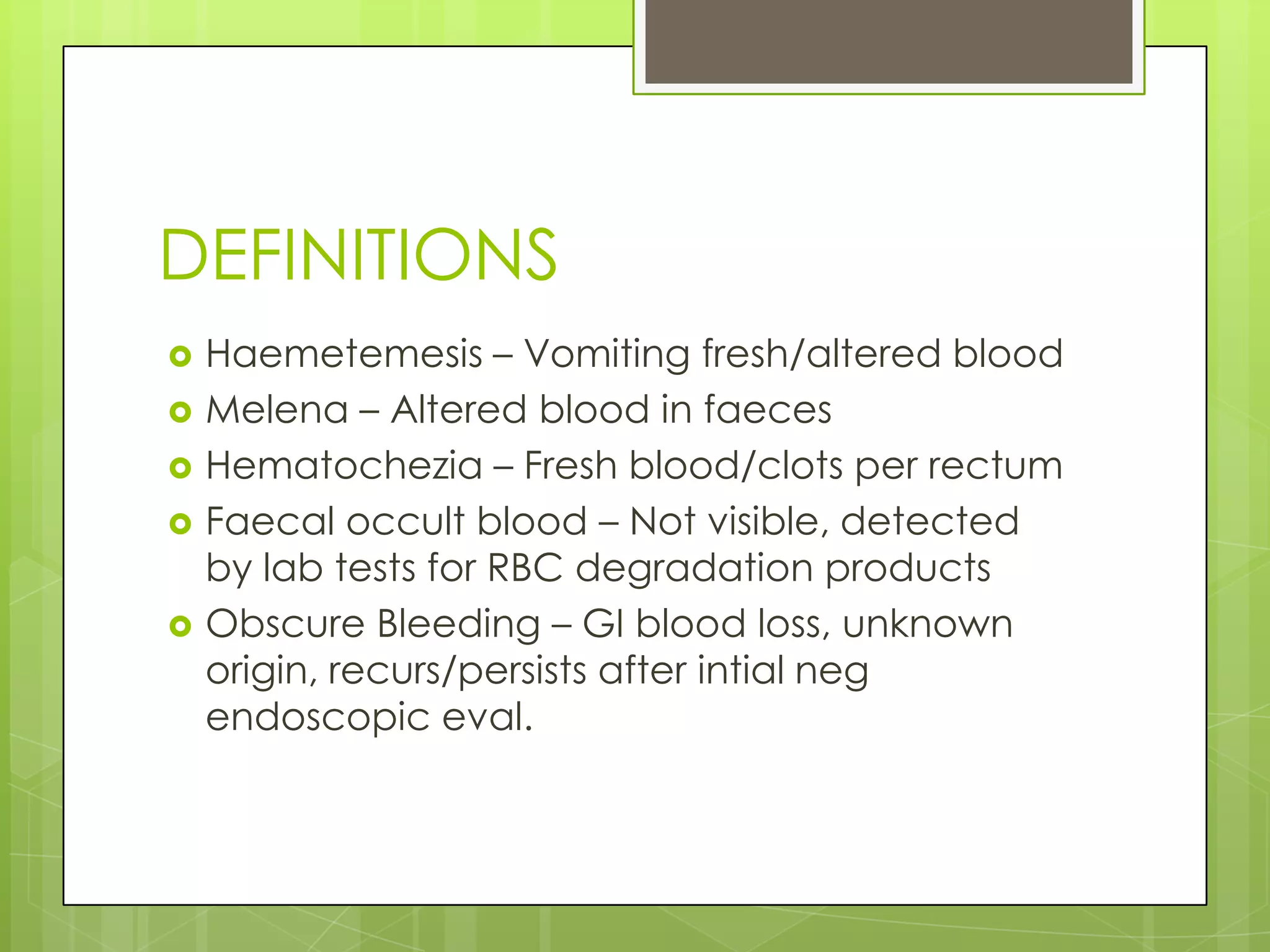
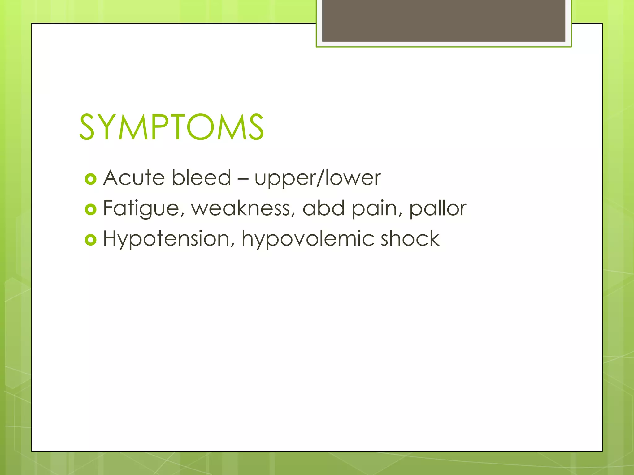
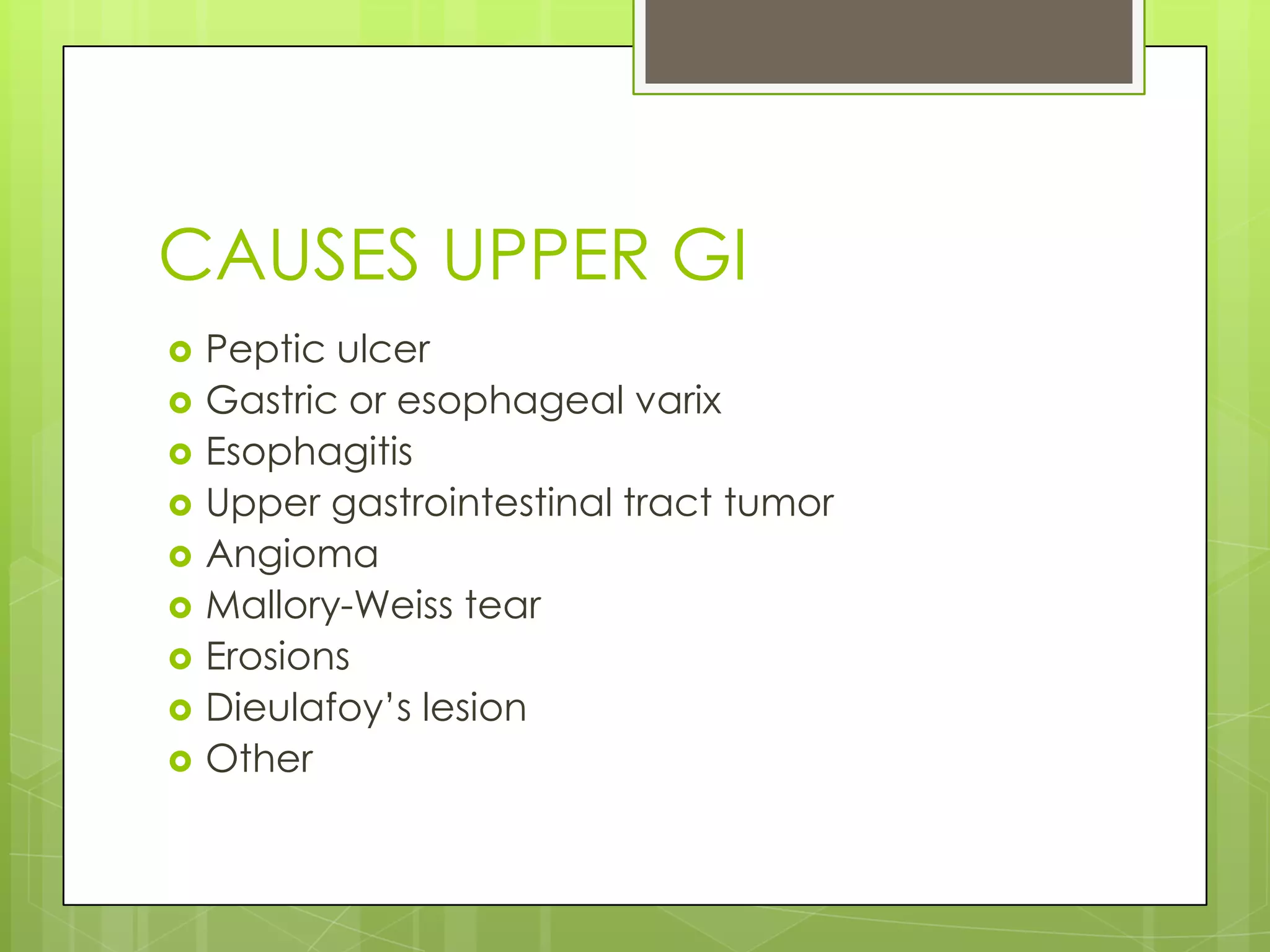
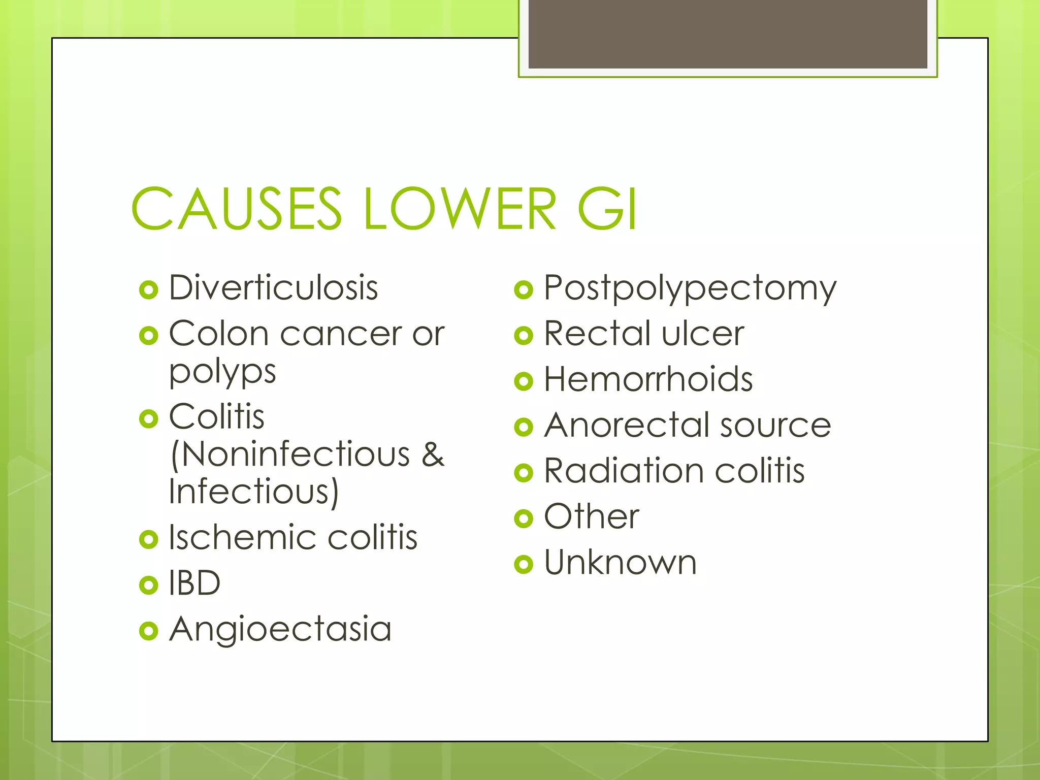
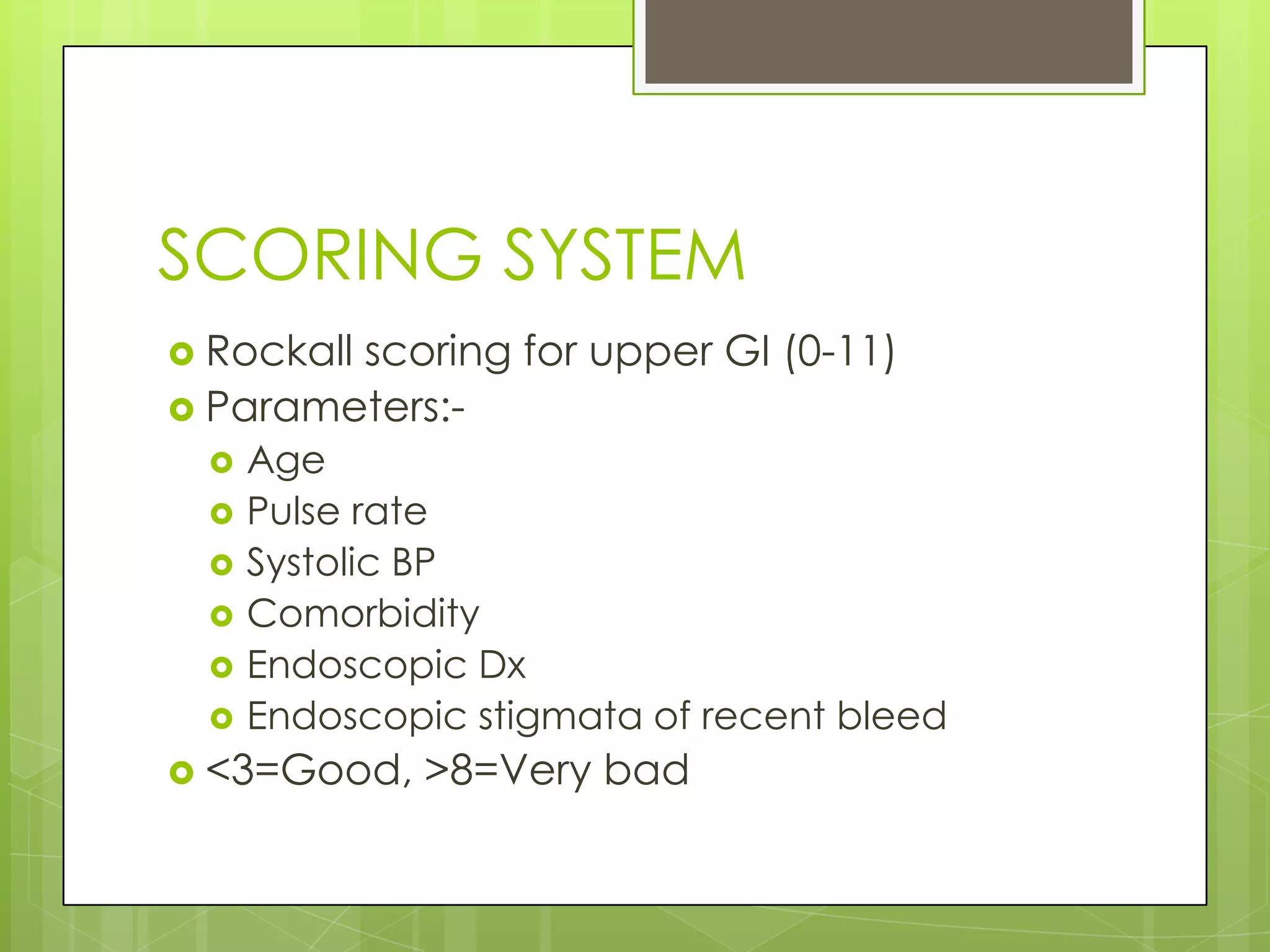
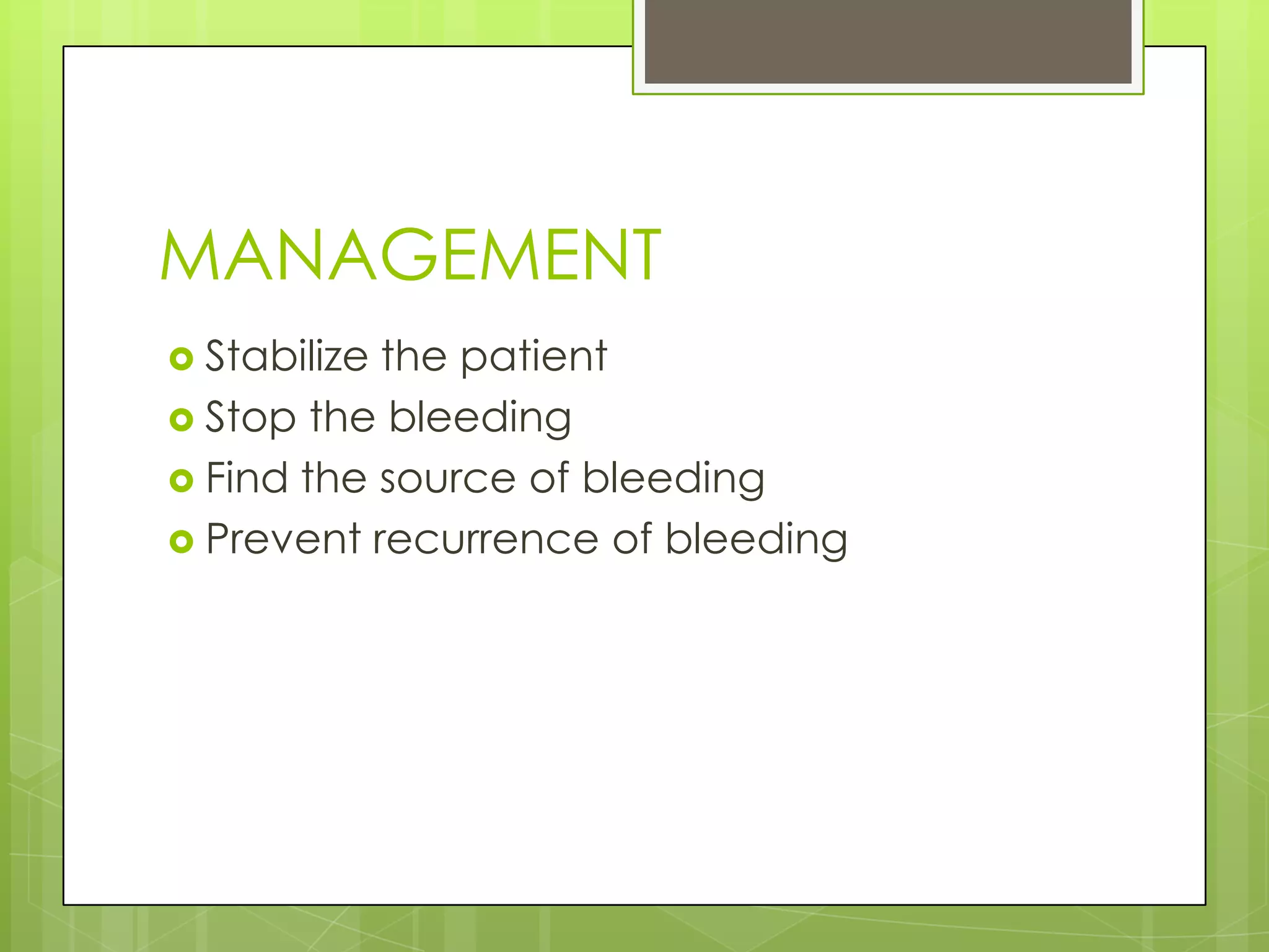
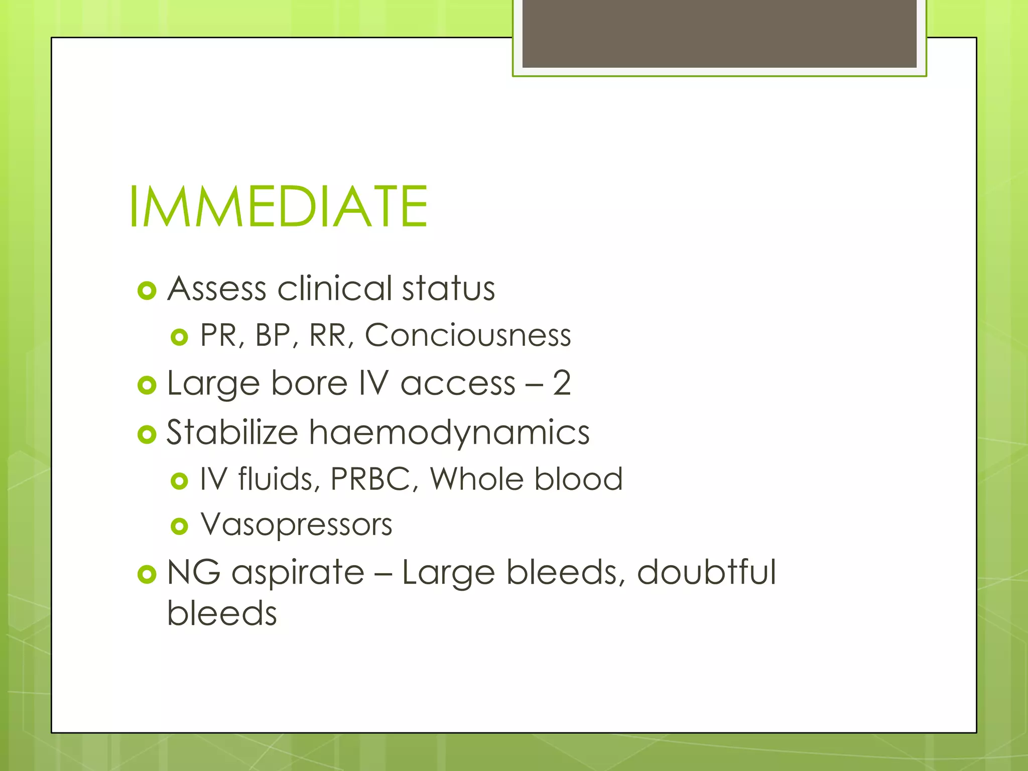
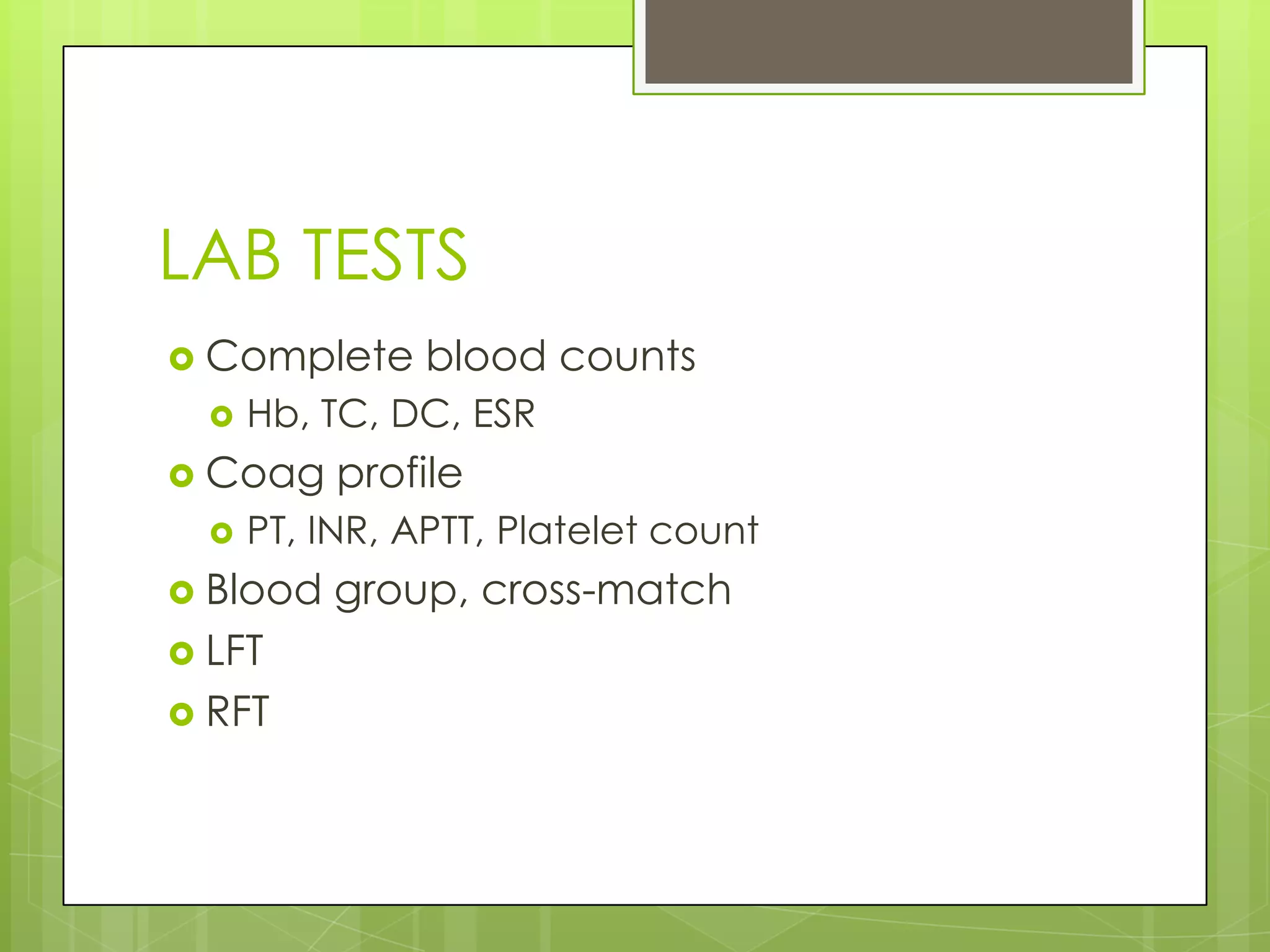
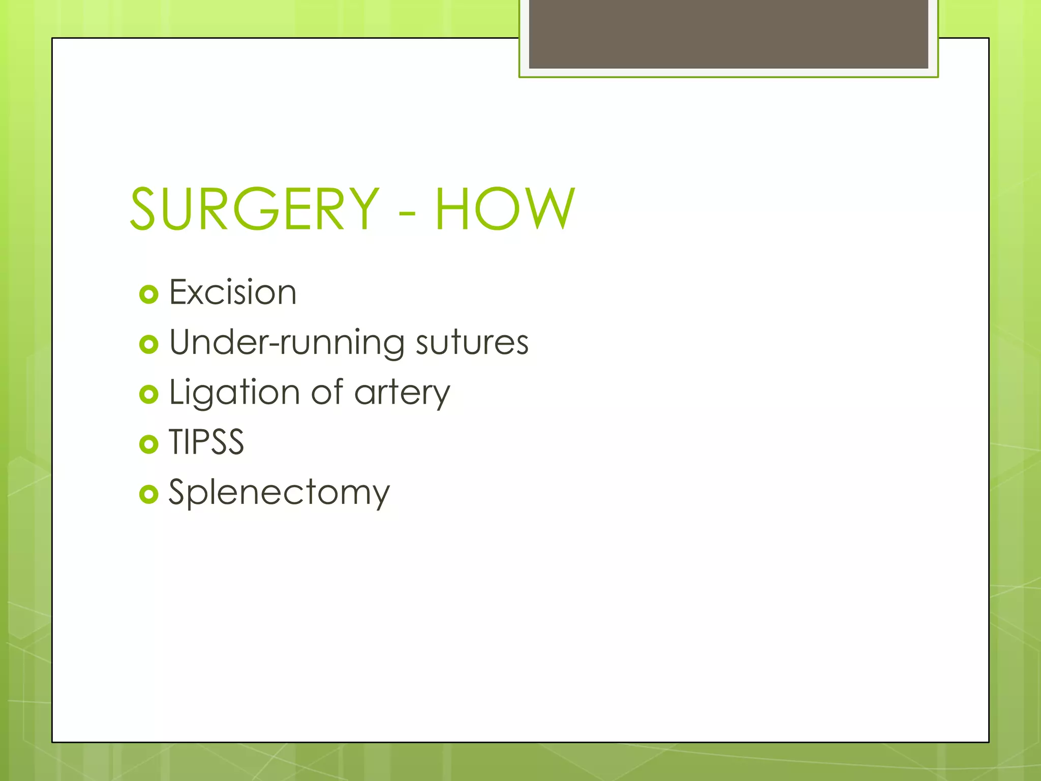
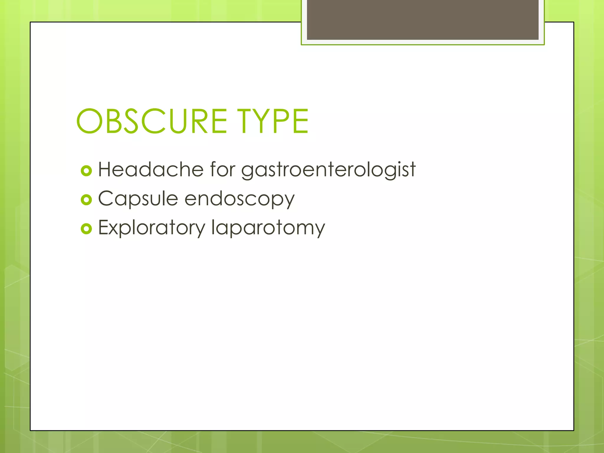
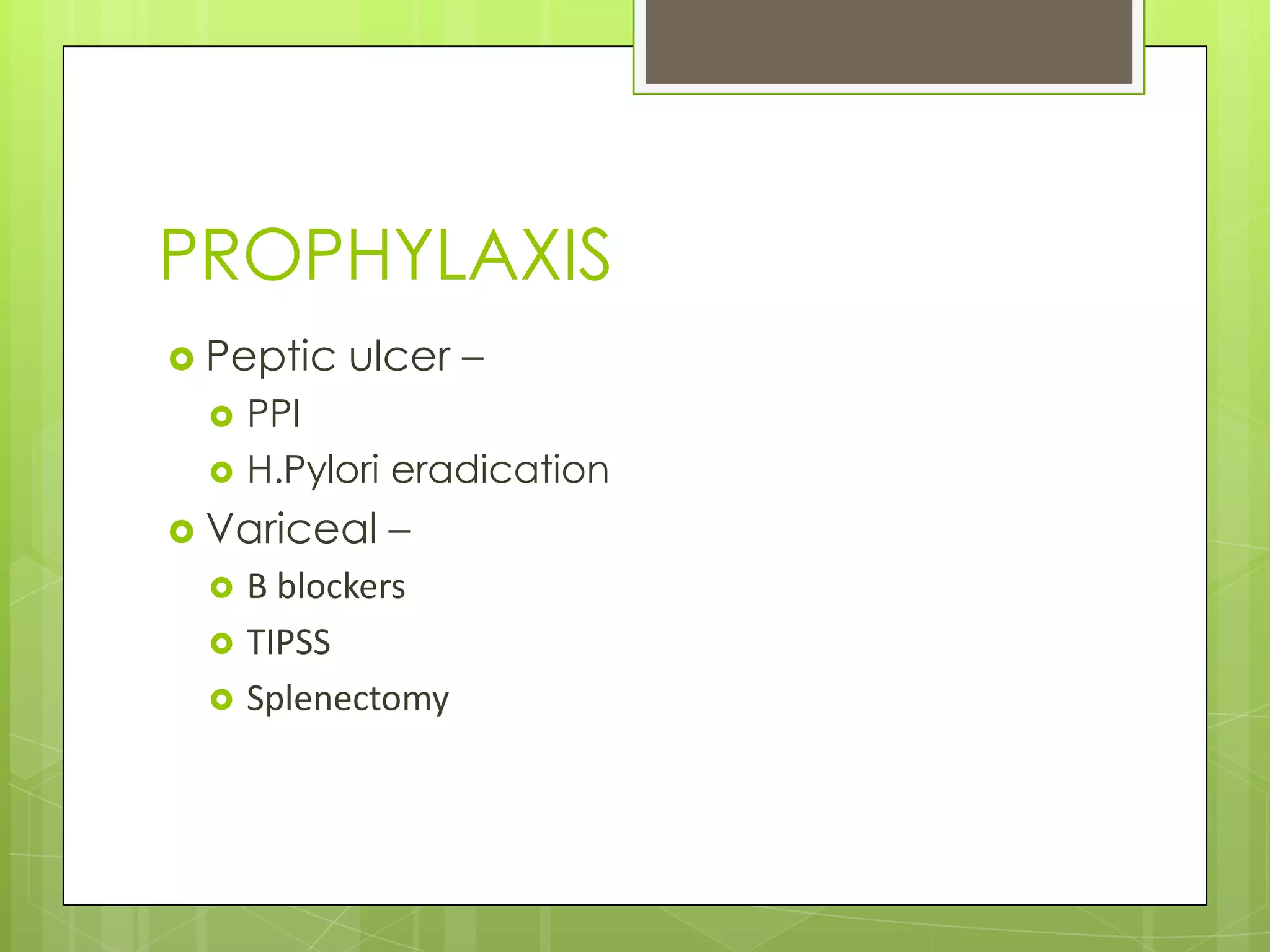
GI BLEED
DEFINITIONS
 Haemetemesis – Vomiting fresh/altered blood
 Melena – Altered blood in faeces
 Hematochezia – Fresh blood/clots per rectum
 Faecal occult blood – Not visible, detected
by lab tests for RBC degradation products
 Obscure Bleeding – GI blood loss, unknown
origin, recurs/persists after intial neg
endoscopic eval.
SYMPTOMS
 Acute bleed – upper/lower
 Fatigue, weakness, abd pain, pallor
 Hypotension, hypovolemic shock
CAUSES UPPER GI
 Peptic ulcer
 Gastric or esophageal varix
 Esophagitis
 Upper gastrointestinal tract tumor
 Angioma
 Mallory-Weiss tear
 Erosions
 Dieulafoy’s lesion
 Other
CAUSES LOWER GI
 Diverticulosis
 Colon cancer or
polyps
 Colitis
(Noninfectious &
Infectious)
 Ischemic colitis
 IBD
 Angioectasia
 Postpolypectomy
 Rectal ulcer
 Hemorrhoids
 Anorectal source
 Radiation colitis
 Other
 Unknown
SCORING SYSTEM
 Rockall scoring for upper GI (0-11)
 Parameters:-
 Age
 Pulse rate
 Systolic BP
 Comorbidity
 Endoscopic Dx
 Endoscopic stigmata of recent bleed
 <3=Good, >8=Very bad
SCORING ALTERNATE
 Glasgow – Blatchford:-
 Whom to Rx as OP
MANAGEMENT
 Stabilize the patient
 Stop the bleeding
 Find the source of bleeding
 Prevent recurrence of bleeding
IMMEDIATE
 Assess clinical status
 PR, BP, RR, Conciousness
 Large bore IV access – 2
 Stabilize haemodynamics
 IV fluids, PRBC, Whole blood
 Vasopressors
 NG aspirate – Large bleeds, doubtful
bleeds
LAB TESTS
 Complete blood counts
 Hb, TC, DC, ESR
 Coag profile
 PT, INR, APTT, Platelet count
 Blood group, cross-match
 LFT
 RFT
SPECIAL INVESTIGATIONS
 Tagged RBC scintigraphy
 Arteriography
HISTORY & EXAMINATION
 Drugs – OAC, NSAID, Aspirin, Doxycycline
 Alcohol, Chronic Liver Disease
 Coagulation disorders
 Retching
 Carcinoma, Polyps
 Radiation, Surgery (abd, aortic)
 H/o embolism
EXTRAS
 Stop anticoagulants
 FFP
 Vit K
 Protamine (for heparin)
 Platelet conc – for low platelet count
 Enema, prokinetics
 ETT – Unconscious, severe bleed
SPECIALS
 Non-variceal – PPI
 Variceal -
 Octreotide (synthetic Somatostatin)
 Vasopressin
 CLD - Cephalosporins
INTERVENTIONS
 Endoscopy :-
 Diagnostic
 Therapeutic – Ligation, Banding, Clipping,
Sclero
 Sengstaken-Blakemore tube :-
 Variceal bleed
 Embolisation of bleeding artery
SURGICAL OPTION
 Indication:-
 Haemodynamic instability
 Clinical deterioration
 >6 units of transfusion
 Persistent/Recurrent bleed
SURGERY - HOW
 Excision
 Under-running sutures
 Ligation of artery
 TIPSS
 Splenectomy
OBSCURE TYPE
 Headache for gastroenterologist
 Capsule endoscopy
 Exploratory laparotomy
PROPHYLAXIS
 Peptic ulcer –
 PPI
 H.Pylori eradication
 Variceal –
 Β blockers
 TIPSS
 Splenectomy
And that is it for now

Gastrointestinal bleed overview

  • 1.
  • 2.
    DEFINITIONS  Haemetemesis –Vomiting fresh/altered blood  Melena – Altered blood in faeces  Hematochezia – Fresh blood/clots per rectum  Faecal occult blood – Not visible, detected by lab tests for RBC degradation products  Obscure Bleeding – GI blood loss, unknown origin, recurs/persists after intial neg endoscopic eval.
  • 3.
    SYMPTOMS  Acute bleed– upper/lower  Fatigue, weakness, abd pain, pallor  Hypotension, hypovolemic shock
  • 4.
    CAUSES UPPER GI Peptic ulcer  Gastric or esophageal varix  Esophagitis  Upper gastrointestinal tract tumor  Angioma  Mallory-Weiss tear  Erosions  Dieulafoy’s lesion  Other
  • 5.
    CAUSES LOWER GI Diverticulosis  Colon cancer or polyps  Colitis (Noninfectious & Infectious)  Ischemic colitis  IBD  Angioectasia  Postpolypectomy  Rectal ulcer  Hemorrhoids  Anorectal source  Radiation colitis  Other  Unknown
  • 6.
    SCORING SYSTEM  Rockallscoring for upper GI (0-11)  Parameters:-  Age  Pulse rate  Systolic BP  Comorbidity  Endoscopic Dx  Endoscopic stigmata of recent bleed  <3=Good, >8=Very bad
  • 7.
    SCORING ALTERNATE  Glasgow– Blatchford:-  Whom to Rx as OP
  • 8.
    MANAGEMENT  Stabilize thepatient  Stop the bleeding  Find the source of bleeding  Prevent recurrence of bleeding
  • 9.
    IMMEDIATE  Assess clinicalstatus  PR, BP, RR, Conciousness  Large bore IV access – 2  Stabilize haemodynamics  IV fluids, PRBC, Whole blood  Vasopressors  NG aspirate – Large bleeds, doubtful bleeds
  • 10.
    LAB TESTS  Completeblood counts  Hb, TC, DC, ESR  Coag profile  PT, INR, APTT, Platelet count  Blood group, cross-match  LFT  RFT
  • 11.
    SPECIAL INVESTIGATIONS  TaggedRBC scintigraphy  Arteriography
  • 12.
    HISTORY & EXAMINATION Drugs – OAC, NSAID, Aspirin, Doxycycline  Alcohol, Chronic Liver Disease  Coagulation disorders  Retching  Carcinoma, Polyps  Radiation, Surgery (abd, aortic)  H/o embolism
  • 13.
    EXTRAS  Stop anticoagulants FFP  Vit K  Protamine (for heparin)  Platelet conc – for low platelet count  Enema, prokinetics  ETT – Unconscious, severe bleed
  • 14.
    SPECIALS  Non-variceal –PPI  Variceal -  Octreotide (synthetic Somatostatin)  Vasopressin  CLD - Cephalosporins
  • 15.
    INTERVENTIONS  Endoscopy :- Diagnostic  Therapeutic – Ligation, Banding, Clipping, Sclero  Sengstaken-Blakemore tube :-  Variceal bleed  Embolisation of bleeding artery
  • 16.
    SURGICAL OPTION  Indication:- Haemodynamic instability  Clinical deterioration  >6 units of transfusion  Persistent/Recurrent bleed
  • 17.
    SURGERY - HOW Excision  Under-running sutures  Ligation of artery  TIPSS  Splenectomy
  • 18.
    OBSCURE TYPE  Headachefor gastroenterologist  Capsule endoscopy  Exploratory laparotomy
  • 19.
    PROPHYLAXIS  Peptic ulcer–  PPI  H.Pylori eradication  Variceal –  Β blockers  TIPSS  Splenectomy
  • 20.
    And that isit for now