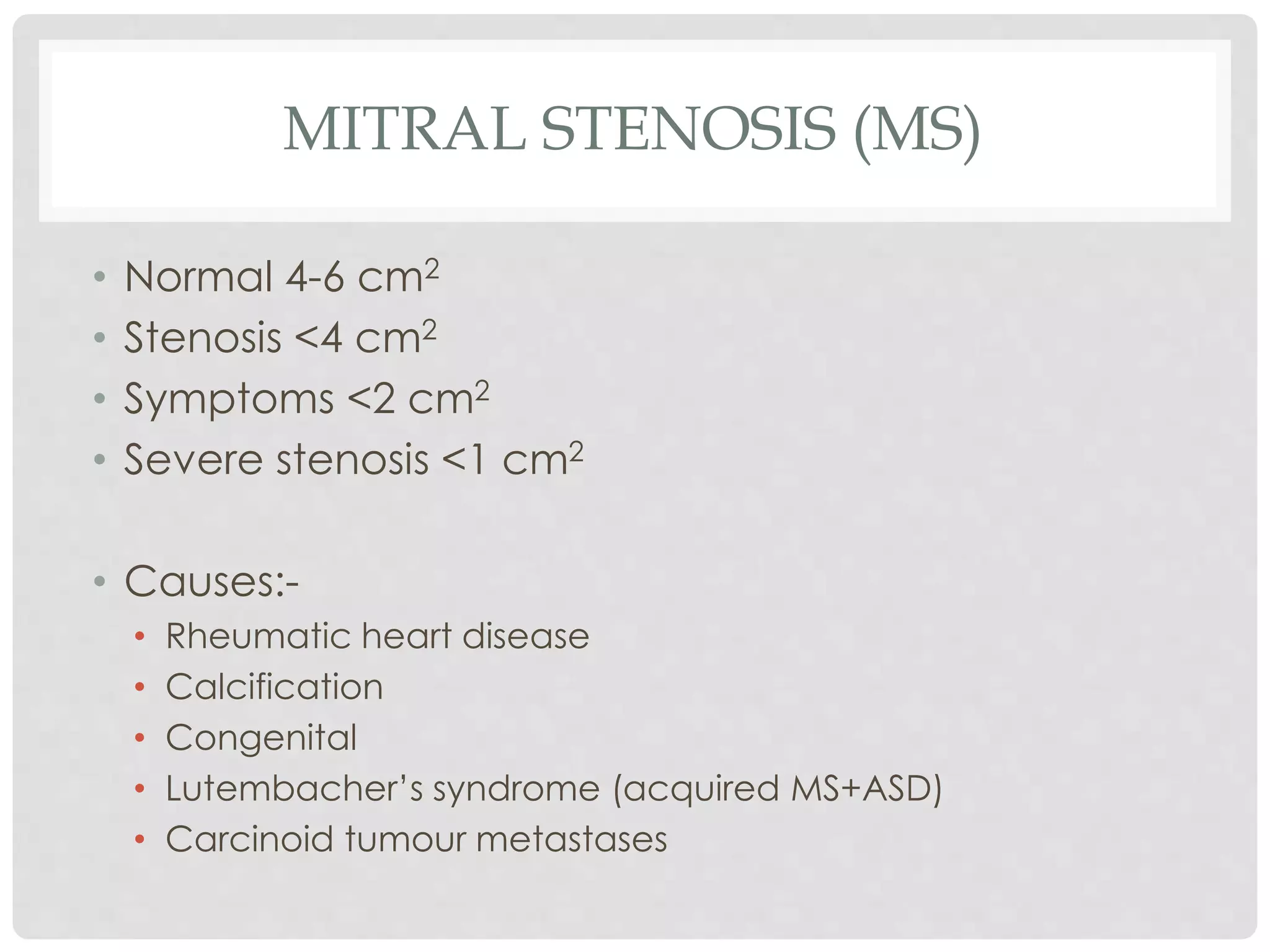
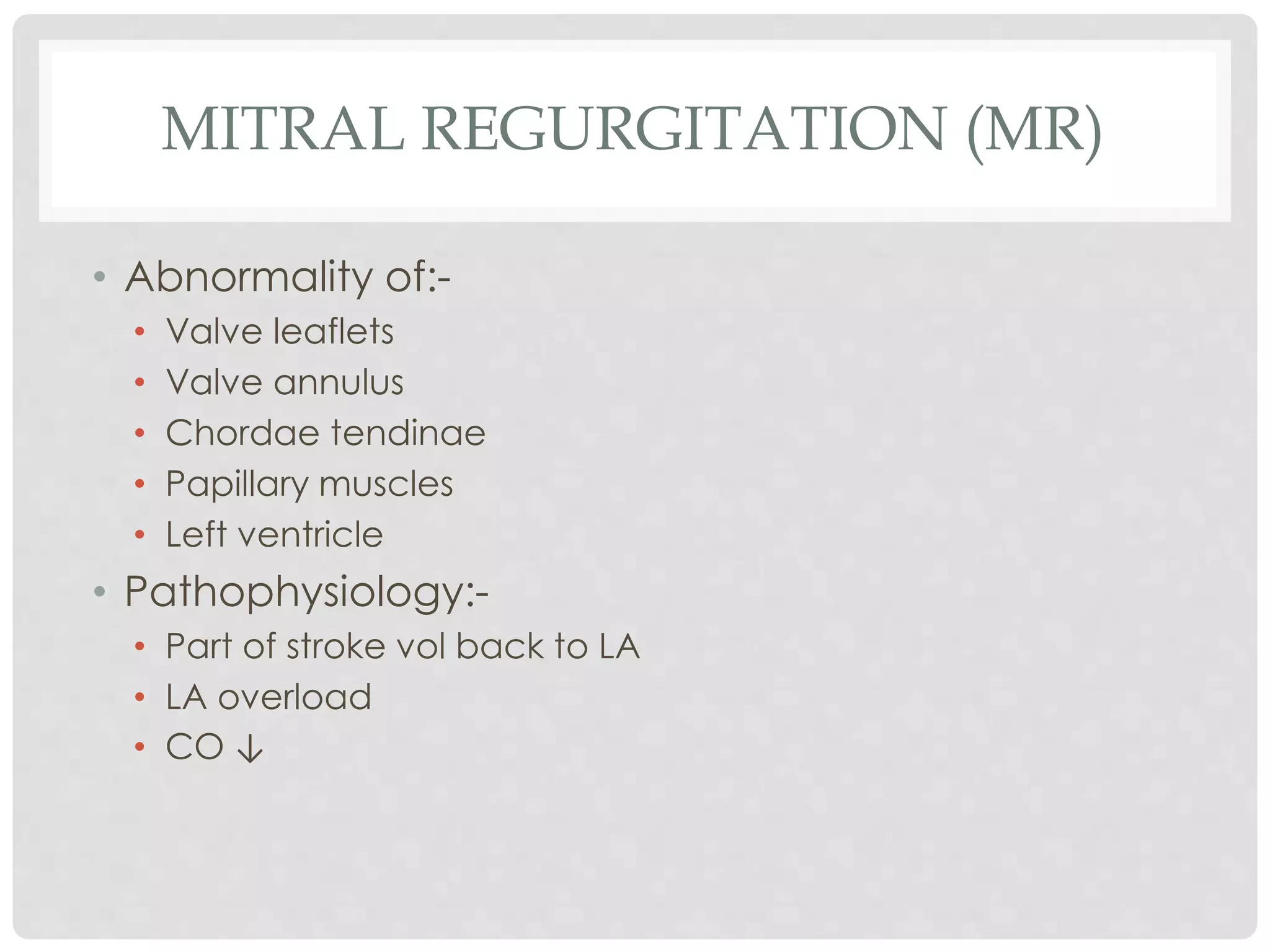
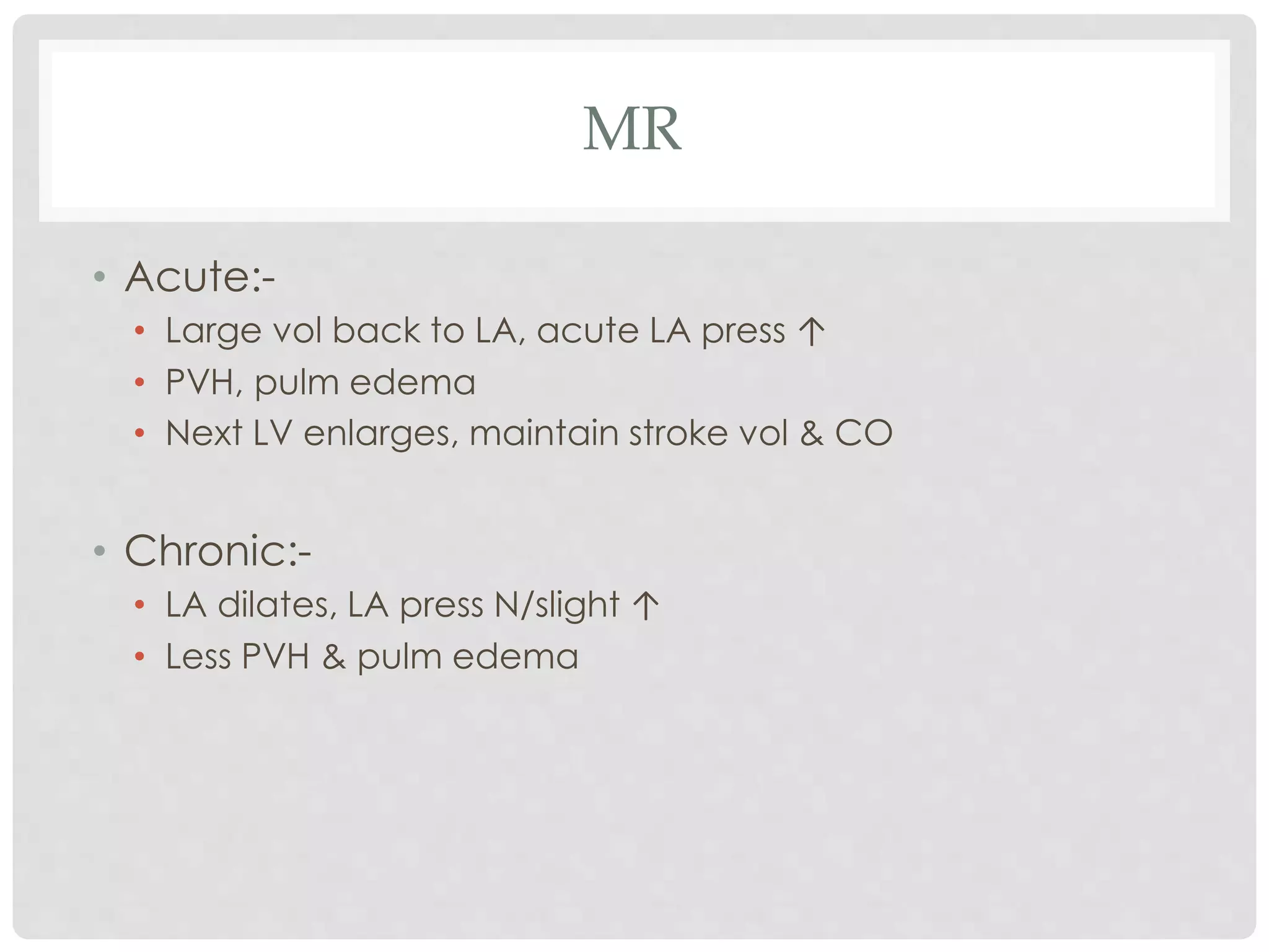
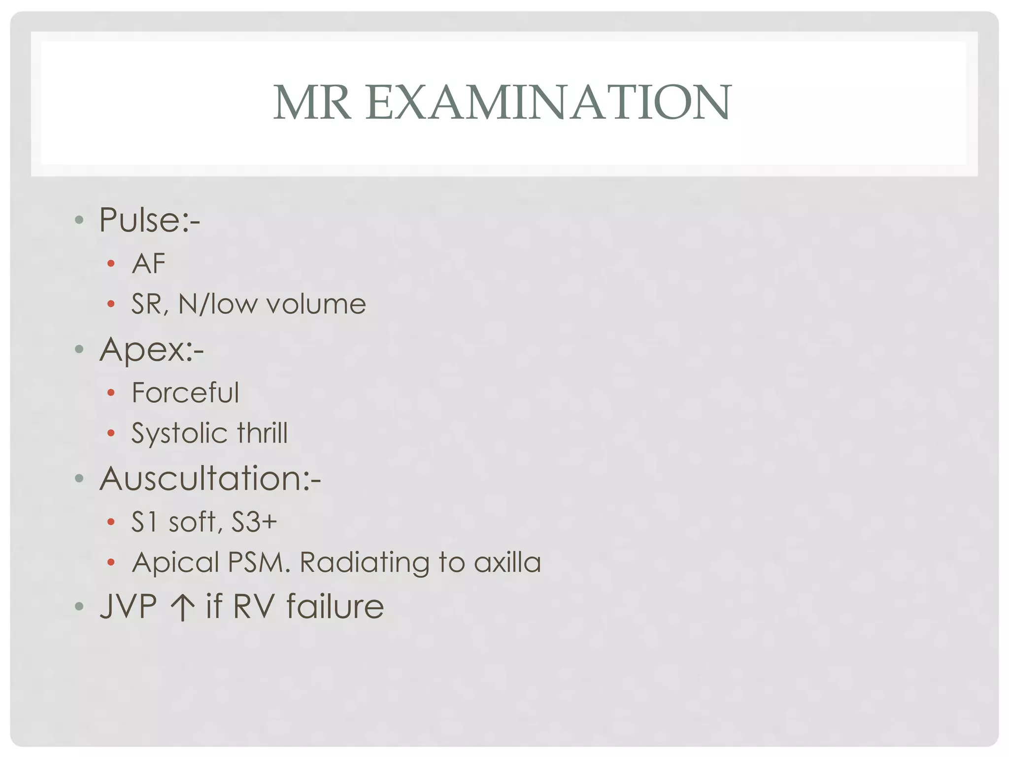
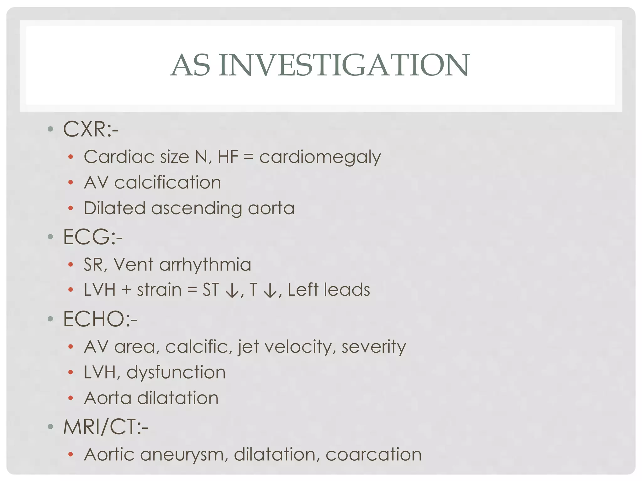
A S Y N O P S I S
VALVULAR HEART
LESIONS
MITRAL STENOSIS (MS)
• Normal 4-6 cm2
• Stenosis <4 cm2
• Symptoms <2 cm2
• Severe stenosis <1 cm2
• Causes:-
• Rheumatic heart disease
• Calcification
• Congenital
• Lutembacher’s syndrome (acquired MS+ASD)
• Carcinoid tumour metastases
MS
• Pathophysiology:-
• LA outlet obstruction
• LA pressure ↑, LA dilatation
• LV filling ↓, dependent on LA contraction
• Symptoms:-
• ↑ing DoE, haemoptysis, cough, chest pain(Pulm cong, PAH)
• Fatigue, pedal edema, ascites (Rt sided HF)
• Palpitation (AF)
• Systemic embolism:- CVA, Renal, Mesenteric, Peripheral
ischaemia
MS EXAMINATION
• Signs:-
• Malar flush (AV anastamoses, venous stasis)
• JVP ↑ (RVH, TR, PAH)
• Pulse – SR, low volume, AF
• Apex – localized tapping
• RV – heaving, sustained (PAH, RVH)
• Loud palpable S1, opening snap, loud P2 (PAH)
• Mid-diastolic, rough, rumbling apical murmur + presystolic
accentuation
• Graham Steele – EDM (func PR)
MS INVESTIGATIONS
• CXR:-
• Enlarged LA
• PVH, PAH, pulm edema
• Calcified mitral valve
• ECG:-
• Bifid P (P mitrale)
• RVH – RAD, tall R V1
• ECHO:-
• Mitral valve – area, severity, calcific, mobility
• PR, TR
• Atria – size, LA thrombus
• Ventricles – size, function
• CAG – prior to MVR
MS TREATMENT
• Diuretics (HF)
• Digoxin, β-blocker, CCB, anticoag (AF)
• Rheumatic fever prophylaxis
• IE prophylaxis
• Surgery – medical fails/not feasible
• Trans-septal balloon valvotomy
• Closed valvotomy
• Open valvotomy
• MVR
MITRAL REGURGITATION (MR)
• Abnormality of:-
• Valve leaflets
• Valve annulus
• Chordae tendinae
• Papillary muscles
• Left ventricle
• Pathophysiology:-
• Part of stroke vol back to LA
• LA overload
• CO ↓
MR
• Acute:-
• Large vol back to LA, acute LA press ↑
• PVH, pulm edema
• Next LV enlarges, maintain stroke vol & CO
• Chronic:-
• LA dilates, LA press N/slight ↑
• Less PVH & pulm edema
MR CAUSES
• Myxomatous valvular degeneration
• Ischaemic HD
• Rheumatic HD
• Infective endocarditis
• DCM, HCM
• Autoimmune – SLE
• Collagen vascular – Marfan’s, Ehler Danlos
• Congenital – 1o ASD
• Drugs – Fenfluramine, cabergoline
MR SYMPTOMATOLOGY
• Slow progress – No symp for years
• Sense forceful heartbeat - ↑ stroke volume
• Fatigue, lethargy, cardiac cachexia - ↓ CO
• Dyspnoea, orthopnoea –PVH, a/c pulm edema
• Later – dyspnoea, ascites, pedal edema, ↑ JVP,
palpitation (RVH, PAH, AF)
• Sub a/c IE – PUO
MR EXAMINATION
• Pulse:-
• AF
• SR, N/low volume
• Apex:-
• Forceful
• Systolic thrill
• Auscultation:-
• S1 soft, S3+
• Apical PSM. Radiating to axilla
• JVP ↑ if RV failure
MR INVESTIGATION
• CXR:-
• Cardiomegaly, LA & LV enlarged, valve calcific
• ECG:-
• AF, SR, bifid P mitrale, LVH
• ECHO:-
• LA & LV dilated
• Valve leaflet motion
• Papillary muscle anatomy & function
• Regurg jet direction, severity
MR TREATMENT
• Mild MR – asymp – conservative
• Vasodilators - ↓ pre & afterload
• Diuretics – HF
• Anticoag, antiarrhythmics – AF
• β blockers – atrial arrhythmia
• ICD – vent arrhythmia
• Endocarditis prophylaxis
• Symptomatic = Sx (prevent LVD)
• MV repair – Sx, clip
• MV replacement
BARLOW’S SYNDROME (MVP)
• Pathophysiology:-
• Large leaflet/ annulus
• Long chordae
• Papillary muscle dysfunction
• Seen max in young females
• Associations:-
• Marfan’s
• Thyrotoxicosis
• RHD, IHD, ASD, HCM
• Findings – early systolic click, PSM +/-
AORTIC STENOSIS (AS)
• LV outflow obstruction
• Causes:-
• Calcific degeneration – elderly, male, DM, HTN, DLP
• Congenital bicuspid vlave
• Rheumatic HD
• Misc –
• CKD
• Paget’s of bone
• Radiation
• Familial hypercholesterolemia
AS PATHOPHYSIOLOGY
• LV emptying obstructed
• LV pressures ↑
• LV hypertrophy (press overload)
• LV ischaemia – angina, arrhythmia, HF
• Exertion –
• CO rises very little
• Worsens angina and fatigue
• Syncope/presyncope
• Later LA press ↑, PVH = dyspnoea
AS SYMPTOMS
• When AV area <⅓ normal
• Exertional symptoms
• Angina = 4 years
• Syncope = 3 years
• Dyspnoea = 2 years
• Heart failure = 1.5 years
• Cachexia, fatigue = end-stage
AS EXAMINATION
• Pulse – SR, low vol, slow rising (parvus et tardus)
• Apex – undisplaced, heaving
• Aortic area – systolic thrill
• Auscultation:-
• Ejection click
• Soft A2, reversed split +/-
• S4
• Systolic, low pitched, ejection (crescendo-decrescendo)
murmur – aortic area, radiating to carotids
AS INVESTIGATION
• CXR:-
• Cardiac size N, HF = cardiomegaly
• AV calcification
• Dilated ascending aorta
• ECG:-
• SR, Vent arrhythmia
• LVH + strain = ST ↓, T ↓, Left leads
• ECHO:-
• AV area, calcific, jet velocity, severity
• LVH, dysfunction
• Aorta dilatation
• MRI/CT:-
• Aortic aneurysm, dilatation, coarcation
AS TREATMENT
• Asymptomatic:-
• Regular follow-up ECHO
• IE prophylaxis
• Valvotomy:-
• Buys time, improves LV temporarily
• Childhood, adolescents mainly
• Surgery:-
• Symptomatic
• Aorta gross/rapidly progressing dilatation
• AV replacement – open, percutaneous
AORTIC REGURGITATION (AR)
• Pathophysology:-
• Blood ejected into aorta in systole
• Leaks back into LV in diastole
• DBP ↓
• LV volume overload
• ↑ Stroke vol to maintain effective CO
• LV dilatation, later dysfunction
AR CAUSES
• Acute:-
• Rheumatic fever
• Infective endocarditis
• Aortic dissection
• Rupture sinus of valslva
• Prosthetic valve failure
• Chronic:-
• Rheumatic heart disease, syphilis, HTN
• Bicuspid valve, valve calcification, subvalvular VSD
• Arthritides – reactive, ankylosing spondylitis, rheumatoid
• Marfan’s, osteogenesis imperfecta
AR SYMPTOMS
• Late in disease, when LV fails
• Pounding of heart
• ↑ Stroke volume & force of contraction
• Angina
• DoE
• Arrhythmias +/-
AR EXAMINATION
• Pulse:-
• SR, large vol, collapsing
• Water hammer/Corrigan’s pulse
• BP:-
• SBP ↑, DBP ↓
• Wide pulse pressure
• Apex:- Displaced down & out (LV dilatation)
• Sounds:-
• High pitched EDM at Lt sternal border
• ESM at aortic area, radiating to carotids
• MDM at apex (Austin-Flint)
AR PERIPHERAL SIGNS
• Light-house – Flushing & blanching forehead
• Landolfi’s – Pupillary size alternation
• Becker’s – Retinal artery pulsation
• De Musset’s – Head nodding
• Muller’s – Uvula pulsation
• Corrigan’s – Dancing carotids
• Quincke’s – Capillary pulsation in nails
• Locomotor brachii
• Rosenbach’s – Liver pulsations
• Gerhardt’s – Spleen pulsations
• Traube’s – Pistol shot sounds over femorals
• Duroziez’s – Systolic & diastolic murmurs over femorals
• Hill’s – SBP popliteal>brachial
AR INVESTIGATIONS
• CXR:-
• LVH
• Ascending aorta dilatation & calcification
• AV calcific
• ECG:- LVH
• Tall R & deep T ↓ in left side leads
• Deep S right side leads
• ECHO:-
• Dilated aortic arch
• LV – dilatation, dysfunction
• Severity of AR
• TEE – Aortic valve & aortic root
• MRI & CT – Assess thoracic aorta & root
AR TREATMENT
• Rx for specific cause
• A/c AR – vasodilators, inotropes
• LV dysfunction – ACEi
• Surgery:-
• Before LVD sets in – not completely reversible
• Before significant symptoms develop
• A/c severe AR
• Symptomatic c/c severe AR
• LVD present
• LV dilatation present
• Along with other cardiac Sx
TRICUSPID STENOSIS (TS)
• Uncommon
• Women > men
• Associated mitral & aortic valve disease
• Causes:-
• RHD
• Carcinoid
• Pathophysiology:-
• RA emptying impaired, CO ↓
• RA press ↑
• Venous congestion (↑JVP, hepatomeg, ascites, pedal
edema)
TS
• Symptoms:-
• Abd pain + swelling
• Pedal edema
• Left sided failure symptoms
• Signs:-
• ↑ JVP, pedal edema
• Pulsatile liver, hepatomegaly
• Rumbling MDM @ lower LSE, louder on inspiration
• Tricuspid OS
TS INVESTIGATIONS
• CXR:-
• Prominent right atrial bulge
• ECG:-
• Peaked, tall P waves (>3 mm) in lead II (RAE)
• ECHO:-
• Thickened & immobile tricuspid valve
TS TREATMENT
• Medical:-
• Diuretic therapy
• Salt restriction
• Surgical:-
• Tricuspid valvotomy
• Tricuspid valve replacement is often necessary
• Other valves usually also need replacement
TRICUSPID REGURGITATION (TR)
• RA pressure overload
• Causes:-
• Functional-
• RV dilatation
• Cor pulmonale, MI, pulmonary HTN
• Organic-
• RHD
• IE
• Carcinoid syndrome
• Congenital - Ebstein’s anomaly
TR
• Symptoms:-
• Right heart failure
• Signs:-
• Large jugular venous c & v waves
• Hepatomegaly + pulsates in systole
• Lt parasternal heave (RVH)
• Blowing PSM @ LSE, best heard on inspiration
• AF common
• ECHO:-
• Dilated RV
• Thickened tricuspid valve
TR TREATMENT
• Functional – Medical Rx
• Severe organic:-
• TV repair – plasty, plication
• TV replacement
PULMONARY STENOSIS (PS)
• Causes:-
• Congenital – most common (isolated, ToF)
• RHD
• Carcinoid
• Pathophysiology:-
• RV press overload
• RVH, then RAH
• Rt heart failure
PS
• Symptoms:-
• Mild – asymptomatic
• Moderate – fatigue, syncope, dyspnoea +/-
• Severe – incompatible
• Signs:-
• JVP – prominent a wave
• Lt parasternal heave
• Delayed, soft P2 + pulmonary ejection click
• Harsh midsystolic ejection murmur @ 2nd Lt ICS, best heard
on inspiration, thrill +
• RV S4
PS
• Investigations:-
• CXR:-
• Prominent pulmonary artery
• ECG:-
• RAH – Tall P right leads
• RVH – Tall R right leads
• ECHO:-
• Doppler – stenotic flow
• RVH
• RA hypertrophy/enlargement
• Treatment:-
• Pulmonary valvotomy (balloon,direct surgery)
PULMONARY REGURGITATION (PR)
• Most common acquired pulm valve defect
• Pulm HTN most common cause (annular dilatation)
• Decrescendo diastolic murmur
• No symptoms
• Treatment rarely needed

Valvular heart lesions

  • 1.
    A S YN O P S I S VALVULAR HEART LESIONS
  • 2.
    MITRAL STENOSIS (MS) •Normal 4-6 cm2 • Stenosis <4 cm2 • Symptoms <2 cm2 • Severe stenosis <1 cm2 • Causes:- • Rheumatic heart disease • Calcification • Congenital • Lutembacher’s syndrome (acquired MS+ASD) • Carcinoid tumour metastases
  • 3.
    MS • Pathophysiology:- • LAoutlet obstruction • LA pressure ↑, LA dilatation • LV filling ↓, dependent on LA contraction • Symptoms:- • ↑ing DoE, haemoptysis, cough, chest pain(Pulm cong, PAH) • Fatigue, pedal edema, ascites (Rt sided HF) • Palpitation (AF) • Systemic embolism:- CVA, Renal, Mesenteric, Peripheral ischaemia
  • 4.
    MS EXAMINATION • Signs:- •Malar flush (AV anastamoses, venous stasis) • JVP ↑ (RVH, TR, PAH) • Pulse – SR, low volume, AF • Apex – localized tapping • RV – heaving, sustained (PAH, RVH) • Loud palpable S1, opening snap, loud P2 (PAH) • Mid-diastolic, rough, rumbling apical murmur + presystolic accentuation • Graham Steele – EDM (func PR)
  • 5.
    MS INVESTIGATIONS • CXR:- •Enlarged LA • PVH, PAH, pulm edema • Calcified mitral valve • ECG:- • Bifid P (P mitrale) • RVH – RAD, tall R V1 • ECHO:- • Mitral valve – area, severity, calcific, mobility • PR, TR • Atria – size, LA thrombus • Ventricles – size, function • CAG – prior to MVR
  • 6.
    MS TREATMENT • Diuretics(HF) • Digoxin, β-blocker, CCB, anticoag (AF) • Rheumatic fever prophylaxis • IE prophylaxis • Surgery – medical fails/not feasible • Trans-septal balloon valvotomy • Closed valvotomy • Open valvotomy • MVR
  • 7.
    MITRAL REGURGITATION (MR) •Abnormality of:- • Valve leaflets • Valve annulus • Chordae tendinae • Papillary muscles • Left ventricle • Pathophysiology:- • Part of stroke vol back to LA • LA overload • CO ↓
  • 8.
    MR • Acute:- • Largevol back to LA, acute LA press ↑ • PVH, pulm edema • Next LV enlarges, maintain stroke vol & CO • Chronic:- • LA dilates, LA press N/slight ↑ • Less PVH & pulm edema
  • 9.
    MR CAUSES • Myxomatousvalvular degeneration • Ischaemic HD • Rheumatic HD • Infective endocarditis • DCM, HCM • Autoimmune – SLE • Collagen vascular – Marfan’s, Ehler Danlos • Congenital – 1o ASD • Drugs – Fenfluramine, cabergoline
  • 10.
    MR SYMPTOMATOLOGY • Slowprogress – No symp for years • Sense forceful heartbeat - ↑ stroke volume • Fatigue, lethargy, cardiac cachexia - ↓ CO • Dyspnoea, orthopnoea –PVH, a/c pulm edema • Later – dyspnoea, ascites, pedal edema, ↑ JVP, palpitation (RVH, PAH, AF) • Sub a/c IE – PUO
  • 11.
    MR EXAMINATION • Pulse:- •AF • SR, N/low volume • Apex:- • Forceful • Systolic thrill • Auscultation:- • S1 soft, S3+ • Apical PSM. Radiating to axilla • JVP ↑ if RV failure
  • 12.
    MR INVESTIGATION • CXR:- •Cardiomegaly, LA & LV enlarged, valve calcific • ECG:- • AF, SR, bifid P mitrale, LVH • ECHO:- • LA & LV dilated • Valve leaflet motion • Papillary muscle anatomy & function • Regurg jet direction, severity
  • 13.
    MR TREATMENT • MildMR – asymp – conservative • Vasodilators - ↓ pre & afterload • Diuretics – HF • Anticoag, antiarrhythmics – AF • β blockers – atrial arrhythmia • ICD – vent arrhythmia • Endocarditis prophylaxis • Symptomatic = Sx (prevent LVD) • MV repair – Sx, clip • MV replacement
  • 14.
    BARLOW’S SYNDROME (MVP) •Pathophysiology:- • Large leaflet/ annulus • Long chordae • Papillary muscle dysfunction • Seen max in young females • Associations:- • Marfan’s • Thyrotoxicosis • RHD, IHD, ASD, HCM • Findings – early systolic click, PSM +/-
  • 15.
    AORTIC STENOSIS (AS) •LV outflow obstruction • Causes:- • Calcific degeneration – elderly, male, DM, HTN, DLP • Congenital bicuspid vlave • Rheumatic HD • Misc – • CKD • Paget’s of bone • Radiation • Familial hypercholesterolemia
  • 16.
    AS PATHOPHYSIOLOGY • LVemptying obstructed • LV pressures ↑ • LV hypertrophy (press overload) • LV ischaemia – angina, arrhythmia, HF • Exertion – • CO rises very little • Worsens angina and fatigue • Syncope/presyncope • Later LA press ↑, PVH = dyspnoea
  • 17.
    AS SYMPTOMS • WhenAV area <⅓ normal • Exertional symptoms • Angina = 4 years • Syncope = 3 years • Dyspnoea = 2 years • Heart failure = 1.5 years • Cachexia, fatigue = end-stage
  • 18.
    AS EXAMINATION • Pulse– SR, low vol, slow rising (parvus et tardus) • Apex – undisplaced, heaving • Aortic area – systolic thrill • Auscultation:- • Ejection click • Soft A2, reversed split +/- • S4 • Systolic, low pitched, ejection (crescendo-decrescendo) murmur – aortic area, radiating to carotids
  • 19.
    AS INVESTIGATION • CXR:- •Cardiac size N, HF = cardiomegaly • AV calcification • Dilated ascending aorta • ECG:- • SR, Vent arrhythmia • LVH + strain = ST ↓, T ↓, Left leads • ECHO:- • AV area, calcific, jet velocity, severity • LVH, dysfunction • Aorta dilatation • MRI/CT:- • Aortic aneurysm, dilatation, coarcation
  • 20.
    AS TREATMENT • Asymptomatic:- •Regular follow-up ECHO • IE prophylaxis • Valvotomy:- • Buys time, improves LV temporarily • Childhood, adolescents mainly • Surgery:- • Symptomatic • Aorta gross/rapidly progressing dilatation • AV replacement – open, percutaneous
  • 21.
    AORTIC REGURGITATION (AR) •Pathophysology:- • Blood ejected into aorta in systole • Leaks back into LV in diastole • DBP ↓ • LV volume overload • ↑ Stroke vol to maintain effective CO • LV dilatation, later dysfunction
  • 22.
    AR CAUSES • Acute:- •Rheumatic fever • Infective endocarditis • Aortic dissection • Rupture sinus of valslva • Prosthetic valve failure • Chronic:- • Rheumatic heart disease, syphilis, HTN • Bicuspid valve, valve calcification, subvalvular VSD • Arthritides – reactive, ankylosing spondylitis, rheumatoid • Marfan’s, osteogenesis imperfecta
  • 23.
    AR SYMPTOMS • Latein disease, when LV fails • Pounding of heart • ↑ Stroke volume & force of contraction • Angina • DoE • Arrhythmias +/-
  • 24.
    AR EXAMINATION • Pulse:- •SR, large vol, collapsing • Water hammer/Corrigan’s pulse • BP:- • SBP ↑, DBP ↓ • Wide pulse pressure • Apex:- Displaced down & out (LV dilatation) • Sounds:- • High pitched EDM at Lt sternal border • ESM at aortic area, radiating to carotids • MDM at apex (Austin-Flint)
  • 25.
    AR PERIPHERAL SIGNS •Light-house – Flushing & blanching forehead • Landolfi’s – Pupillary size alternation • Becker’s – Retinal artery pulsation • De Musset’s – Head nodding • Muller’s – Uvula pulsation • Corrigan’s – Dancing carotids • Quincke’s – Capillary pulsation in nails • Locomotor brachii • Rosenbach’s – Liver pulsations • Gerhardt’s – Spleen pulsations • Traube’s – Pistol shot sounds over femorals • Duroziez’s – Systolic & diastolic murmurs over femorals • Hill’s – SBP popliteal>brachial
  • 26.
    AR INVESTIGATIONS • CXR:- •LVH • Ascending aorta dilatation & calcification • AV calcific • ECG:- LVH • Tall R & deep T ↓ in left side leads • Deep S right side leads • ECHO:- • Dilated aortic arch • LV – dilatation, dysfunction • Severity of AR • TEE – Aortic valve & aortic root • MRI & CT – Assess thoracic aorta & root
  • 27.
    AR TREATMENT • Rxfor specific cause • A/c AR – vasodilators, inotropes • LV dysfunction – ACEi • Surgery:- • Before LVD sets in – not completely reversible • Before significant symptoms develop • A/c severe AR • Symptomatic c/c severe AR • LVD present • LV dilatation present • Along with other cardiac Sx
  • 28.
    TRICUSPID STENOSIS (TS) •Uncommon • Women > men • Associated mitral & aortic valve disease • Causes:- • RHD • Carcinoid • Pathophysiology:- • RA emptying impaired, CO ↓ • RA press ↑ • Venous congestion (↑JVP, hepatomeg, ascites, pedal edema)
  • 29.
    TS • Symptoms:- • Abdpain + swelling • Pedal edema • Left sided failure symptoms • Signs:- • ↑ JVP, pedal edema • Pulsatile liver, hepatomegaly • Rumbling MDM @ lower LSE, louder on inspiration • Tricuspid OS
  • 30.
    TS INVESTIGATIONS • CXR:- •Prominent right atrial bulge • ECG:- • Peaked, tall P waves (>3 mm) in lead II (RAE) • ECHO:- • Thickened & immobile tricuspid valve
  • 31.
    TS TREATMENT • Medical:- •Diuretic therapy • Salt restriction • Surgical:- • Tricuspid valvotomy • Tricuspid valve replacement is often necessary • Other valves usually also need replacement
  • 32.
    TRICUSPID REGURGITATION (TR) •RA pressure overload • Causes:- • Functional- • RV dilatation • Cor pulmonale, MI, pulmonary HTN • Organic- • RHD • IE • Carcinoid syndrome • Congenital - Ebstein’s anomaly
  • 33.
    TR • Symptoms:- • Rightheart failure • Signs:- • Large jugular venous c & v waves • Hepatomegaly + pulsates in systole • Lt parasternal heave (RVH) • Blowing PSM @ LSE, best heard on inspiration • AF common • ECHO:- • Dilated RV • Thickened tricuspid valve
  • 34.
    TR TREATMENT • Functional– Medical Rx • Severe organic:- • TV repair – plasty, plication • TV replacement
  • 35.
    PULMONARY STENOSIS (PS) •Causes:- • Congenital – most common (isolated, ToF) • RHD • Carcinoid • Pathophysiology:- • RV press overload • RVH, then RAH • Rt heart failure
  • 36.
    PS • Symptoms:- • Mild– asymptomatic • Moderate – fatigue, syncope, dyspnoea +/- • Severe – incompatible • Signs:- • JVP – prominent a wave • Lt parasternal heave • Delayed, soft P2 + pulmonary ejection click • Harsh midsystolic ejection murmur @ 2nd Lt ICS, best heard on inspiration, thrill + • RV S4
  • 37.
    PS • Investigations:- • CXR:- •Prominent pulmonary artery • ECG:- • RAH – Tall P right leads • RVH – Tall R right leads • ECHO:- • Doppler – stenotic flow • RVH • RA hypertrophy/enlargement • Treatment:- • Pulmonary valvotomy (balloon,direct surgery)
  • 38.
    PULMONARY REGURGITATION (PR) •Most common acquired pulm valve defect • Pulm HTN most common cause (annular dilatation) • Decrescendo diastolic murmur • No symptoms • Treatment rarely needed