Downloaded 301 times

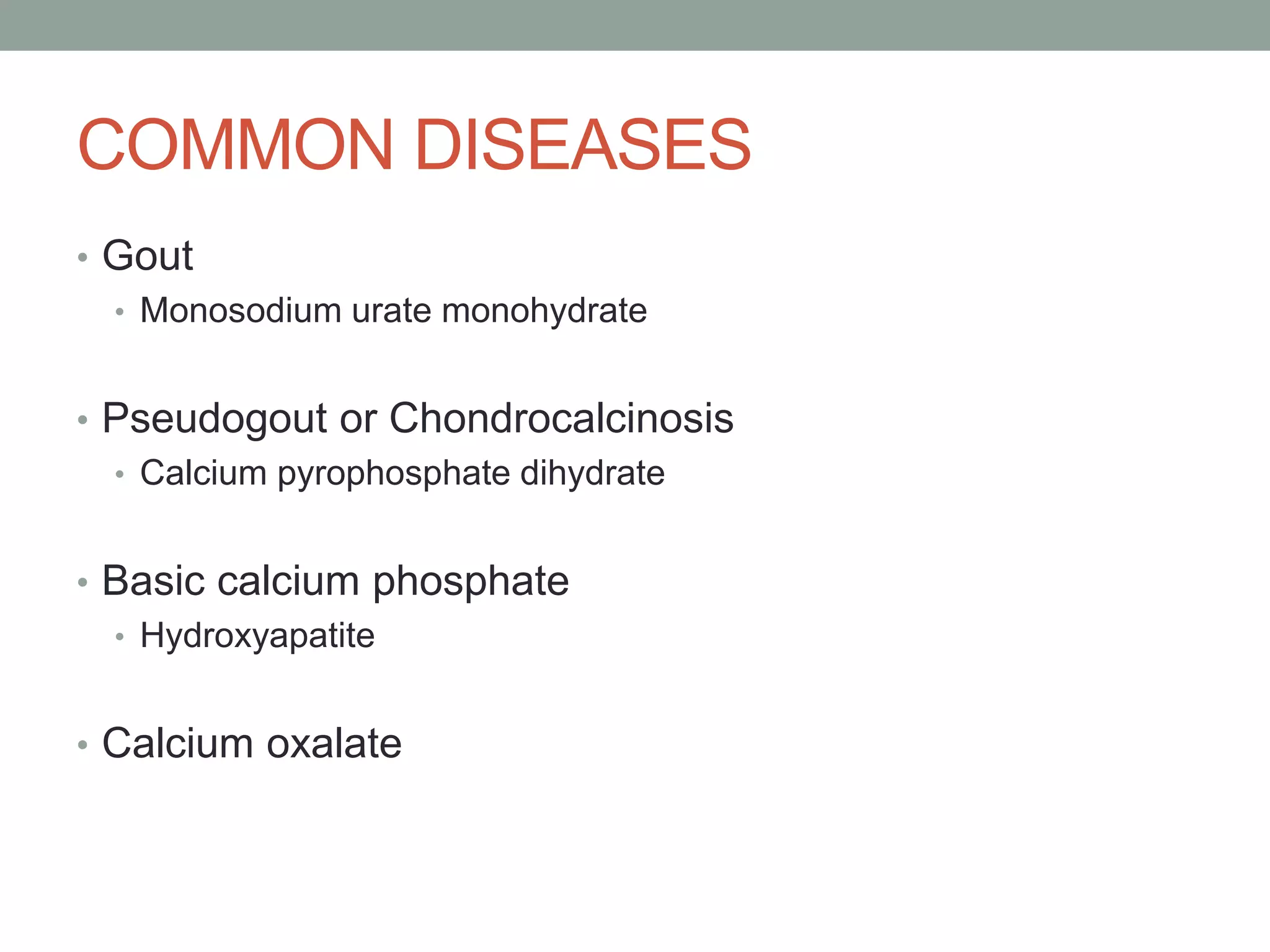


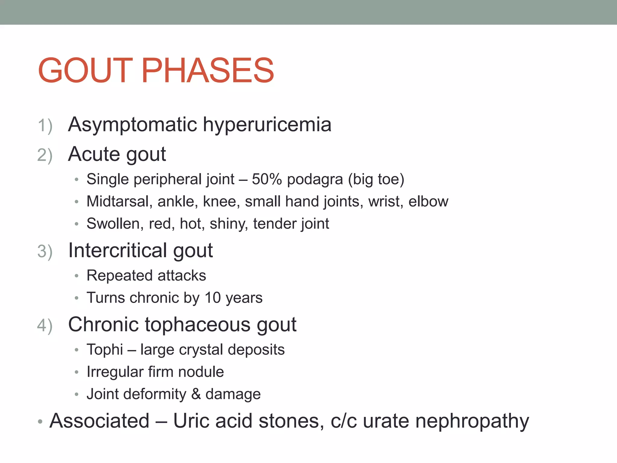


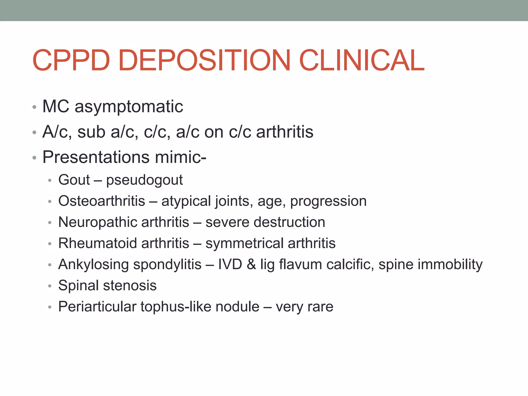


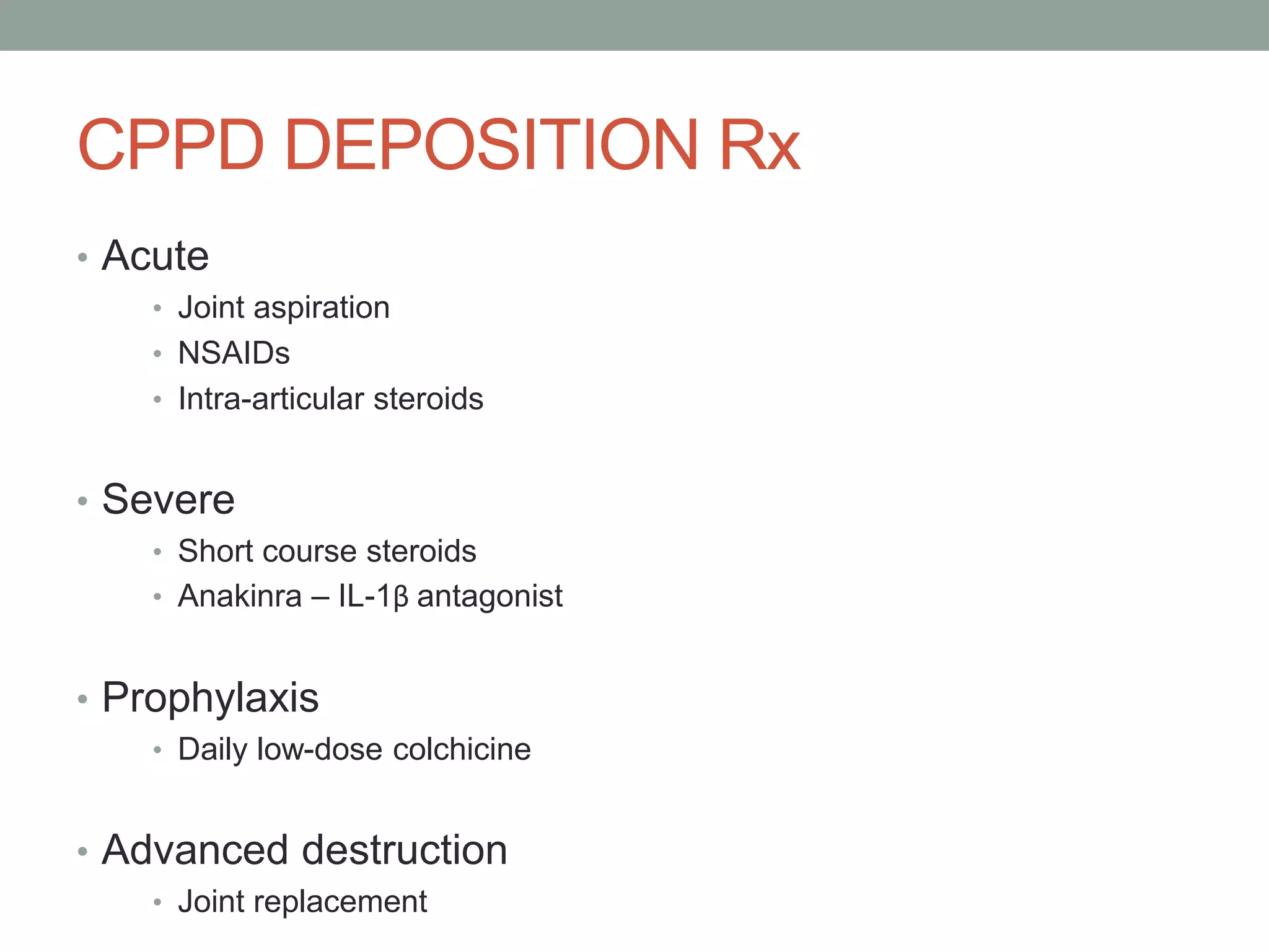

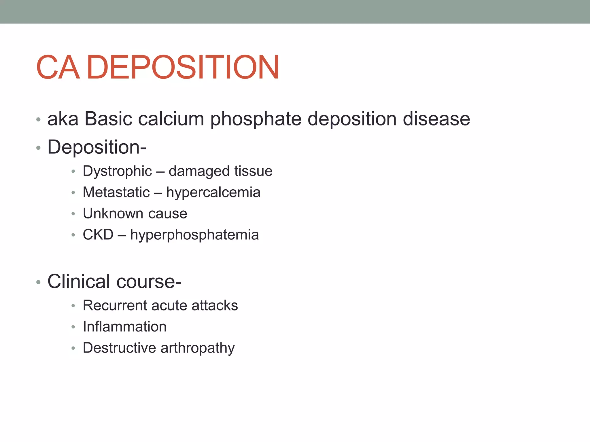
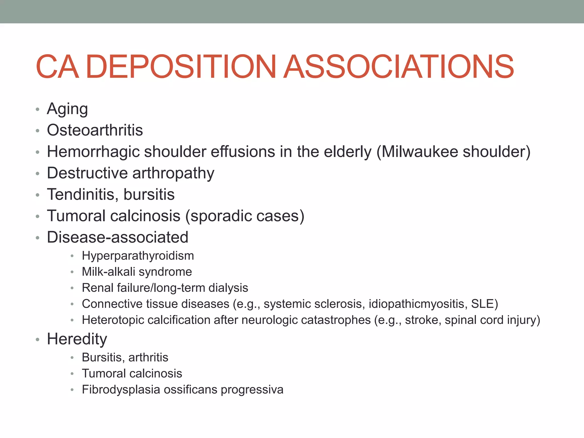


This document discusses various types of crystal-associated arthropathies including: - Monosodium urate monohydrate crystals which cause gout. Gout typically affects middle-aged males and causes painful inflammation in joints. - Calcium pyrophosphate dihydrate crystals which cause pseudogout/chondrocalcinosis. This tends to affect the elderly and can mimic many other joint diseases. - Calcium-containing crystals like hydroxyapatite which are associated with aging, osteoarthritis, and renal disease and can cause acute attacks of inflammation.


