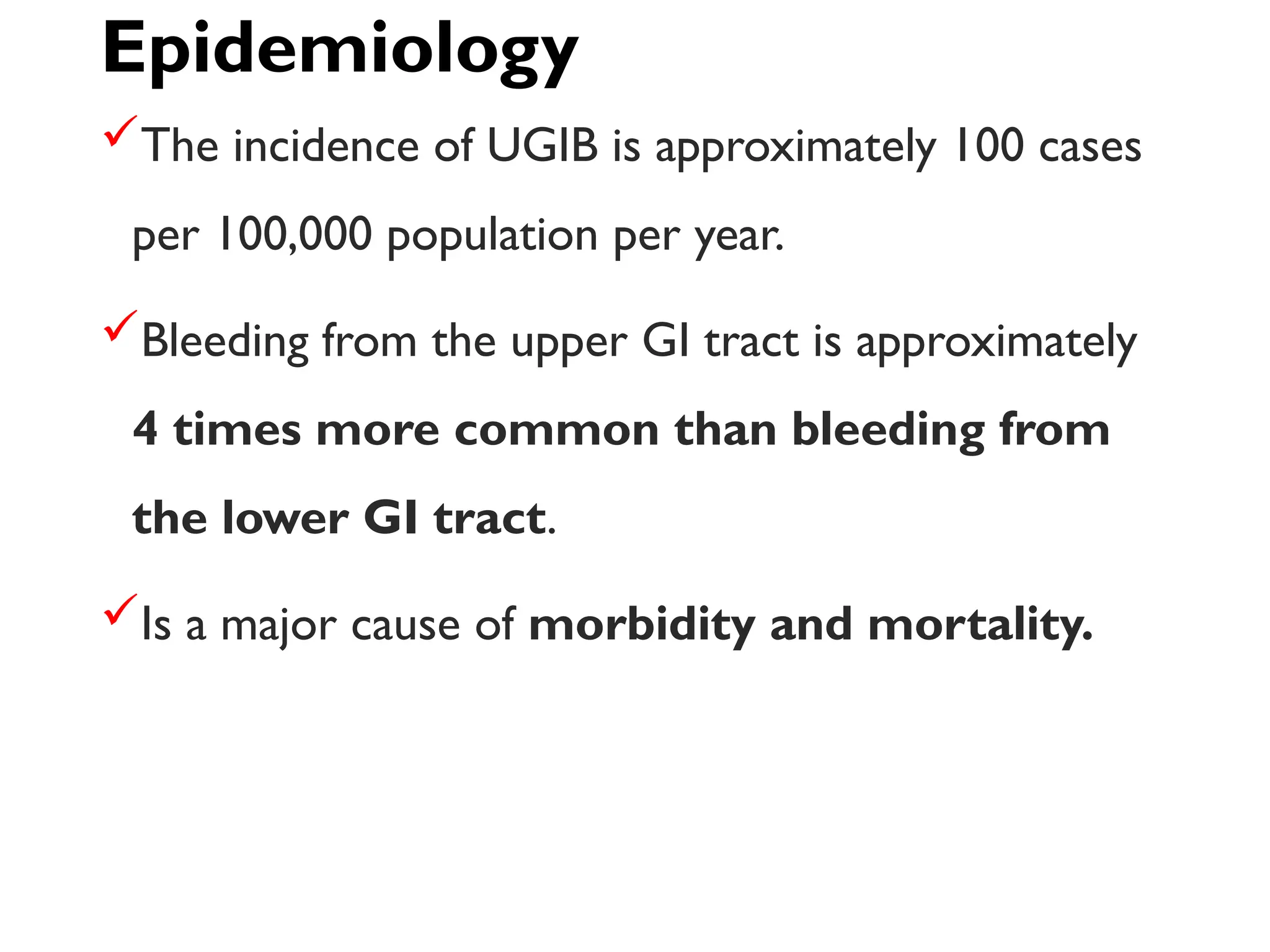
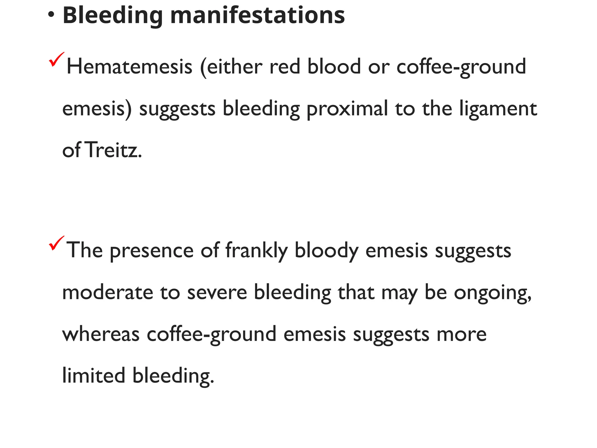
Upper and Lower
Gastrointestinal
Bleeding
Internal medicine
Class.BMS.4.1
DR:Abdullahi hussein
Upper Gastrointestinal
Bleeding
Definition
Upper GI bleeding (UGIB) is defined as bleeding
derived from a source proximal to the ligament of
Treitz.
Acute gastrointestinal (GI) bleeding is a potentially
life-threatening abdominal emergency that remains
a common cause of hospitalization.
Epidemiology
The incidence of UGIB is approximately 100 cases
per 100,000 population per year.
Bleeding from the upper GI tract is approximately
4 times more common than bleeding from
the lower GI tract.
Is a major cause of morbidity and mortality.
Causes of Upper Gi Bleeding
Gastric and/or duodenal ulcers
Severe or erosive gastritis/duodenitis
Severe or erosive esophagitis
Esophagogastric varices
Portal hypertensive gastropathy
Angiodysplasia (also known as vascular ectasia)
Mallory-Weiss syndrome
Mass lesions (polyps/cancers)
Hemophila
• Bleeding manifestations
Hematemesis (either red blood or coffee-ground
emesis) suggests bleeding proximal to the ligament
ofTreitz.
The presence of frankly bloody emesis suggests
moderate to severe bleeding that may be ongoing,
whereas coffee-ground emesis suggests more
limited bleeding.
The majority of melena (black, tarry stool)
originates proximal to the ligament of Treitz.
Melena may be seen with variable degrees
of blood loss, being seen with as little as 50
mL of blood.
Hematochezia can occur with massive
upper GI bleeding.
Associated with orthostatic hypotension.
Symptom assessment
Patients should be asked about symptoms as part of
the assessment of the severity of the bleed.
As a part of the evaluation for potential bleeding
sources.
Symptoms that suggest the bleeding is severe include:
orthostatic dizziness.
Confusion.
Angina.
severe palpitations and cold/clammy extremities.
Specific causes of upper GI bleeding may be suggested by the
patient's symptoms:
 Peptic ulcer – Upper abdominal pain
 Esophageal ulcer – Odynophagia, gastroesophageal reflux,
dysphagia
 Mallory-Weiss tear – Emesis, retching, or coughing prior to
hematemesis
 Variceal hemorrhage or portal hypertensive gastropathy:
Jaundice, abdominal distention (ascites)
 Malignancy – Dysphagia, early satiety, involuntary weight
loss, cachexia
Physical examination
Mild to moderate hypovolemia – Resting
tachycardia.
Blood volume loss – Orthostatic hypotension
Blood volume loss of at least 40 percent – Supine
hypotension.
Abdominal pain with rebound tenderness or
involuntary guarding, raises concern for perforation.
Laboratory data
• Laboratory tests that should be obtained in
patients with acute upper gastrointestinal
bleeding include:
• Complete blood count.
• Serum chemistries.
• Liver tests.
• Coagulation studies.
Upper endoscopy:
Upper endoscopy is the diagnostic modality of
choice for acute upper GI bleeding
Endoscopy has a high sensitivity and specificity for
locating and identifying bleeding lesions in the upper
GI tract.
Early endoscopy (within 24 hours) is recommended
for most patients with acute upper GI bleeding.
For patients with suspected variceal bleeding, we
perform endoscopy within 12 hours of presentation.
It may be helpful to give a
prokinetic agent such as
erythromycin or to irrigate the
stomach prior to endoscopy
to help remove residual blood
and other gastric contents.
Other diagnostic tests for acute upper GI bleeding
include
Ct angiography (CTA), which can detect active
bleeding.
Deep small bowel enteroscopy.
Intraoperative enteroscopy.
A colonoscopy is generally required for patients
with hematochezia and a negative upper endoscopy
Factors associated with rebleeding identified in a
meta-analysis included:
Hemodynamic instability (systolic blood pressure
less than 100 mmHg, heart rate greater than 100
beats per minute)
Hemoglobin less than 10 g/L
Active bleeding at the time of endoscopy
Large ulcer size (greater than 1 to 3 cm in various
studies)
Ulcer location (posterior duodenal bulb or high
lesser gastric curvature)
GENERAL MANAGEMENT
Adequate peripheral access should be attained
with two 18 gauge.
Larger intravenous catheters.
Large-bore, single-lumen central cordis.
Patients should receive supplemental oxygen by
nasal cannula
initially should receive nothing per mouth in the
event urgent upper endoscopy is needed.
Fluid resuscitation
 Fluid resuscitation should begin immediately
should not be delayed pending transfer of the
patient to an intensive care unit.
Patients with active bleeding should receive
intravenous fluids (eg, 500 mL of normal saline or
lactated Ringer's solution over 30 minutes)
Transfusion
Patients with active bleeding and hypovolemia may
require red blood cell transfusion.
Particularly if the patient remains hemodynamically
unstable despite appropriate fluid resuscitation.
Should receive a blood transfusion if the
hemoglobin is <9 g/dL (90 g/L).
However, it is important to avoid overtransfusion
in patients with suspected variceal bleeding.
The blood transfusion goals for variceal bleeding
are to transfuse if the hemoglobin is <7 g/dL (70
g/L).
Transfusion can precipitate worsening of the
bleeding (Cerqueira,Andrade, Correia, Fernandes, & Manso, 2012).
Transfusion of platelets may be required if the platelet count
is <50,000/microL.
prothrombin complex concentrate is indicated if the INR is
>2.
Treatment with low dose vitamin K or fresh frozen plasma if
hemodynamically unstable.
Fresh frozen plasma and platelets after every four units of
blood.
Acid suppression
 give a high-dose bolus (eg, esomeprazole 80 mg)
a second dose of an IV PPI should be given 12
hours later (eg, esomeprazole 40 mg).
For patients who may have stopped bleeding (eg,
patients who are hemodynamically stable with
melena), we give an IV PPI every 12 hours (eg,
esomeprazole 40 mg).
Vasoactive medications
Somatostatin, its analog octreotide and terlipressin
are used in the treatment of variceal bleeding.
octreotide is given as an intravenous bolus of 50
mcg, followed by a continuous infusion at a rate of
50 mcg per hour.
THERAPEUTIC ROLE OF ENDOSCOPY
• Methods of endoscopic hemostasis for acute UGIB
and LGIB include:
• Injection (usually diluted epinephrine or a
special sclerosing agent).
• Contact and non-contact thermal devices
(unipolar or bipolar electrocoagulation, heater
probes, and argon plasma coagulation).
• Mechanical devices (endoscopic clips and band
ligation) (Jung & Moon, 2019).
Lower Gastrointestinal
bleeding
Definition
lower gastrointestinal bleeding was defined
as bleeding originating distal to the
ligament of Treitz.
 acute lower gastrointestinal bleeding is
defined as the onset of hematochezia
originating from either the colon or the
rectum.
Epidemiology
Lower gastrointestinal bleeding (LGIB) that
requires hospitalization represents less than 1% of
all hospital admissions in the United States.
Worldwide, acute LBIG accounts for 1%-2% of
hospital emergencies.
Colonic diverticular disease is the most common
cause of LGIB.
Hemorrhoids are the most common cause of LGIB
in patients younger than 50 years.
The causes of acute lower GI bleeding may
be grouped into several categories:
Anatomic (diverticulosis).
Vascular (angiodysplasia, ischemic,
radiation-induced).
Inflammatory (infectious, inflammatory
bowel disease).
Neoplastic.
Clinical Manifestations
A patient with lower gastrointestinal (GI) bleeding
typically reports hematochezia
Blood originating from the left colon tends to be
bright red in color,
Bleeding from the right side of the colon usually
appears dark or maroon colored.
Rarely, bleeding from the right side of the colon
will present with melena.
Patients should also be asked about symptoms that
may suggest a particular etiology for the bleeding.
Painless hematochezia with diverticular bleeding.
Change in bowel habits with malignancy.
Abdominal pain with colitis.
Patients should be asked about medication use,
Particularly agents that are associated with
bleeding or that may impair coagulation, such as.
 Nonsteroidal antiinflammatory agents,
 Anticoagulants, and
Antiplatelet agents.
Physical examination:Signs of hypovolemia include:
 Mild to moderate hypovolemia: Resting tachycardia
 Blood volume loss of at least 15 percent: Orthostatic
hypotension.
 Blood volume loss of at least 40 percent: Supine
hypotension
 The presence of abdominal pain suggests
the presence of an inflammatory bleeding
source such as ischemic or infectious colitis
or a perforation
Diagnosis
Colonoscopy is the initial examination of choice for
the diagnosis and treatment of acute lower GI
bleeding.
Computed tomographic (CT) angiography.
Mesenteric angiography.
These radiographic procedures require active
bleeding at the time of examination
Colonoscopy
Potential to precisely localize the site of the
bleeding
The ability to collect pathologic specimens, and the
potential for therapeutic intervention.
Disadvantages of colonoscopy include
the need for bowel preparation.
poor visualization in an unprepared or poorly prepared
colon.
the risks of sedation in an acutely bleeding patient.
Treatment of the bleeding site
In many cases, the bleeding can be controlled with
therapies applied at the time of colonoscopy or
angiography.
Rarely, patients with exsanguinating lower GI
bleeding will need immediate surgery.
Diverticular bleeding: Colonoscopy with bipolar
probe coagulation, epinephrine injection, or metallic
clips
Recurrent bleeding: Resection of the affected bowel
segment
Angiodysplasia:Thermal therapy (eg,
electrocoagulation, argon plasma coagulation)
Conservative management, including nothing by
mouth (NPO) and IV hydration in patients with
ischemic colitis
Indications for surgery include the following:
oActive persistent bleeding with hemodynamic
instability that is refractory to aggressive
resuscitation
oPersistent, recurrent bleeding
oTransfusion of more than 4 units of packed red
blood cells (PRBCs) in a 24-hour period, with active
or recurrent bleeding
oTransfusion of more than 6 units of PRBCs during
the same hospitalization
Recurrent lower Gi bleeding
A repeat colonoscopy should be performed with
endoscopic hemostasis if indicated
Factors associated with rebleeding include
the presence of underlying comorbidities,
antiplatelet/anticoagulant/NSAID use.
source of bleeding.
the initial modality of hemostasis.
•thank you
•END

GI bleeding Gastrointestinal presentation.pptx

  • 1.
    Upper and Lower Gastrointestinal Bleeding Internalmedicine Class.BMS.4.1 DR:Abdullahi hussein
  • 2.
  • 3.
    Definition Upper GI bleeding(UGIB) is defined as bleeding derived from a source proximal to the ligament of Treitz. Acute gastrointestinal (GI) bleeding is a potentially life-threatening abdominal emergency that remains a common cause of hospitalization.
  • 4.
    Epidemiology The incidence ofUGIB is approximately 100 cases per 100,000 population per year. Bleeding from the upper GI tract is approximately 4 times more common than bleeding from the lower GI tract. Is a major cause of morbidity and mortality.
  • 5.
    Causes of UpperGi Bleeding Gastric and/or duodenal ulcers Severe or erosive gastritis/duodenitis Severe or erosive esophagitis Esophagogastric varices Portal hypertensive gastropathy Angiodysplasia (also known as vascular ectasia) Mallory-Weiss syndrome Mass lesions (polyps/cancers) Hemophila
  • 6.
    • Bleeding manifestations Hematemesis(either red blood or coffee-ground emesis) suggests bleeding proximal to the ligament ofTreitz. The presence of frankly bloody emesis suggests moderate to severe bleeding that may be ongoing, whereas coffee-ground emesis suggests more limited bleeding.
  • 7.
    The majority ofmelena (black, tarry stool) originates proximal to the ligament of Treitz. Melena may be seen with variable degrees of blood loss, being seen with as little as 50 mL of blood. Hematochezia can occur with massive upper GI bleeding. Associated with orthostatic hypotension.
  • 8.
    Symptom assessment Patients shouldbe asked about symptoms as part of the assessment of the severity of the bleed. As a part of the evaluation for potential bleeding sources. Symptoms that suggest the bleeding is severe include: orthostatic dizziness. Confusion. Angina. severe palpitations and cold/clammy extremities.
  • 9.
    Specific causes ofupper GI bleeding may be suggested by the patient's symptoms:  Peptic ulcer – Upper abdominal pain  Esophageal ulcer – Odynophagia, gastroesophageal reflux, dysphagia  Mallory-Weiss tear – Emesis, retching, or coughing prior to hematemesis  Variceal hemorrhage or portal hypertensive gastropathy: Jaundice, abdominal distention (ascites)  Malignancy – Dysphagia, early satiety, involuntary weight loss, cachexia
  • 10.
    Physical examination Mild tomoderate hypovolemia – Resting tachycardia. Blood volume loss – Orthostatic hypotension Blood volume loss of at least 40 percent – Supine hypotension. Abdominal pain with rebound tenderness or involuntary guarding, raises concern for perforation.
  • 11.
    Laboratory data • Laboratorytests that should be obtained in patients with acute upper gastrointestinal bleeding include: • Complete blood count. • Serum chemistries. • Liver tests. • Coagulation studies.
  • 12.
    Upper endoscopy: Upper endoscopyis the diagnostic modality of choice for acute upper GI bleeding Endoscopy has a high sensitivity and specificity for locating and identifying bleeding lesions in the upper GI tract. Early endoscopy (within 24 hours) is recommended for most patients with acute upper GI bleeding. For patients with suspected variceal bleeding, we perform endoscopy within 12 hours of presentation.
  • 19.
    It may behelpful to give a prokinetic agent such as erythromycin or to irrigate the stomach prior to endoscopy to help remove residual blood and other gastric contents.
  • 20.
    Other diagnostic testsfor acute upper GI bleeding include Ct angiography (CTA), which can detect active bleeding. Deep small bowel enteroscopy. Intraoperative enteroscopy. A colonoscopy is generally required for patients with hematochezia and a negative upper endoscopy
  • 21.
    Factors associated withrebleeding identified in a meta-analysis included: Hemodynamic instability (systolic blood pressure less than 100 mmHg, heart rate greater than 100 beats per minute) Hemoglobin less than 10 g/L Active bleeding at the time of endoscopy Large ulcer size (greater than 1 to 3 cm in various studies) Ulcer location (posterior duodenal bulb or high lesser gastric curvature)
  • 22.
    GENERAL MANAGEMENT Adequate peripheralaccess should be attained with two 18 gauge. Larger intravenous catheters. Large-bore, single-lumen central cordis. Patients should receive supplemental oxygen by nasal cannula initially should receive nothing per mouth in the event urgent upper endoscopy is needed.
  • 23.
    Fluid resuscitation  Fluidresuscitation should begin immediately should not be delayed pending transfer of the patient to an intensive care unit. Patients with active bleeding should receive intravenous fluids (eg, 500 mL of normal saline or lactated Ringer's solution over 30 minutes)
  • 24.
    Transfusion Patients with activebleeding and hypovolemia may require red blood cell transfusion. Particularly if the patient remains hemodynamically unstable despite appropriate fluid resuscitation. Should receive a blood transfusion if the hemoglobin is <9 g/dL (90 g/L).
  • 25.
    However, it isimportant to avoid overtransfusion in patients with suspected variceal bleeding. The blood transfusion goals for variceal bleeding are to transfuse if the hemoglobin is <7 g/dL (70 g/L). Transfusion can precipitate worsening of the bleeding (Cerqueira,Andrade, Correia, Fernandes, & Manso, 2012).
  • 26.
    Transfusion of plateletsmay be required if the platelet count is <50,000/microL. prothrombin complex concentrate is indicated if the INR is >2. Treatment with low dose vitamin K or fresh frozen plasma if hemodynamically unstable. Fresh frozen plasma and platelets after every four units of blood.
  • 27.
    Acid suppression  givea high-dose bolus (eg, esomeprazole 80 mg) a second dose of an IV PPI should be given 12 hours later (eg, esomeprazole 40 mg). For patients who may have stopped bleeding (eg, patients who are hemodynamically stable with melena), we give an IV PPI every 12 hours (eg, esomeprazole 40 mg).
  • 28.
    Vasoactive medications Somatostatin, itsanalog octreotide and terlipressin are used in the treatment of variceal bleeding. octreotide is given as an intravenous bolus of 50 mcg, followed by a continuous infusion at a rate of 50 mcg per hour.
  • 29.
    THERAPEUTIC ROLE OFENDOSCOPY • Methods of endoscopic hemostasis for acute UGIB and LGIB include: • Injection (usually diluted epinephrine or a special sclerosing agent). • Contact and non-contact thermal devices (unipolar or bipolar electrocoagulation, heater probes, and argon plasma coagulation). • Mechanical devices (endoscopic clips and band ligation) (Jung & Moon, 2019).
  • 30.
  • 31.
    Definition lower gastrointestinal bleedingwas defined as bleeding originating distal to the ligament of Treitz.  acute lower gastrointestinal bleeding is defined as the onset of hematochezia originating from either the colon or the rectum.
  • 32.
    Epidemiology Lower gastrointestinal bleeding(LGIB) that requires hospitalization represents less than 1% of all hospital admissions in the United States. Worldwide, acute LBIG accounts for 1%-2% of hospital emergencies. Colonic diverticular disease is the most common cause of LGIB. Hemorrhoids are the most common cause of LGIB in patients younger than 50 years.
  • 33.
    The causes ofacute lower GI bleeding may be grouped into several categories: Anatomic (diverticulosis). Vascular (angiodysplasia, ischemic, radiation-induced). Inflammatory (infectious, inflammatory bowel disease). Neoplastic.
  • 35.
    Clinical Manifestations A patientwith lower gastrointestinal (GI) bleeding typically reports hematochezia Blood originating from the left colon tends to be bright red in color, Bleeding from the right side of the colon usually appears dark or maroon colored. Rarely, bleeding from the right side of the colon will present with melena.
  • 36.
    Patients should alsobe asked about symptoms that may suggest a particular etiology for the bleeding. Painless hematochezia with diverticular bleeding. Change in bowel habits with malignancy. Abdominal pain with colitis.
  • 37.
    Patients should beasked about medication use, Particularly agents that are associated with bleeding or that may impair coagulation, such as.  Nonsteroidal antiinflammatory agents,  Anticoagulants, and Antiplatelet agents.
  • 38.
    Physical examination:Signs ofhypovolemia include:  Mild to moderate hypovolemia: Resting tachycardia  Blood volume loss of at least 15 percent: Orthostatic hypotension.  Blood volume loss of at least 40 percent: Supine hypotension  The presence of abdominal pain suggests the presence of an inflammatory bleeding source such as ischemic or infectious colitis or a perforation
  • 39.
    Diagnosis Colonoscopy is theinitial examination of choice for the diagnosis and treatment of acute lower GI bleeding. Computed tomographic (CT) angiography. Mesenteric angiography. These radiographic procedures require active bleeding at the time of examination
  • 40.
    Colonoscopy Potential to preciselylocalize the site of the bleeding The ability to collect pathologic specimens, and the potential for therapeutic intervention. Disadvantages of colonoscopy include the need for bowel preparation. poor visualization in an unprepared or poorly prepared colon. the risks of sedation in an acutely bleeding patient.
  • 41.
    Treatment of thebleeding site In many cases, the bleeding can be controlled with therapies applied at the time of colonoscopy or angiography. Rarely, patients with exsanguinating lower GI bleeding will need immediate surgery.
  • 42.
    Diverticular bleeding: Colonoscopywith bipolar probe coagulation, epinephrine injection, or metallic clips Recurrent bleeding: Resection of the affected bowel segment Angiodysplasia:Thermal therapy (eg, electrocoagulation, argon plasma coagulation) Conservative management, including nothing by mouth (NPO) and IV hydration in patients with ischemic colitis
  • 43.
    Indications for surgeryinclude the following: oActive persistent bleeding with hemodynamic instability that is refractory to aggressive resuscitation oPersistent, recurrent bleeding oTransfusion of more than 4 units of packed red blood cells (PRBCs) in a 24-hour period, with active or recurrent bleeding oTransfusion of more than 6 units of PRBCs during the same hospitalization
  • 44.
    Recurrent lower Gibleeding A repeat colonoscopy should be performed with endoscopic hemostasis if indicated Factors associated with rebleeding include the presence of underlying comorbidities, antiplatelet/anticoagulant/NSAID use. source of bleeding. the initial modality of hemostasis.
  • 45.