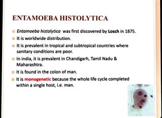
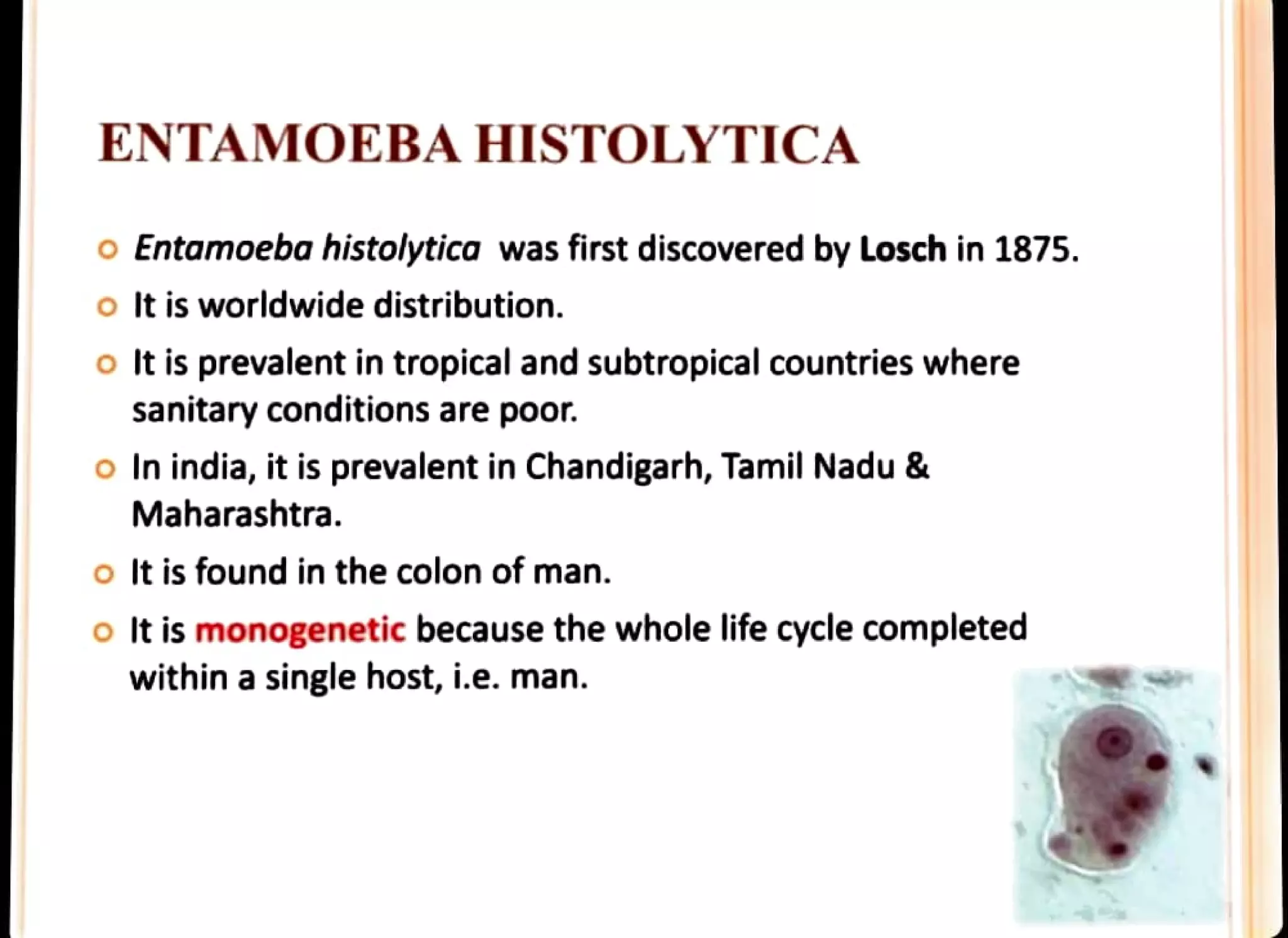
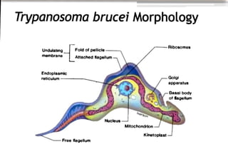
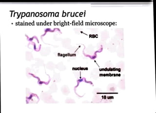
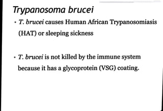
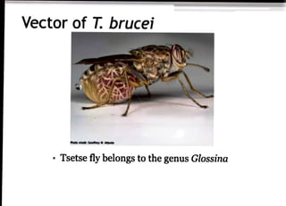
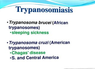
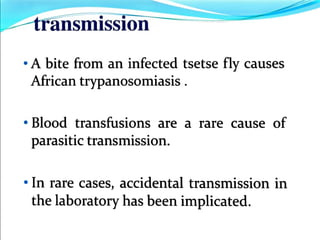
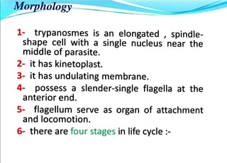
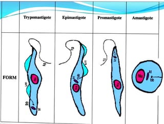
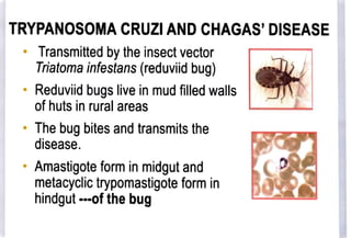
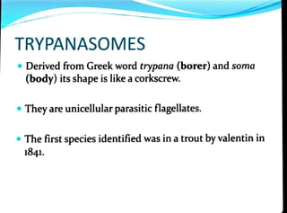
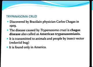
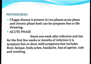
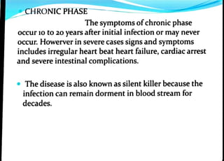
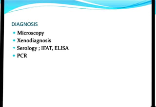
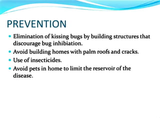
ENTAMOEBA HISTOLYTICA
o Entamoeba histolytica was first discovered by Losch in 1875.
oIt is worldwide distribution.
oIt is prevalent in tropicaland subtropical countries where
sanitary conditions are poor.
o In india, it is prevalent in Chandigarh, Tamil Nadu &
Maharashtra.
oIt is found in the colon of man.
oIt is monogenetic because the whole life cycle completed
within a single host, i.e. man.
o The locomotory organ is pseudopodia.
oThe typical amoeboid motility is crawling or gliding
movement.
o The nucleus contain central karyosome surrounded by clear
halo and it gives cartwheel appearance.
o They divide by binary fission.
oThey are killed by drying, heat and chemical sterilisation.
2. Pre-cyst
The pre-cyst formation occurs in intestinal lumen.
It is oval in shape.
It contain glycogen vacoule and two
chromatid bar.
Chromatoidal body
Cyst
Nucleus
3. Cyst
It is the mature cyst.
It is spherical in shape.
It contain 4 nuclei, hence it is called quadrinucleate.
Glycogen vacoule and chromatid bars are absent.
It is resistant to gastric juice.
Pathogenesis & Clinical Features
IntestinalAmoebiasis
Here the infection is limited to large intestine.
The metacystic trophozoites enters the intestinal epithelium
through Crypts of Liberkuhn.
The trophozoites releases histolysin enzyme which brings
about the destruction, necrosis and abscess of the tissue.
It results amoebic ulcer.
Clinical features
* The stool is large, foul-smelling, brownish black with mucus
and blood and it is called Amoebic dysentery.
* The incubation period is 1-4 months.
2. Extra Intestinal Amoebiasis
a) Hepatic Amoebiasis
It is the inflammation of the liver.
It occurs due to the repeated invasion of amoeba from
ulcerated gut through blood stream.
Liver contain thick chocolate brown pus.
b)Pulmonary Amoebiasis
It is the inflammation of the right lung.
It occurs due to the repeated invasion of amoeba from liver
through blood stream.
It results chocolate brown sputum.
c) Metastatic Amoebiasis
It involves kidney, brain, spleen &adrenals.
d) Cutaneous Amoebiasis
It involves destruction of skin around anus.
e) Genitourinary Amoebiasis
It involves amoebic vaginitis or amoebic ulcer on penis.
uclei
(n)
metacystic
trophozoites
(intective stage)
excystic
form or
metacyst
tetranucleate form
emerging as
cytoplasmic
Protrusion
yt walt
mature trophozoite
EXCYSTATION
endopiasn
ectoplasm
cetranucleate yst
plasmalemma
trophozoite
young
yst wall
ENCYSTATION
chromatold slycogen
bodies
pseudopodium
binucleate cyst
BINARY
FISSION
daughter
cells
precystic or
minuta forn
nucleus
uninucleate
yst
Fig. 1.13 :Reproductionand life-history of Entamohr ktstolytics.
1.
2
3.
4
2.
DIAGNOSIS
Microscopic examination of stool, pus &sputum.
Liver biopsy
Serodiagnosis include IHA Test, Latex Agglutination test and ELISA.
Demonstration of ghost cells, pyknotic bodies and CL crystals.
Their presence indicates that the immune response arised due to
parasitic infection.
TREATMENT
Administration of Metronidazole, Tinidazole, Paromomycin,
lodoquinal.
Oral rehydration &electrolyte replacement should be done
wherever necessary.
Trypanosoma brucei Morphology
Undulat1ng
membrane
Endoplasmic
reticulum
Fold of pellicle
Attached flagellum
-Freeflagellum
Nucleus
hverson
Mitochondrion
Kinetoplast
-Ribosomes
Golgi
apparatus
Basal body
of flageHum
Trypanosoma brucei
stained under bright-field microscope:
flagellum
nucleus
RBC
undulating
membrane
10 um
Trypanosoma brucei
"T. brucei causes Human African Trypanosomiasis
(HAT)orsleeping sickness
"T. brucei is not killed by the immunesystem
because it has aglycoprotein (VSG) coating.
Vector of T. brucei
htsoedt Gesthey MAae
" Tsetse fly belongs to the genus Glossina
Trypanosomiasis
"Trypanosoma brucei(African
trypanosomes)
"sleeping sickness
"Trypanosoma cruzi (American
trypanosomes)
"Chagas' disease
"S. and Central America
transmission
"A bite from an infected tsetse fly causes
African trypanosomiasis
" Blood transfusions are a rare cause of
parasitic transmission.
"In rare cases, accidental transmission in
the laboratory has been implicated.
Morphology
1- trypanosmes is an elongated , spindle
shape cell with asingle nucleus near the
middle of parasite.
2- it has kinetoplast.
3- it has undulating membrane.
4- possess a slender-single flagella at the
anterior end.
5- flagellum serve as organ of attachment
and locomotion.
6- there are four stages in life cycle :
FORM
Trypomastigote Epimastigote Promastigote
9
Amastigote
Nu
TRYPANOSOMA CRUZI
CHAGAS' DISEASE
ROBIN T. VAVACHAN
TRYPANOSOMA CRUZI AND CHAGAS'DISEASE
Transmitted by theinsectvector
Triatoma infestans (reduviid bug)
Reduviid bugs live in mud filled walls
of huts in rural areas
" The bug bites and transmits the
disease.
" Amastigote form in midgut and
metacyclic trypomastigote form in
hindgut ofthe bug
TRYPANASOMES
"Derived from Greek word trypana (borer) and soma
(body) its shape is like acorkscrew.
Theyare unicellularparasitic flagellates.
The first species identified was in atrout byvalentin in
1841.
TRYPANASOMA CRUZI
Discovered by Brazilain physician Carlos Chagas in
1909.
Thedisease caused by Trypanasoma cruzi is chagas
disease also called as American trypanasomiasis.
" It is transmitted toanimals and people by insectvector
(reduviid bug)
It is found only in America.
PATHOGENESIS
Chagas disease is present in two phases acute phase
and chronic phase both can be symptom free or life
threating.
ACUTE PHASE
Starts one week after infection and last
for the first few weeks or months of infection it is
symptom freeorshow mild symptoms that includes
fever, fatique, bodyaches, headache, loss ofapetite, rash
and vomiting
CHRONIC PHASE
The symptoms ofchronic phase
occur 10 to 20 years after initial infection or may never
occur. Howerver in severe cases signs and symptoms
includes irregular heart beat heart failure, cardiac arrest
and severe intestinal complications.
The disease is also known as silent killer because the
infection can remain dorment in blood stream for
decades.
DIAGNOSIS
" Microscopy
Xenodiagnosis
Serology ;IFAT, ELISA
PCR
TREATMENT
Chagas disease can be treated with benzidazole and
also nifurtimox. Both medicines are 1o0% effective in
curing the disease ifgiven soonafterthe infectionat
the onset of acute phase including the cases of
congenital transmission.
PREVENTION
Elimination ofkissing bugs by building structures that
discourage bug inhibiation.
" Avoid building homes with palm roofs and cracks.
Use of insecticides.
" Avoid pets in home to limit the reservoir ofthe
disease.

fungi.pdf

  • 1.
    ENTAMOEBA HISTOLYTICA o Entamoebahistolytica was first discovered by Losch in 1875. oIt is worldwide distribution. oIt is prevalent in tropicaland subtropical countries where sanitary conditions are poor. o In india, it is prevalent in Chandigarh, Tamil Nadu & Maharashtra. oIt is found in the colon of man. oIt is monogenetic because the whole life cycle completed within a single host, i.e. man.
  • 2.
    o The locomotoryorgan is pseudopodia. oThe typical amoeboid motility is crawling or gliding movement. o The nucleus contain central karyosome surrounded by clear halo and it gives cartwheel appearance. o They divide by binary fission. oThey are killed by drying, heat and chemical sterilisation. 2. Pre-cyst The pre-cyst formation occurs in intestinal lumen. It is oval in shape. It contain glycogen vacoule and two chromatid bar. Chromatoidal body Cyst Nucleus
  • 3.
    3. Cyst It isthe mature cyst. It is spherical in shape. It contain 4 nuclei, hence it is called quadrinucleate. Glycogen vacoule and chromatid bars are absent. It is resistant to gastric juice. Pathogenesis & Clinical Features IntestinalAmoebiasis Here the infection is limited to large intestine. The metacystic trophozoites enters the intestinal epithelium through Crypts of Liberkuhn. The trophozoites releases histolysin enzyme which brings about the destruction, necrosis and abscess of the tissue. It results amoebic ulcer.
  • 4.
    Clinical features * Thestool is large, foul-smelling, brownish black with mucus and blood and it is called Amoebic dysentery. * The incubation period is 1-4 months. 2. Extra Intestinal Amoebiasis a) Hepatic Amoebiasis It is the inflammation of the liver. It occurs due to the repeated invasion of amoeba from ulcerated gut through blood stream. Liver contain thick chocolate brown pus.
  • 5.
    b)Pulmonary Amoebiasis It isthe inflammation of the right lung. It occurs due to the repeated invasion of amoeba from liver through blood stream. It results chocolate brown sputum. c) Metastatic Amoebiasis It involves kidney, brain, spleen &adrenals. d) Cutaneous Amoebiasis It involves destruction of skin around anus. e) Genitourinary Amoebiasis It involves amoebic vaginitis or amoebic ulcer on penis.
  • 6.
    uclei (n) metacystic trophozoites (intective stage) excystic form or metacyst tetranucleateform emerging as cytoplasmic Protrusion yt walt mature trophozoite EXCYSTATION endopiasn ectoplasm cetranucleate yst plasmalemma trophozoite young yst wall ENCYSTATION chromatold slycogen bodies pseudopodium binucleate cyst BINARY FISSION daughter cells precystic or minuta forn nucleus uninucleate yst Fig. 1.13 :Reproductionand life-history of Entamohr ktstolytics.
  • 7.
    1. 2 3. 4 2. DIAGNOSIS Microscopic examination ofstool, pus &sputum. Liver biopsy Serodiagnosis include IHA Test, Latex Agglutination test and ELISA. Demonstration of ghost cells, pyknotic bodies and CL crystals. Their presence indicates that the immune response arised due to parasitic infection. TREATMENT Administration of Metronidazole, Tinidazole, Paromomycin, lodoquinal. Oral rehydration &electrolyte replacement should be done wherever necessary.
  • 8.
    Trypanosoma brucei Morphology Undulat1ng membrane Endoplasmic reticulum Foldof pellicle Attached flagellum -Freeflagellum Nucleus hverson Mitochondrion Kinetoplast -Ribosomes Golgi apparatus Basal body of flageHum
  • 9.
    Trypanosoma brucei stained underbright-field microscope: flagellum nucleus RBC undulating membrane 10 um
  • 10.
    Trypanosoma brucei "T. bruceicauses Human African Trypanosomiasis (HAT)orsleeping sickness "T. brucei is not killed by the immunesystem because it has aglycoprotein (VSG) coating.
  • 11.
    Vector of T.brucei htsoedt Gesthey MAae " Tsetse fly belongs to the genus Glossina
  • 12.
    Trypanosomiasis "Trypanosoma brucei(African trypanosomes) "sleeping sickness "Trypanosomacruzi (American trypanosomes) "Chagas' disease "S. and Central America
  • 13.
    transmission "A bite froman infected tsetse fly causes African trypanosomiasis " Blood transfusions are a rare cause of parasitic transmission. "In rare cases, accidental transmission in the laboratory has been implicated.
  • 14.
    Morphology 1- trypanosmes isan elongated , spindle shape cell with asingle nucleus near the middle of parasite. 2- it has kinetoplast. 3- it has undulating membrane. 4- possess a slender-single flagella at the anterior end. 5- flagellum serve as organ of attachment and locomotion. 6- there are four stages in life cycle :
  • 15.
  • 16.
  • 17.
    TRYPANOSOMA CRUZI ANDCHAGAS'DISEASE Transmitted by theinsectvector Triatoma infestans (reduviid bug) Reduviid bugs live in mud filled walls of huts in rural areas " The bug bites and transmits the disease. " Amastigote form in midgut and metacyclic trypomastigote form in hindgut ofthe bug
  • 18.
    TRYPANASOMES "Derived from Greekword trypana (borer) and soma (body) its shape is like acorkscrew. Theyare unicellularparasitic flagellates. The first species identified was in atrout byvalentin in 1841.
  • 19.
    TRYPANASOMA CRUZI Discovered byBrazilain physician Carlos Chagas in 1909. Thedisease caused by Trypanasoma cruzi is chagas disease also called as American trypanasomiasis. " It is transmitted toanimals and people by insectvector (reduviid bug) It is found only in America.
  • 20.
    PATHOGENESIS Chagas disease ispresent in two phases acute phase and chronic phase both can be symptom free or life threating. ACUTE PHASE Starts one week after infection and last for the first few weeks or months of infection it is symptom freeorshow mild symptoms that includes fever, fatique, bodyaches, headache, loss ofapetite, rash and vomiting
  • 21.
    CHRONIC PHASE The symptomsofchronic phase occur 10 to 20 years after initial infection or may never occur. Howerver in severe cases signs and symptoms includes irregular heart beat heart failure, cardiac arrest and severe intestinal complications. The disease is also known as silent killer because the infection can remain dorment in blood stream for decades.
  • 22.
  • 23.
    TREATMENT Chagas disease canbe treated with benzidazole and also nifurtimox. Both medicines are 1o0% effective in curing the disease ifgiven soonafterthe infectionat the onset of acute phase including the cases of congenital transmission.
  • 24.
    PREVENTION Elimination ofkissing bugsby building structures that discourage bug inhibiation. " Avoid building homes with palm roofs and cracks. Use of insecticides. " Avoid pets in home to limit the reservoir ofthe disease.