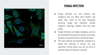
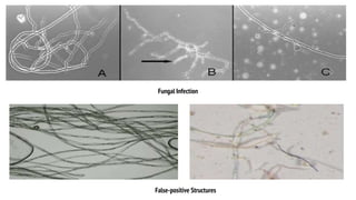
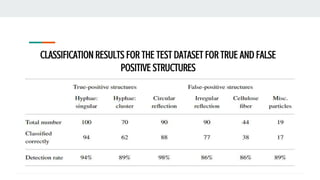
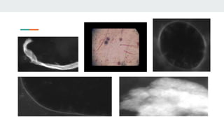
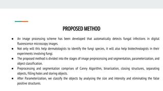
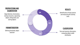
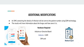
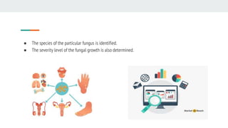
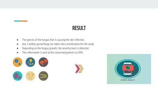
FUNGAL INFECTION DETECTION AND SEVERITY
LEVEL MEASUREMENT
By : Deekshitha S
FUNGAL INFECTION
● Fungal infections are very common skin
conditions that can affect both children and
adults alike. Some of the most frequently
occurring fungal skin infections include
ringworm, intertrigo, athlete’s foot and tinea
capitis.
● Fungal infections are highly contagious and can
be transmitted from person to person very easily,
but they can also be found in communal spaces.
● The most common and noticeable symptom of a
fungal skin infection is itching. You may
experience itching before you see any kind of
rash that shows an infection is present.
● The incidence of superficial fungal infections is
assumed to be 20 to 25% of the global human
population. Fluorescence microscopy of extracted
skin samples is frequently used for a swift
assessment of infections.
● Fluorescence staining increases sample contrast
and therefore further facilitates the detection of
fungi. A drawback of microscopy is that no
information on the fungal species can be
obtained.
● Hence, additional methods such as fungal culture
or DNA-based polymerase chain reaction
methods have to be performed, whenever
information about the fungal species is
important.
DRAWBACKS OF FLUORESCENCE MICROSCOPY
● Depending on sample condition, and sample size, it may still
be time-consuming to evaluate complete samples. Diagnosing
multiple samples at once may therefore be a tedious task that
could lead to classification errors and increased intra- and
interobserver variability.
● False-positive structures may tamper with the results that
look mostly like the described fungi.
● False-positive structures that are present in the samples are
mainly cellulose fibers of clothing, circular and irregular
reflections of the illumination unit occurring at air inclusions,
and other miscellaneous objects such as plastic particles and
dirt.
Fungal Infection
False-positive Structures
On My Topics
IMAGE PROCESSING BASED DETECTION OF FUNGAL DISEASES IN PLANTS
One of the paper presents a study on the image processing techniques used to identify and classify fungal
disease symptoms affected on different agriculture/horticulture crops.
RESULT
The comparisons of image processing techniques applied for identification and classification of fungal
disease affected on different agriculture/horticulture crops. The database is created to store the outputs of
feature extraction. The database is used to keep track of disorders of fungal disease symptoms affected on
vegetable crops , fruit crops , cereal crops and commercial crops the have been processed.
IMAGE PROCESSING METHODS USED TO DETECT FUNGAL DISEASES
IMAGE PROCESSING SCHEME TO DETECT SUPERFICIAL FUNGAL INFECTIONS OF
THE SKIN
To support the dermatologist, an image-analysis scheme has developed that evaluates digital microscopic
images to detect fungal hyphae. The aim of the study was to increase diagnostic quality and to shorten the
time-to-diagnosis.
A test dataset of hyphae and false-positive objects was created to evaluate the algorithm.
The results comprised of the classification for the test dataset of true and false-positive structures. In the
paper “classified correctly” means that objects are recognized as hyphae for true-positives and that objects
are not recognized as hyphae for false-positives.
CLASSIFICATION RESULTS FOR THE TEST DATASET FOR TRUE AND FALSE
POSITIVE STRUCTURES
DRAWBACKS OF THE PREVIOUS METHODS
● Previous techniques could only identify the fungus and
not it’s species. The accuracy was not that promising.
● Most of the studies are done on the fungus that affects
plants.
PROPOSED METHOD
● An image processing scheme has been developed that automatically detects fungal infections in digital
fluorescence microscopy images.
● Not only will this help dermatologists to identify the fungi species, it will also help biotechnologists in their
experiments involving fungi.
● The proposed method is divided into the stages of image preprocessing and segmentation, parameterization, and
object classification.
● Preprocessing and segmentation comprises of Canny Algorithm, binarization, closing structures, separating
objects, filling holes and storing objects.
● After Parameterization, we classify the objects by analysing the size and intensity and eliminating the false
positive structures.
01
02
03
04
PREPROCESSING AND
SEGMENTATION
Edge detection, binarization,
closing structures, separating
objects, filling holes and
storing objects
PARAMETERIZATION
Defining threshold, object
selection
RESULTS
Identification of fungi species
and measuring the severity
level
CLASSIFICATION
Size and intensity information,
evaluating object width
distribution
MATERIALS
AND
METHODS
✓ Sample Material and Preparation
✓ Imaging Device
✓ Description of Images and Other
Structures
✓ Image Analysis
✓ Specification of Classification
Parameters
✓ Evaluation
ADDITIONAL MODIFICATIONS
● An SMS containing the details of infection will be sent to the patient number using GSM technology.
● The results will have information about the fungus and how sever it is.
Hardware Required
Arduino or Genuino Board
Arduino + GSM
SIM card
● The species of the particular fungus is identified.
● The severity level of the fungal growth is also determined.
RESULT
● The species of the fungus that is causing the skin infection.
● Any 3 widely spread fungi are taken into consideration for the study.
● Depending on the fungus growth, the severity level is detected.
● This information is sent to the concerned patient via SMS.
THANK YOU

Fungal infection detection

  • 1.
    FUNGAL INFECTION DETECTIONAND SEVERITY LEVEL MEASUREMENT By : Deekshitha S
  • 2.
    FUNGAL INFECTION ● Fungalinfections are very common skin conditions that can affect both children and adults alike. Some of the most frequently occurring fungal skin infections include ringworm, intertrigo, athlete’s foot and tinea capitis. ● Fungal infections are highly contagious and can be transmitted from person to person very easily, but they can also be found in communal spaces. ● The most common and noticeable symptom of a fungal skin infection is itching. You may experience itching before you see any kind of rash that shows an infection is present.
  • 3.
    ● The incidenceof superficial fungal infections is assumed to be 20 to 25% of the global human population. Fluorescence microscopy of extracted skin samples is frequently used for a swift assessment of infections. ● Fluorescence staining increases sample contrast and therefore further facilitates the detection of fungi. A drawback of microscopy is that no information on the fungal species can be obtained. ● Hence, additional methods such as fungal culture or DNA-based polymerase chain reaction methods have to be performed, whenever information about the fungal species is important.
  • 4.
    DRAWBACKS OF FLUORESCENCEMICROSCOPY ● Depending on sample condition, and sample size, it may still be time-consuming to evaluate complete samples. Diagnosing multiple samples at once may therefore be a tedious task that could lead to classification errors and increased intra- and interobserver variability. ● False-positive structures may tamper with the results that look mostly like the described fungi. ● False-positive structures that are present in the samples are mainly cellulose fibers of clothing, circular and irregular reflections of the illumination unit occurring at air inclusions, and other miscellaneous objects such as plastic particles and dirt.
  • 5.
  • 6.
  • 7.
    IMAGE PROCESSING BASEDDETECTION OF FUNGAL DISEASES IN PLANTS One of the paper presents a study on the image processing techniques used to identify and classify fungal disease symptoms affected on different agriculture/horticulture crops. RESULT The comparisons of image processing techniques applied for identification and classification of fungal disease affected on different agriculture/horticulture crops. The database is created to store the outputs of feature extraction. The database is used to keep track of disorders of fungal disease symptoms affected on vegetable crops , fruit crops , cereal crops and commercial crops the have been processed.
  • 8.
    IMAGE PROCESSING METHODSUSED TO DETECT FUNGAL DISEASES
  • 9.
    IMAGE PROCESSING SCHEMETO DETECT SUPERFICIAL FUNGAL INFECTIONS OF THE SKIN To support the dermatologist, an image-analysis scheme has developed that evaluates digital microscopic images to detect fungal hyphae. The aim of the study was to increase diagnostic quality and to shorten the time-to-diagnosis. A test dataset of hyphae and false-positive objects was created to evaluate the algorithm. The results comprised of the classification for the test dataset of true and false-positive structures. In the paper “classified correctly” means that objects are recognized as hyphae for true-positives and that objects are not recognized as hyphae for false-positives.
  • 10.
    CLASSIFICATION RESULTS FORTHE TEST DATASET FOR TRUE AND FALSE POSITIVE STRUCTURES
  • 12.
    DRAWBACKS OF THEPREVIOUS METHODS ● Previous techniques could only identify the fungus and not it’s species. The accuracy was not that promising. ● Most of the studies are done on the fungus that affects plants.
  • 13.
    PROPOSED METHOD ● Animage processing scheme has been developed that automatically detects fungal infections in digital fluorescence microscopy images. ● Not only will this help dermatologists to identify the fungi species, it will also help biotechnologists in their experiments involving fungi. ● The proposed method is divided into the stages of image preprocessing and segmentation, parameterization, and object classification. ● Preprocessing and segmentation comprises of Canny Algorithm, binarization, closing structures, separating objects, filling holes and storing objects. ● After Parameterization, we classify the objects by analysing the size and intensity and eliminating the false positive structures.
  • 14.
    01 02 03 04 PREPROCESSING AND SEGMENTATION Edge detection,binarization, closing structures, separating objects, filling holes and storing objects PARAMETERIZATION Defining threshold, object selection RESULTS Identification of fungi species and measuring the severity level CLASSIFICATION Size and intensity information, evaluating object width distribution
  • 15.
    MATERIALS AND METHODS ✓ Sample Materialand Preparation ✓ Imaging Device ✓ Description of Images and Other Structures ✓ Image Analysis ✓ Specification of Classification Parameters ✓ Evaluation
  • 16.
    ADDITIONAL MODIFICATIONS ● AnSMS containing the details of infection will be sent to the patient number using GSM technology. ● The results will have information about the fungus and how sever it is. Hardware Required Arduino or Genuino Board Arduino + GSM SIM card
  • 17.
    ● The speciesof the particular fungus is identified. ● The severity level of the fungal growth is also determined.
  • 18.
    RESULT ● The speciesof the fungus that is causing the skin infection. ● Any 3 widely spread fungi are taken into consideration for the study. ● Depending on the fungus growth, the severity level is detected. ● This information is sent to the concerned patient via SMS.
  • 19.