This study evaluated the antibacterial effects of copper on microorganisms isolated from bovine mastitis. Milk samples were collected from dairy farms in central and southern Chile between March and September 2013. A total of 327 microorganisms were isolated and identified, with the most common being Escherichia coli, Staphylococcus aureus, Staphylococcus uberis, and coagulase-negative Staphylococci. Antibiotic susceptibility testing found 34% of isolates were resistant to at least one antibiotic. Minimum inhibitory concentration (MIC) assays found that 250 ppm copper inhibited the growth of 65% of isolates. The remaining isolates were inhibited by concentrations between 375-1000 ppm copper. Copper

![Reyes-Jara et al. Antibacterial Effect of Copper
FIGURE 1 | Location in Chile of dairy farms sampled (López, 2014).
Chile is divided into 15 Regions, each one is identified by a Roman numeral
(I to XV). Regions sampled during the study are enclosed in circles.
5% sheep blood agar plates, McConkey agar plates (Oxoid Ltd,
Basingstoke, Hampshire, England), tryptic soy agar plates (BBL,
Becton Dickinson, USA) and Sabouraud dextrose agar. Ten
micro liter of pellet were streaked per agar plate. The agar plates
were incubated at 37◦C for 18–24 h; those with no growth were
left up to 48 h. The results obtained in the culture medium were
interpreted and recorded according to the following criteria: (a)
positive culture: growth of five or more colonies of the same
type or two types of colony with a minimum eight colonies per
type. (b) Negative culture: no growth or growth of less than
five colonies of the same type. (c) Contaminated sample: growth
of three or more different types of colonies. Isolates were sub-
cultured to obtain pure cultures. The identification of bacteria
was performed according to colony morphology, Gram staining,
and biochemical tests.
Phenotypic differentiation of bacterial species was conducted
as follows: Staphylococci and Streptococci were identified
presumptively based on colony morphology, Gram stain
and catalase test. Coagulase-positive Staphylococci (S. aureus,
Staphylococcus intermedius, and Staphylococcus hyicus) were
identified based on morphology, pigmentation, coagulase test,
Voges Proskauer, and the ONPG test (Capurro et al., 1999).
Specific polymerase chain reaction (PCR) tests were used to
confirm S. aureus strains using primers NUC1 and NUC2
described by (Gandra et al., 2011) (Supplementary Figure S1A).
All coagulase-negative Staphylococci were considered as CNS
(coagulase-negative Staphylococci) after showing resistance to
bacitracin (0.04U). The Staphylococci sensitive to bacitracin
(0.04U) were considered as Micrococcus spp. (Falk and Guering,
1983). Streptococcus species were differentiated based on
hemolysis, esculin, CAMP and the inulin test. S. uberis was
confirmed by PCR with oligonucleotide primers designed for the
16S rRNA gene (Hassan et al., 2001) (Supplementary Figure
S1B).
Corynebacterium spp. were identified presumptively based on
colony morphology, hemolysis, esculin, and the APIR
Coryne test
(Biomerieux). Results were confirmed by PCR amplification of
the rpoB gene (Khamis et al., 2004) (Supplementary Figure S1C).
Yeasts and Enterobacteriaceae were presumptively identified
based on colony morphology, Gram stain and APIR
20C
Aux (Biomerieux) for yeasts and APIR
20E (Biomerieux) for
Enterobacteriaceae. E. coli was confirmed by PCR amplification
of the uspA gene (Chen and Griffiths, 1998) (Supplementary
Figure S1D). All isolates were stored at −80◦C until further use.
Antimicrobial Susceptibility Testing
The antimicrobial susceptibility tests included six antibiotics:
amoxicillin/clavulanic Acid (AMC) 20/10 µg (BBL, Becton
Dickinson, USA), sulphamethoxazole/trimethoprim (SXT)
25 µg (Oxoid Ltd., Basingstoke, Hampshire, England),
cefotaxime (CTX) 30 µg (BBL, Becton Dickinson, USA),
tetracycline (TET) 30 µg (BBL, Becton Dickinson, USA),
gentamicin (GN) 10 µg (BBL, Becton Dickinson, USA), and
neomycin (NE) 30 µg, (Oxoid Ltd., Basingstoke, Hampshire,
England).
The susceptibility to antibiotics was tested against all bacterial
isolates (n = 324) using the diffusion test proposed by Kirby–
Bauer following the recommendations of the Clinical Laboratory
Standards Institute guidelines (Clinical Laboratory Standards
Institute [CLSI], 2012b) and the BSAC guide (Andrews and
BSAC Working Party on Susceptibility Testing, 2008). In this
analysis only the three Candida spp. were excluded. Organisms
were incubated on Mueller Hinton (MH) agar plates at 35◦C
for 18–24 h in aerobic atmosphere. E. coli (ATCC-25922) and
S. aureus (ATCC-25923) were used as quality control. The CLSI
breakpoints were used for the interpretation of susceptibility
to all microbial agents. The BSAC standard was applied to
determine the breakpoint value of Neomycin (NE).
Determination of Minimum Inhibitory
Concentration of Copper (MIC-Cu)
The minimum inhibitory concentration of copper (MIC-Cu)
was determined by the agar dilution method against 327
microorganisms isolated, applying standard bacteriological
methods (Clinical Laboratory Standards Institute [CLSI],
2012a). Briefly, MH agar plates were supplemented with
seven concentrations of copper (II) sulfate pentahydrate
(CuSO4x5H2O) (Merck Millipore, Germany): 50, 125, 250,
375, 500, 750, and 1000 ppm of copper. Overnight culture of
each microorganism was diluted to 1x10⁶ CFU/ml; 5 µL of this
dilution was inoculated as spots with a microplate replicator.
Each assay was performed in triplicate. The MIC-Cu was defined
as the lowest concentration of copper at which no growth was
observed following overnight incubation at 37◦C.
Frontiers in Microbiology | www.frontiersin.org 3 April 2016 | Volume 7 | Article 626](https://image.slidesharecdn.com/2b78bc9f-87cf-4ae2-9d35-7ecbe700ec42-161018174842/85/FRONTIER-PUBLICACTION-2016-3-320.jpg)
![Reyes-Jara et al. Antibacterial Effect of Copper
Inactivation of Microorganisms by
Copper
The antibacterial effect of copper over time was evaluated in
the four most prevalent strains [coagulase-negative Staphylococci
(CNS) (MT-163), E. coli (Al-563), S. aureus (MT-359), and
S. uberis (MT-360)] isolated from milk with clinical bovine
mastitis. For this, an overnight culture for each bacteria was
refreshed and grown until it reached an optical density of 1.0
(600 nm) in TSAYE broth (BBL, Becton Dickinson, USA) at 37◦C.
For the inactivation treatment, the same concentration of MIC-
Cu for each microorganism (previously stated) and double the
MIC-Cu were prepared in phosphate buffered saline PBS buffer
(pH = 7.0) and inoculated with the above refreshed culture with
concentration ca. 107–108 CFU/ml.
One-hundred micro liter of the suspension were taken at 0, 15,
30, 60, and 90 min after copper exposition and mixed with 900 µl
of sterile PBS to stop the effect of the copper. One-hundred micro
liter of this dilution as well as the same volume of serial dilutions
were seeded in TSAYE agar and incubated at 37◦C for 24 h for
survivor counting. Experiments were performed in triplicate and
sterile PBS was used as control medium.
Statistical Analysis
To evaluate the significant relationships between species of
microorganisms isolated and geographical region, we used the
two sample proportion test (Stata V11.0). The level of significance
used was p < 0.05.
The Dc value, defined as the time (min) required to decrease
the microbial population by 1 log CFU/ml at a specific copper
concentration, was calculated for all the assayed conditions
mentioned in the previous section. For this purpose, the decimal
logarithm of the number of survivors was plotted versus the
time (min) to obtain the survival curves for each experimental
condition. The curves were fitted using the freeware tool GInaFiT
Add-In Microsoft Excel (Geeraerd et al., 2005). The Dc values
were calculated from the inactivation rate (kmax) from the best
fit provided by GInaFiT (Dc = 2.303/kmax). A one way ANOVA
was conducted to compare Dc values. ANOVA assumptions were
evaluated using the Bartlett test (Zar, 1999) with StatGraphics
Plus 5.0.
RESULTS
Microorganisms Isolated from Bovine
Mastitis
A total of 386 milk samples were collected, 274 from the southern
region where 199 milk samples presented signs of subclininal
mastitis and 75 of clinical mastitis. In the central region 112
samples were collected, 49 with subclinical mastitis and 63
samples with clinical mastitis.
According to the microbiological analysis of samples, the milk
from cattle with mastitis was negative in 123 samples (32%); nine
samples were excluded for contamination with three or more
microorganisms. Of the remaining samples (n = 254) a total of
327 microorganisms were isolated (Figure 2); three of them were
identified as Candida spp. from subclinical mastitis (one from
the central region and two from the southern region). Different
bacterial species were identified; the distribution related to the
origin and the types of mastitis is shown in Figure 2.
In animals from the central region, CNS, Corynebacterium
spp. and S. uberis were the most frequent agents detected
in subclinical mastitis samples; 13 (31%), 10 (22%), and 7
(17%), respectively (Figure 2A). In the animals with clinical
mastitis, E. coli and CNS (33%) were the agents most frequently
identified (Figure 2B). For the samples from the southern region,
S. aureus, CNS, and S. uberis were the most frequent bacteria,
from both clinical and subclinical mastitis (Figures 2C,D). It
can be observed in the same figure that E. coli was identified
only in the central region with 4 and 33% in subclinical and
clinical bovine mastitis, respectively. In addition, S. aureus
had greater presence in the southern region, with 30 and
21% in subclinical and clinical subclinical bovine mastitis,
respectively.
The frequency of S. uberis isolation was greater in the southern
sample than in the central region (22 versus 5%, respectively)
in milk samples from clinical bovine mastitis; however, in
subclinical samples it was slightly greater in the central region
with 17% than in the southern region with 13%. Other bacteria
such as Corynebacterium spp. isolates were also identified in
samples with frequencies of 22 and 15% in the central regions
and 4 and 7% in the southern region from subclinical and clinical
mastitis, respectively.
In order to find potential strategies to control bovine mastitis
we evaluated the susceptibility of all microorganisms isolated to
antibiotics and to copper.
Antibiotic Susceptibility
Antibiotic susceptibility was evaluated for 324 bacteria isolated
(Candida spp. were excluded, since antifungal agents were not
tested). The results of the susceptibility testing showed that
215 out of 324 (66%) isolates were sensitive to all antibiotics
tested. A larger number of these susceptible isolates belonged to
cows with subclinical mastitis (n = 140 isolates). The frequency
of antimicrobial susceptibility to antibiotics is presented in
Supplementary Figure S2. We conclude that in the central
region, the antibiotics CTX, AMC, TET, and NE presented
a resistance percentage greater than 10% in the isolates with
clinical mastitis (intermediate and resistant). The AMC, NE,
and GN antibiotics showed similar results in the isolates
obtained in bovines with clinical mastitis from the southern
region.
The results of antibiotic susceptibility for microorganisms
with higher frequency of isolation in bovines with clinical
mastitis showed that E. coli isolated from the central region
had a variable frequency of resistance to the different antibiotics
tested (Table 1). In particular, 7/13 (54%) isolates showed
resistance to NE. S. uberis isolated from the southern region
showed a high percentage of resistance to aminoglycosides GN
and NE (47 and 58%). Some S. uberis were also resistant
to CTX (26%). S. aureus and CNS isolates presented a
low frequency of resistance to almost all antibiotics tested
(Table 1).
Frontiers in Microbiology | www.frontiersin.org 4 April 2016 | Volume 7 | Article 626](https://image.slidesharecdn.com/2b78bc9f-87cf-4ae2-9d35-7ecbe700ec42-161018174842/85/FRONTIER-PUBLICACTION-2016-4-320.jpg)
![Reyes-Jara et al. Antibacterial Effect of Copper
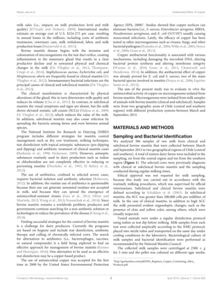
FIGURE 2 | Bacterial species distribution isolated from milk samples. Microorganisms identified in subclinical (A, central; C, southern) and clinical (B, central;
D, southern) mastitis milk samples. The central region includes 82 isolates and the southern region includes 245 isolates. The label “Others” includes
microorganisms isolated with frequency ≤2% of the total. # indicate that the frequency of isolation of microorganisms, from subclinical mastitis, between the regions
is significantly different (p ≤ 0.05). ∗ indicate that the frequency of isolation of microorganisms, from clinical mastitis, between the regions is significantly different
(p ≤ 0.05). Others included: Enterobacter cloacae, Enterococcus faecium, Streptococcus spp., Bacillus, non-fermenting Bacillus, Aeromona, Yersinia sp., Serratia
sp., and Candida spp.
TABLE 1 | Percentage of antibiotic resistance for the most frequently identified isolates from clinical bovine mastitis milk samples.
Central region South region
Resistance to Escherichia coli CNS Staphylococcus aureus Streptococcus uberis CNS
(n:13) (n:13) (n:18) (n:19) (n:16)
GN 15.4% 0 0 47.4% 0
NE 53.8% 15.4% 5.6% 57.9% 6.3%
CTX 15.4% 0 0 26.3% 6.3%
AMC 23.1% 0 0 0 0
TET 23.1% 7.7% 0 0 0
SXT 15.4% 0 0 0 0
Activity of Antimicrobial Copper
To assess the antimicrobial activity of copper, MIC-Cu was
evaluated for all microorganisms isolated from milk with
clinical and subclinical bovine mastitis. The results showed
(Supplementary Figure S3) that a majority of isolates from
clinical and subclinical samples were inhibited by 125 ppm
or less copper (90/327) and 250 ppm of copper (122/327);
however, there were two isolates that required 700 ppm [Candida
spp. (subclinical) in the central region and Streptococcus spp.
(clinical) in the southern region]. No growth was observed
Frontiers in Microbiology | www.frontiersin.org 5 April 2016 | Volume 7 | Article 626](https://image.slidesharecdn.com/2b78bc9f-87cf-4ae2-9d35-7ecbe700ec42-161018174842/85/FRONTIER-PUBLICACTION-2016-5-320.jpg)
![Reyes-Jara et al. Antibacterial Effect of Copper
for any microorganism at 1000 ppm. Subclinical isolates
showed slightly lower MIC-Cu values than those observed
for clinical mastitis isolates in both regions sampled. The
different behavior of bacteria species to MIC-Cu was apparently
not related to origin (central or southern region) of milk
samples (Supplementary Figure S3); in both cases nearly
93% of isolates were inhibited with less than 500 ppm of
copper.
The results for the bacteria identified most frequently in
clinical mastitis showed that 250 ppm of copper inhibited 77
and 62% of isolates of E. coli and CNS from the central region,
respectively (Figure 3A), likewise, S. aureus, CNS, and S. uberis
isolates (Figure 3B) from the southern region were inhibited 39,
50, and 63%, respectively (clinical mastitis). At 500 ppm copper
all the isolates were completely inhibited (Figure 3).
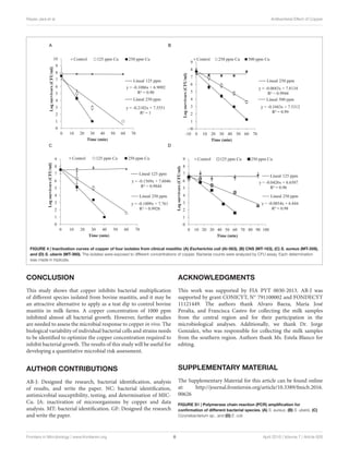
Figure 4 shows the inactivation curves of four strains
belonging to the four most frequent pathogenic species which
were isolated from clinical mastitis [E. coli (Al-563), CNS (MT-
163), S. aureus (MT-359), and S. uberis (MT-360)]. It was
observed that with less than 60 min of exposure to a copper
concentration of 250 ppm E. coli, S. aureus, and S. uberis reduced
their viability by almost 7 log CFU/ml, while CNS reduced its
viability ca. 7 log CFU/ml after 60 min when it was exposed
to 500 ppm of copper. The selected E. coli strain was the most
susceptible to copper.
A log linear relationship was found between the logarithm
of viable bacteria number and time of inactivation. A log-linear
model (equations in Figure 4) of the experimental data resulted
in high coefficients of determination (R2 > 0.90).
The Dc values are shown in Table 2. It can be observed that for
the same intensity of stress (250 ppm Cu), E. coli (Dc = 4.6 min)
was the least resistant, followed by S. aureus (Dc = 6.37 min) and
S. uberis (Dc = 11.71 min). CNS showed the highest resistance
value (Dc = 14.64 min), meaning that it needs a longer time to
reduce the population by 1 log.
The within-strain comparisons (Table 2) for different
intensities of stress (ppm copper) showed that the Dc values were
significantly different in all cases (p < 0.05). Comparison between
strains subjected to the same concentration of copper showed that
the Dc values of E. coli and S. aureus (125 and 250 ppm) were
not significantly different (p > 0.05). The Dc value for 500 ppm
copper in CNS was not significantly different from the Dc value
of 125 ppm for E. coli and S. aureus.
DISCUSSION
Bovine mastitis is a serious problem for dairy production
worldwide. In this study we evaluated in vitro the antimicrobial
effectiveness of copper solutions to inactivate microorganisms
prevailing in bovine mastitis. The results showed that the
prevailing agents are related to the geographical region. One
study reported by Kalmus et al. (2011) indicated that S. uberis and
E. coli are the main pathogens associated with clinical mastitis
in Estonia. However, in Sweden S. aureus has high prevalence
(21.3%) causing this pathology (Ericsson Unnerstad et al., 2009).
In India, cows with subclinical mastitis showed a highly variable
TABLE 2 | Dc values (min) for strains isolated from clinical bovine mastitis
exposed to different copper concentrations.
Dc values (min)
Copper (ppm)
125 250 500
E. coli 9.21a (0.03) 4.6c (0.04) –
CNS – 14.64d (0.012) 9.6a (0.01)
S. aureus 9.91a (0.01) 6.37c (0.03) –
S. uberis 23.47b (0.01) 11.71d (0.02) –
a,b,c,dThe same letter indicates no significant difference (p > 0.05) under the same
copper concentration. Values in brackets are standard error.
microbiota. Metagenomic analysis of the affected milk revealed
that E. coli was the predominant microorganism in two cattle
breeds studied, while S. aureus stood out in a third breed (Bhatt
et al., 2012).
The present study explored the current frequency and
diversity of pathogens involved in bovine mastitis in two
geographical regions of Chile. The isolation frequency of E. coli
was high in the central region, probably due to the permanent
confinement system applied in this region. S. aureus was
more frequently identified in the southern region where a free
pasturage system prevails, as previously described (San Martin
et al., 2002). These authors reported a similar prevalence of
S. uberis in the two regions (near 3%). In contrast, in our study the
frequency of detection of S. uberis was higher (22% clinical bovine
mastitis) in the southern region under pasture feeding (Figure 2).
These differences may imply that the etiology of bovine mastitis
in Chile has undergone modification after 10 years. This change
could be related to the type of feeding, the use of antibiotic
therapy, environmental modifications or other factors as have
been described in other countries (Zadoks and Fitzpatrick, 2009).
The use of antibiotics is frequent in the dairy industry to
control bacteria-causing bovine mastitis (Oliver and Murinda,
2012; Royster and Wagner, 2015). Antibiotic-resistant bacteria
can be favored as a result, becoming a serious problem for
dairy farms. A number of studies have been undertaken to
determine the frequency of antibiotic resistance in bovine
mastitis (Tenhagen et al., 2006; Bengtsson et al., 2009; Oliver
and Murinda, 2012; Saini et al., 2012b; Rato et al., 2013). In our
study we also evaluated the antibiotic susceptibility of isolated
microorganisms to antibiotics commonly used against bovine
mastitis (Supplementary Figure S2). The results showed that
on average 34% of the strains isolated were resistant to at least
one antibiotic evaluated. In particular, E. coli isolates showed
the highest resistance rate for almost all antibiotics tested. In
addition, 69% of E. coli from the central region and clinical
bovine mastitis strains were resistant to one or more antibiotics;
similar results were recently reported (Saini et al., 2012a). In the
current study many bacterial isolates showed resistance against
neomycin (63/324; 19%); this antibiotic is commonly used as an
intramammary antiseptic to treat bovine mastitis in Chile.
The use of iodine as teat dip is common in the national and
international dairy industry; the main advantage being the low
Frontiers in Microbiology | www.frontiersin.org 6 April 2016 | Volume 7 | Article 626](https://image.slidesharecdn.com/2b78bc9f-87cf-4ae2-9d35-7ecbe700ec42-161018174842/85/FRONTIER-PUBLICACTION-2016-6-320.jpg)
![Reyes-Jara et al. Antibacterial Effect of Copper
FIGURE S2 | Frequency of antimicrobial susceptibility of bacteria isolated
from the central region (A, n = 41; B, n = 40) and the southern region (C,
n = 157; D, n = 86) of subclinical and clinical bovine mastitis, respectively.
White bars are susceptible, gray bars are intermediate and black bars resistant
bacteria. Breakpoints (susceptible, intermediate, and resistant) were defined
according to CLSI.
FIGURE S3 | Frequency of MIC-Cu values for microorganisms isolated
from milk samples obtained from: (A) Central region (40 clinical and 42
subclinical mastitis) and (B) Southern region (86 clinical and 159
subclinical mastitis). Gray bars represent the frequency of microorganism
isolated from subclinical mastitis and black bars are from clinical mastitis. Each
determination was performed in triplicate.
REFERENCES
Aguirre, J. S., Pin, C., Rodríguez, M. R., and García de Fernando, G. D.
(2009). Analysis of the variability in the number of viable bacteria after
mild heat treatment of food. Appl. Environ. Microbiol. 75, 6992–6997. doi:
10.1128/AEM.00452-09
Andrews, J. M., and BSAC Working Party on Susceptibility Testing (2008). BSAC
standardized disc susceptibility testing method (version 7). J. Antimicrob.
Chemother. 62, 256–278. doi: 10.1093/jac/dkn194
Aspridou, Z., and Koutsoumanis, K. P. (2015). Individual cell heterogeneity
as variability source in population dynamics of microbial inactivation. Food
Microbiol. 45, 216–221. doi: 10.1016/j.fm.2014.04.008
Barkema, H. W., Schukken, Y. H., Lam, T. J., Beiboer, M. L., Benedictus, G.,
and Brand, A. (1998). Management practices associated with low, medium,
and high somatic cell counts in bulk milk. J. Dairy Sci. 81, 1917–1927. doi:
10.3168/jds.S0022-0302(98)75764-9
Bengtsson, B., Unnerstad, H. E., Ekman, T., Artursson, K., Nilsson-Ost, M., and
Waller, K. P. (2009). Antimicrobial susceptibility of udder pathogens from
cases of acute clinical mastitis in dairy cows. Vet. Microbiol. 136, 142–149. doi:
10.1016/j.vetmic.2008.10.024
Bhatt, V. D., Ahir, V. B., Koringa, P. G., Jakhesara, S. J., Rank, D. N., Nauriyal,
D. S., et al. (2012). Milk microbiome signatures of subclinical mastitis-affected
cattle analysed by shotgun sequencing. J. Appl. Microbiol. 112, 639–650. doi:
10.1111/j.1365-2672.2012.05244.x
Bradley, A. (2002). Bovine mastitis: an evolving disease. Vet. J. 164, 116–128. doi:
10.1053/tvjl.2002.0724
Capurro, A., Concha, C., Nilsson, L., and Ostensson, K. (1999). Identification of
coagulase-positive staphylococci isolated from bovine milk. Acta Vet. Scand.
40, 315–321.
Cavaco, L. M., Hasman, H., and Aarestrup, F. M. (2011). Zinc resistance
of Staphylococcus aureus of animal origin is strongly associated with
methicillin resistance. Vet. Microbiol. 150, 344–348. doi: 10.1016/j.vetmic.2011.
02.014
Cha, E., Bar, D., Hertl, J. A., Tauer, L. W., Bennett, G., González, R. N., et al.
(2011). The cost and management of different types of clinical mastitis in dairy
cows estimated by dynamic programming. J. Dairy Sci. 94, 4476–4487. doi:
10.3168/jds.2010-4123
Chaturvedi, K. S., and Henderson, J. P. (2014). Pathogenic adaptations to
host-derived antibacterial copper. Front. Cell. Infect. Microbiol. 4:3. doi:
10.3389/fcimb.2014.00003
Chen, J., and Griffiths, M. W. (1998). PCR differentiation of Escherichia coli
from other Gram-negative bacteria using primers derived from the nucleotide
sequences flanking the gene encoding the universal stress protein. Lett. Appl.
Microbiol. 27, 369–371. doi: 10.1046/j.1472-765X.1998.00445.x
Cheng, G., Hao, H., Xie, S., Wang, X., Dai, M., Huang, L., et al. (2014).
Antibiotic alternatives: the substitution of antibiotics in animal husbandry?
Front. Microbiol. 5:217. doi: 10.3389/fmicb.2014.00217
Clinical Laboratory Standards Institute [CLSI] (2012a). Clinical Laboratory
Standards Institute. Methods for Dilution Antimicrobial Susceptibility Tests
for Bacteria That Grow Aerobically; Approved Standard—Ninth Edition. CLSI
Document M07-A9. Wayne, PA: Clinical Laboratory Standards Institute.
Clinical Laboratory Standards Institute [CLSI] (2012b). Clinical Laboratory
Standards Institute. Performance Standards for Antimicrobial Susceptibility
Testing; Twenty-Second Informational Supplement. CLSI Documents M100-S22.
Wayne, PA: Clinical Laboratory Standards Institute.
De Vliegher, S., Fox, L. K., Piepers, S., McDougall, S., and Barkema, H. W. (2012).
Invited review: mastitis in dairy heifers: nature of the disease, potential impact,
prevention, and control. J. Dairy Sci. 95, 1025–1040. doi: 10.3168/jds.2010-4074
Drechsler, P. A., O’Neil, J. K., Murdough, P. A., Lafayette, A. R., Wildman,
E. E., and Pankey, J. W. (1993). Efficacy evaluations on five chlorhexidine
teat dip formulations. J. Dairy Sci. 76, 2783–2788. doi: 10.3168/jds.S0022-
0302(93)77616-X
El Behiry, A., Schlenker, G., Szabo, I., and Roesler, U. (2012). In vitro
susceptibility of Staphylococcus aureus strains isolated from cows with
subclinical mastitis to different antimicrobial agents. J. Vet. Sci. 13, 153–161.
doi: 10.4142/jvs.2012.13.2.153
Elguindi, J., Wagner, J., and Rensing, C. (2009). Genes involved in copper
resistance influence survival of Pseudomonas aeruginosa on copper
surfaces. J. Appl. Microbiol. 106, 1448–1455. doi: 10.1111/j.1365-2672.2009.
04148.x
Ericsson Unnerstad, H., Lindberg, A., Persson Waller, K., Ekman, T., Artursson, K.,
Nilsson-Ost, M., et al. (2009). Microbial aetiology of acute clinical
mastitis and agent-specific risk factors. Vet. Microbiol. 137, 90–97. doi:
10.1016/j.vetmic.2008.12.005
Espírito Santo, C., Lam, E. W., Elowsky, C. G., Quaranta, D., Domaille, D. W.,
Chang, C. J., et al. (2011). Bacterial killing by dry metallic copper surfaces. Appl.
Environ. Microbiol. 77, 794–802. doi: 10.1128/AEM.01599-10
Espírito Santo, C., Morais, P. V., and Grass, G. (2010). Isolation and
characterization of bacteria resistant to metallic copper surfaces. Appl. Environ.
Microbiol. 76, 1341–1348. doi: 10.1128/AEM.01952-09
Espírito Santo, C., Taudte, N., Nies, D. H., and Grass, G. (2008). Contribution of
copper ion resistance to survival of Escherichia coli on metallic copper surfaces.
Appl. Environ. Microbiol. 74, 977–986. doi: 10.1128/AEM.01938-07
Falk, D., and Guering, S. J. (1983). Differentiation of Staphylococcus and
Micrococcus spp. with the Taxo A bacitracin disk. J. Clin. Microbiol. 18, 719–721.
Faúndez, G., Troncoso, M., Navarrete, P., and Figueroa, G. (2004). Antimicrobial
activity of copper surfaces against suspensions of Salmonella enterica and
Campylobacter jejuni. BMC Microbiol. 4:19. doi: 10.1186/1471-2180-4-19
Galton, D. M., Petersson, L. G., Merrill, W. G., Bandler, D. K., and Shuster,
D. E. (1984). Effects of premilking udder preparation on bacterial population,
sediment, and iodine residue in milk. J. Dairy Sci. 67, 2580–2589. doi:
10.3168/jds.S0022-0302(84)81616-1
Gandra, E., Fernandez, M., Silva, J., and da Silva, W. (2011). Standardization of a
multiplex PCR for the identification of coagulase-positive Staphylococcus. Ciên.
Tecnol. Ali. 31, 946–949. doi: 10.1590/S0101-20612011000400019
Gao, J., Yu, F. Q., Luo, L. P., He, J. Z., Hou, R. G., Zhang, H. Q., et al. (2012).
Antibiotic resistance of Streptococcus agalactiae from cows with mastitis. Vet. J.
194, 423–424. doi: 10.1016/j.tvjl.2012.04.020
Geeraerd, A. H., Valdramidis, V. P., and Van Impe, J. F. (2005). GInaFiT, a freeware
tool to assess non-log-linear microbial survivor curves. Int. J. Food Microbiol.
102, 95–105. doi: 10.1016/j.ijfoodmicro.2004.11.038
Gomes, F., and Henriques, M. (2016). Control of bovine mastitis: old and recent
therapeutic approaches. Curr. Microbiol. 72, 377–382. doi: 10.1007/s00284-015-
0958-8
Grass, G., Rensing, C., and Solioz, M. (2011). Metallic copper as an antimicrobial
surface. Appl. Environ. Microbiol. 77, 1541–1547. doi: 10.1128/AEM.02766-10
Halasa, T., Nielen, M., De Roos, A. P., Van Hoorne, R., de Jong, G., Lam,
T. J., et al. (2009). Production loss due to new subclinical mastitis in Dutch
dairy cows estimated with a test-day model. J. Dairy Sci. 92, 599–606. doi:
10.3168/jds.2008-1564
Hassan, A. A., Khan, I. U., Abdulmawjood, A., and Lämmler, C. (2001). Evaluation
of PCR methods for rapid identification and differentiation of Streptococcus
uberis and Streptococcus parauberis. J. Clin. Microbiol. 39, 1618–1621. doi:
10.1128/JCM.39.4.1618-1621.2001
Kalmus, P., Aasmäe, B., Kärssin, A., Orro, T., and Kask, K. (2011). Udder pathogens
and their resistance to antimicrobial agents in dairy cows in Estonia. Acta Vet.
Scand. 53, 4. doi: 10.1186/1751-0147-53-4
Khamis, A., Raoult, D., and La Scola, B. (2004). rpoB gene sequencing for
identification of Corynebacterium species. J. Clin. Microbiol. 42, 3925–3931. doi:
10.1128/JCM.42.9.3925-3931.2004
Frontiers in Microbiology | www.frontiersin.org 9 April 2016 | Volume 7 | Article 626](https://image.slidesharecdn.com/2b78bc9f-87cf-4ae2-9d35-7ecbe700ec42-161018174842/85/FRONTIER-PUBLICACTION-2016-9-320.jpg)