Fluorescence in vivo Endomicroscopy: Imaging and Analysis (Co-Hosted w/Invicro)

Advanced benchtop microscopes can deliver resolution on the order of nanometers. However, they are highly invasive and require hours to days of tedious sample preparation, increasing the possibility of artifacts. Imagine a microscope that can capture images in real time directly from a living animal! Befitting this, laser confocal endomicroscopy (LCE), with its probe-based imaging approach, delivers cellular resolution, while being minimally invasive. Many scientists and imaging core facility managers refer to it as “virtual histology”; it captures real time microscale images like classical histology, in a non-destructive manner. It is a cutting-edge imaging modality with endless possibilities in preclinical imaging. The FIVE2 (ViewnVivo) from Optiscan Imaging is the latest and most competent model of LCE designed for effortless, real-time preclinical imaging in numerous animal models. This live webinar from two different imaging experts provided scientists and postdocs with the theoretical and practical knowledge about the FIVE2 (ViewnVivo) and its preclinical research applications. Former product manager of FIVE2 technology, Dr. Mohammedayaz Rangrez, gave a hands-on demonstration of the technology, imaging in different tissue types, Z sectioning and other key capabilities of the system. Dr. Howard Dobson from Invicro discussed some of its applications and image analysis illustrated with examples from normal and diseased rodents. This included images of the liver, kidneys and colon. Imaging of the colon was demonstrated using an intraabdominal approach in both rats and mice, as well as through a rectal approach in rats, making it ideally suited for longitudinal studies. In addition, the use of the FIVE2 (ViewnVivo) to image and quantify blood flow in small vessels was illustrated. Examples of quantitative image analysis approaches were included for each organ. Topics to be discussed in this webinar included: Hardware functionality and software operation of the FIVE2 (ViewnVivo) – fluorescence in vivo endomicroscope How to use the FIVE2 (ViewnVivo) for imaging tissue architecture with cellular resolution and for optical sectioning Examples of normal and abnormal tissues in the abdominal cavity using a variety of fluorophores Overview of the approach to image analysis An understanding of applications of the FIVE2 (ViewnVivo) in preclinical research

Recommended
Recommended
More Related Content
What's hot
What's hot (11)
Similar to Fluorescence in vivo Endomicroscopy: Imaging and Analysis (Co-Hosted w/Invicro)
Similar to Fluorescence in vivo Endomicroscopy: Imaging and Analysis (Co-Hosted w/Invicro) (20)
More from Scintica Instrumentation
More from Scintica Instrumentation (20)
Recently uploaded
Recently uploaded (20)
Fluorescence in vivo Endomicroscopy: Imaging and Analysis (Co-Hosted w/Invicro)
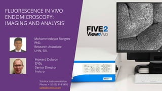
- 1. Mohammedayaz Rangrez PhD. Research Associate UHN, SRI. Scintica Instrumentation Phone: +1 (519) 914 5495 sales@scintica.com FLUORESCENCE IN VIVO ENDOMICROSCOPY: IMAGING AND ANALYSIS Howard Dobson DVSc Senior Director Invicro
- 2. WEBINAR Dr. Mohammedayaz Rangrez Introduction to the Technology FIVE2 instrument and functionality Imaging with FIVE2 X Y imaging Optical sectioning Dr. Howard Dobson In vivo image acquisition Image analysis Summary
- 3. BACKGROUND
- 4. MODERN MICROSCOPES AND IMAGING
- 5. WHAT ARE THE CHALLENGES ASSOCIATED WITH CLASSICAL MICROSCOPY?
- 6. Benchtop Microscopy: high resolution, trouble with in vivo imaging, possibilities of artifacts!
- 7. Intravital/Multiphoton Microscopy: high resolution, low flexibility, complicated protocols! Limited only to mice
- 8. PET/MRI: in vivo imaging, but no cellular details!
- 9. INVASIVE - - - - MINIMALLY INVASIVE - - - NON INVASIVE nm---µm----RESOLUTION----mm---- In vivo Ex vivo MRI CLSM
- 10. INVASIVE - - - - MINIMALLY INVASIVE - - - NON INVASIVE nm---µm----RESOLUTION----mm---- In vivo Ex vivo MRI CLSM
- 11. RAT BRAIN GLIOBLASTOMA • Invasive! Fluorescence/Confocal Microscope? • Insufficient Resolution! MRI PET? • FIVE - ViewnVivo Laser Confocal Endomicroscopy!
- 12. LCE – FIVE2 is a simple user-friendly tool!
- 13. Laser Objective lens Tissue Detector Optical fiber Imaging plane Scan mechanism Ex = 488nm Em > 515 nm FIBER OPTIC CONFOCAL SYSTEM Transformation and Miniaturization
- 14. 14 • The confocal endomicroscope is an “optical sectioning” device (Out of focus material turns black, instead of blurry) • It isolates A thin plane of cells, viewed en face (en face = parallel to tissue surface, unlike typical cut sections) • One of the most important user operations is the control of the imaging plane depth • Imaging depth up to 400um • Tissue dependent THE ‘Z’ DIMENSION Capturing 3D Volumes and Enabling 3D Visualization X Axis Y Axis Z Axis
- 15. 15 EN FACE IMAGING VS. T.S. HISTOLOGY
- 17. Image does not show included 3-way footswitch, or optional items Confocal Processor PC, Monitor, Keyboard and Mouse Probe Animal Stage and Probe Holder Imager Application + Fiji (ImageJ) FIVE2 Components
- 18. 0.1% acriflavin for 30 minutes
- 19. 30 minutes wash
- 25. Courtesy of Dr. Amanda Howard, Bitplane
- 26. Courtesy of Dr. Amanda Howard, Bitplane
- 27. 1. General Non-specific Or Semi-specific Fluorophores • Mainly intercellular compartment 2. Acridines • Nuclear/Acidic Organelle Stains • Acriflavine, Proflavine, Acridine Orange… 3. Cycline Antibiotics • Tetracycline, Doxycycline, more.. 4. Membrane Dyes • Good for nerve tracing • Dii, DiO, 4-Di-2-Asp TYPES OF CONTRAST AGENTS 5. Specific Dyes • Conjugated Peptides • Conjugated Antibodies 6. Functional Dyes • Calcium Markers (G-CAMP, Fluo-3, Fluo-4) • Metabolic Markers • pH Markers • Live/Dead Markers 7. Transgenic Fluorophores • Produced by the living cells when a certain gene is expressed • GFP, eGFP, YFP can be imaged
- 28. Fluorescein Sodium (10ml of 10%): Intravenously InjectedTopically Applied Acriflavine 0.05% Images courtesy Prof Adrian Polglase, Cabrini Monash University Academic Surgical Unit, Melbourne Australia CONTRAST AGENT COMPARISON (COLONIC MUCOSA)
- 30. Courtesy of Dr Philip Currie Cancer Fibrous Tissue Normal Tissue FIVE2 Images: Instant, easy, live and in vivo! Classical Histology: Tedious prep, slow, possible artifacts and ex vivo!
- 31. While we switch between our speakers, tell us more about your research • What animal models are you working with, • What types of instruments do you work with, and • What are your research goals? Please type your answers in the Q&A box below:
- 32. HOWARD DOBSON: Exploring FIVE2 for animal imaging Objective was to gain experience acquiring images and explore the image analysis options Used normal mice and rats and acute disease models
- 33. Disease models • Liver • Kidney • Colon • Normal brain vasculature
- 34. Fluorophores • Fluorescein– topical and intravenous • Acriflavine – topical and intravenous • Fluorescein isothiocyanate – Dextran 150 (FITC) – intravenous • Annexin Vivo- intravenous
- 35. Liver – thioacetamide acute liver necrosis model • Necrosis present at 24 hours post single 200mg/kg dose thioacetamide • Images acquired at laparotomy
- 36. Columns of cells Sinusoids – channels between cells through which blood flows Image of normal liver acquired after the intravenous administration of FITC followed by topical application of acriflavine.
- 37. Image of normal liver (Left) and abnormal liver (right) demonstrating swelling of cells resulting in compression of the sinusoids
- 38. Image of abnormal mouse liver acquired two hours following the intravenous administration of Annexin Vivo which binds to the surface of apoptotic cells which are distributed throughout the liver
- 39. Sinusoid segmentation is performed using filtration and adaptive thresholding with additional morphological processing. Sinusoid segmentation is performed using the same parameters for both images.
- 40. Normal Sinusoid Area : 16.9% of FOV Abnormal Sinusoid Area: 8.9% of FOV
- 42. Kidney – folic acid model of acute renal disease 1 . N ec rosis of ren al t u b u les p resent at 2 4 h ou rs p ost 2 5 0 mg / kg d ose folic ac id 2 . Images ac q u ired at lap arotomy 3 . S mall in c ision of t h e kid n ey capsu le allows g reater d epth of p en et rat ion
- 43. Image of a normal kidney tubules acquired following intravenous administration of fluorescein and topical acriflavine Small amounts of fluid within the tubules can be identified (Red arrow)
- 44. Image of abnormal kidney showing dilated tubules (Red arrows)
- 45. Two image analysis approaches • Both segment tubules from background based on signal intensity • Adaptive histogram with adaptive thresholding • Distance to background transform • U s e d a s a p rox y f o r t u b u l e d i a m e t e r
- 46. AbnormalNormal Fluorescence Image Tubule Segmentation Distance-to-BackgroundSkeleton Overlay 70µm 0µm
- 47. Plots of the results from the two tubule segmentation methods demonstrating tubule dilation in the abnormal kidney, but different results based on the two methods.
- 48. Colon – dextran sodium sulfate (DSS) model A c u t e c o l i t i s a p p r o x i m a t e l y s e v e n d a y s p o s t 5 % D S S i n t h e d r i n k i n g w a t e r • A l f a l f a f r e e d i e t t o a v o i d a u t o f l u o r e s c e n c e i n t h e g u t • I m a g e s a c q u i r e d b y l a p a r o t o m y i n t h e m o u s e a n d r a t • I m a g e s c a n b e a c q u i r e d p e r r e c t u m i n t h e r a t g r e a t e r t h a n 2 5 0 g b o d y w e i g h t
- 49. Images of the normal (Left) and abnormal (Right) rat colon following intravenous FITC. The abnormal images demonstrates marked hypervascularity (Red arrow) and distortion of the tissue architecture
- 50. Topical administration of acriflavine demonstrates marked distortion of the tissue architecture
- 51. Vascular imaging of brain surface in normal animals • Probe applied to the surface of the organ via a laparotomy • Craniotomy and small incision into the meninges to allow direct access to the brain surface • Intravenous fluorescein immediately prior to imaging • Image frames collected at 1.34±0.1s
- 53. Processing Algorithm • Image patches of 51 pixel edge length are registered between temporally adjacent images using normalized cross correlation • The calculated displacements for each image patch are then translated to a velocity • A speed and a velocity map are calculated for each frame
- 54. Original Image Flow Direction
- 55. Original Image Flow Speed
- 56. Image of the surface of the colon acquired after intravenous administration of fluorescein demonstrating vessels of various sizes
- 57. In abnormal tissue the vascularity becomes tortuous. This can be quantified using various vessel branching metrics
- 58. Branch Metrics • Radius (R) = average radius of the branch skeleton • Length (L) = length of the smoothed centerline • Aspect Ratio (AR) = Length/Radius • Vessel Branching Fraction (VBF) = 1/Length • 4 Measures of Tortuosity Analysis Methodology - Branch Analysis
- 59. Poll Question: Which of the following would you like to learn more about? (check all that apply) The FIVE2 technology Having an on-site demonstration of the FIVE2 Invicro’s image analysis capabilities All the above
- 60. Conclusions • The ViewnVivo Five2 system is very easy to use with little training required • Image analysis routines can be developed for multiple specific applications – beyond the simple examples presented • Prototype analysis routines developed in MATLAB or using Python
- 61. SUMMARY
- 64. To ask a question, click the Q&A Button, type your question and click send. Any questions that are not addressed during the live webinar will be answered following the event. Please indicate whom you want to address your question. Thank you for participating! Q&A SESSION: Mohammedayaz Rangrez PhD. Research Associate UHN, SRI. Howard Dobson DVSc Senior Director Invicro