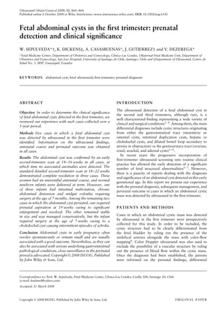
1) The study reviewed 5 cases of fetal abdominal cysts detected by ultrasound in the first trimester between 10-13 weeks of gestation.
2) In 3 cases, the cyst resolved spontaneously by the detailed second trimester scan, though 1 infant later required surgery for intestinal malrotation.
3) In the remaining 2 cases, 1 cyst was aspirated at 19 weeks for enlargement and the other remained stable but the infant later required surgery for a choledochal cyst.
This study evaluated platelet parameters in 100 subjects with type 2 diabetes and 100 non-diabetic controls. Mean platelet volume (MPV) and platelet distribution width (PDW) were significantly higher in diabetic subjects compared to controls. Among diabetics, those with HbA1c >7% had significantly higher MPV and PDW than those with HbA1c ≤7%, indicating poorer glycemic control is associated with increased platelet activity. MPV was also higher in diabetics with disease duration >10 years compared to those with duration ≤10 years, suggesting longer diabetes duration impacts platelet function. The study concludes that MPV can serve as a prognostic marker for cardiovascular risk in diabetes.
Primary biliary cirrhosis and autoimmune hepatitis 20120902 Chien-Wei Su
This document summarizes a case of primary biliary cirrhosis (PBC) in a 66-year-old man who presented with intermittent pruritus for 8 years. Laboratory tests showed elevated liver enzymes and positive anti-mitochondrial antibodies, confirming the diagnosis of PBC. Liver biopsy showed features characteristic of PBC including destruction of bile duct epithelium and lymphocyte infiltration. The document then provides information on the epidemiology, risk factors, pathogenesis, natural history, diagnosis, treatment and prognosis of PBC.
Nonalcoholic fatty liver disease (NAFLD) refers to hepatic steatosis without other causes like heavy alcohol use. It ranges from nonalcoholic fatty liver (NAFL) to nonalcoholic steatohepatitis (NASH) with inflammation. NAFLD prevalence is increasing worldwide in line with obesity, diabetes, and metabolic syndrome. Weight loss through lifestyle changes and possibly bariatric surgery is the only treatment with evidence of benefit for NAFLD/NASH.
This document provides information on alcoholic liver disease, including its incidence, progression, risk factors, pathogenesis, clinical findings, diagnostic testing, differential diagnosis, prognosis, and screening tools. Some key points:
- Heavy alcohol use can lead to fatty liver in 90-100% of people within 10 years, while only 10-35% will develop alcoholic steatohepatitis and 8-20% will progress to cirrhosis.
- Risk is increased by factors like younger age of onset, female sex, certain ethnicities, coinfection with hepatitis B or C, iron overload, and obesity.
- Alcohol causes liver injury through mechanisms like centrilobular hypoxia, neutrophil infiltration, antigen formation,
This document presents a case of ulcerative colitis in a 20-year-old male. Examination found bloody stools, abdominal pain, and fever. Tests confirmed the presence of Entamoeba histolytica in stool and detected ulceration and crypt abscess on biopsy. The patient was diagnosed with ulcerative colitis and amoebiasis. Treatment included antibiotics, a PPI, antispasmodics, and supplements. The patient was counseled on his condition, medications, and recommended dietary changes.
A 20-year-old girl presented with chronic diarrhea, fever, weight loss, and loss of appetite for 4 weeks. Colonoscopy and biopsy revealed diffuse large B-cell lymphoma of the duodenum and stomach. Imaging showed thickening and nodularity of the third part of the duodenum with enlarged lymph nodes. The patient was diagnosed with primary diffuse large B-cell lymphoma of the duodenum and stomach, stage II, and started on CHOP chemotherapy.
This document discusses cardiovascular toxicity that can result from cancer treatments. It covers 9 main categories of cardiovascular complications and focuses on myocardial dysfunction and heart failure, which are among the most common side effects. Several chemotherapy agents, targeted therapies, and radiotherapy can cause heart failure by damaging heart tissue through oxidative stress or other mechanisms. Regular cardiac monitoring is important for early detection of issues so they can be managed to prevent further deterioration.
1) The document discusses atrial fibrillation (AF), its increasing prevalence, and its association with increased risk of stroke.
2) It reviews stroke risk assessment tools like CHADS2 and CHA2DS2-VASc scores and guidelines for stroke prevention in AF patients using anticoagulation or the newer oral anticoagulants (NOACs).
3) It also discusses left atrial appendage closure with the Watchman device as an alternative for stroke prevention in patients who cannot tolerate long-term anticoagulation. The Watchman trials demonstrated the device's safety and efficacy in reducing stroke risk comparable to warfarin.
Hypertension is very common in patients with chronic kidney disease (CKD), affecting 67-92% of patients. Control of hypertension is important for slowing the loss of kidney function and reducing the risk of further kidney damage. The document discusses several risk factors for hypertension in CKD patients including older age, African descent, overweight or obesity, and concurrent diabetes or heart disease. It also reviews guidelines for treating hypertension in CKD, which generally recommend a target blood pressure under 140/90 mmHg and use of renin-angiotensin system blocking agents along with monitoring of kidney function and potassium levels.
A 45-year-old female patient with a history of systemic lupus erythematosus (SLE) was admitted to the hospital with a cough producing sputum for 8 days and fever for 5 days. On examination, the patient exhibited rashes, lesions, and nail abnormalities consistent with SLE. Laboratory tests showed slightly elevated albumin and globulin levels. She was diagnosed with SLE and treated with corticosteroids, hydroxychloroquine, proton pump inhibitors, and other medications. The patient was counseled on adherence to treatment, lifestyle changes like avoiding sunlight, and reporting any adverse effects.
This document provides an overview of Hodgkin lymphoma (HL), including:
- HL is a malignancy of mature B lymphocytes that represents 10% of lymphomas diagnosed annually.
- Classical HL has high cure rates of over 85% with radiation and chemotherapy. However, late therapy-related toxicities are a new challenge.
- Staging is important for selecting appropriate therapy intensity, with early stage patients having a better prognosis than advanced stage.
- Treatment depends on stage and risk factors, and may involve chemotherapy alone or with radiation for early stage disease. Advanced stage is treated with chemotherapy only.
- Late effects of therapy include secondary cancers and heart disease, so reducing radiation exposure is a focus of research.
Update on Patterns of Study in ANCA Associated Vasculitis presented at regional Northern Ireland Nephrology Meeting with Dr David Jayne as guest speaker..
This patient is a 54-year-old female with type 2 diabetes presenting with worsening gastrointestinal symptoms of early satiety, heartburn and nausea despite dietary modifications. Investigations including EGD revealed mild antral erythema and food residue in her stomach, with a hiatal hernia present but no esophagitis or Barrett's esophagus. H. pylori testing was negative. The primary diagnosis is likely gastroparesis, and recommended treatment includes a proton pump inhibitor along with modifying her diabetes medications.
This document summarizes the PARADIGM-HF clinical trial which compared the angiotensin receptor-neprilysin inhibitor LCZ696 to the ACE inhibitor enalapril in patients with heart failure and reduced ejection fraction. The trial found that LCZ696 was superior to enalapril in reducing cardiovascular death and hospitalization for heart failure. Specifically, LCZ696 reduced the risk of the primary composite outcome of death from cardiovascular causes or hospitalization for heart failure by 16% compared to enalapril. LCZ696 also reduced deaths from any cause by 16% compared to enalapril.
1) Diabetes is now the leading cause of end-stage renal disease (ESRD) in the United States, surpassing other causes like hypertension.
2) Diabetic nephropathy follows a typical progression from increased kidney function to protein in the urine to declining kidney function over many years.
3) Tight control of blood pressure, blood sugar, and use of angiotensin-converting enzyme inhibitors (ACEIs) or angiotensin receptor blockers (ARBs) can slow the progression of diabetic nephropathy.
An 86-year-old male presented to the emergency room with dizziness and was admitted to the ICU for dehydration. Medical workup showed malignant neoplasms, hypovolemia, pancytopenia, and a bloodstream infection. Laboratory results indicated anemia, thrombocytopenia, and abnormalities consistent with liver and kidney disease. Stool and blood cultures grew Clostridium difficile and Escherichia coli. The patient was diagnosed with myelodysplastic syndrome and received multiple blood transfusions.
CASE PRESENTATION ONCIRRHOSIS OF LIVER WITH PORTAL HYPERTENSION, HEPATIC EN...Akhil Joseph
A DETAIL CASE PRESENTATION ON CIRRHOSIS OF LIVER WITH PORTAL HYPERTENSION, HEPATIC ENCEPHALOPATHY AND GRADE II OESOPHAGEAL VARICES WITH CONGESTIVE GASTROPATHY. LIVER CIRRHOSIS AND ALL ITS COMPLICATION IN A PATIENT.
This document summarizes the current status of pancreaticobiliary endoscopic ultrasound (EUS) at Delhi Apollo Hospital. It finds that EUS, with or without fine needle aspiration (FNA), provides accurate diagnosis and staging of pancreaticobiliary lesions. In a study of 67 patients, EUS identified lesions in the pancreas, bile duct, ampulla, and detected cancers, cysts, stones. No complications occurred. The results are similar to other established centers and confirm EUS as a safe, accurate method for evaluating pancreaticobiliary diseases.
The document discusses non-alcoholic fatty liver disease (NAFLD), which includes a spectrum of conditions from simple steatosis to non-alcoholic steatohepatitis (NASH) and cirrhosis. NAFLD is strongly associated with obesity and metabolic syndrome. The prevalence of NAFLD is increasing globally and varies from 5-30% in different regions. Diagnosis requires imaging and liver biopsy. Treatment focuses on lifestyle modifications and medications to improve insulin resistance.
The document discusses the relationship between hypertension and diabetes, noting that they often occur together and worsen each other's effects on target organs like the vasculature. Both conditions should be treated to reduce cardiovascular risks, with a target blood pressure under 140/90 mmHg for diabetic hypertensives. Achieving this often requires two or more antihypertensive drugs, especially agents that block the renin-angiotensin-aldosterone system like ACE inhibitors.
This document provides information on Coronary Artery Disease (CAD) and acute gastroenteritis. CAD is caused by plaque buildup in the coronary arteries which can lead to chest pain, shortness of breath, or heart attack. Symptoms of acute gastroenteritis include vomiting, diarrhea, abdominal pain and cramps, fever, and dizziness. The document then provides details on the case of a 58-year-old male patient presenting with vomiting, abdominal pain, chest pain, and general weakness. His medical history and examination results indicate diagnoses of CAD, hypertension, hypothyroidism, and acute gastroenteritis. He is started on treatment including aspirin, statins, beta blockers, antiemetics
Cardio-Renal Protection Through Renin–Angiotensin–Aldosterone System Inhibitionmagdy elmasry
Physiological and detrimental roles of RAAS molecules in cardiac, vascular tissues and kidneys.‘cardiovascular continuum’ Barriers In Optimizing RAAS Inhibition.The effects of angiotensin II inhibition and improvement in bradykinin availability
Anti-Diabetics For Cardiac Patients The Proper Selectionmagdy elmasry
Cardiovascular Disease and Type 2 Diabetes.Tight glycaemic control can reduce microvascular complications of T2DM, but does not lower CV risk sufficiently.
Multifactorial intervention, comprising of lowering lipid levels and BP, and use of aspirin, has been shown to reduce vascular complications and mortality.Shifting the Paradigm in Diabetes Care
Treating Diabetes Beyond A1C :Considerations for Cardiovascular Protection.
Autoimmune hemolytic anemia (AIHA) can be primary or secondary. Primary AIHA has no obvious cause, while secondary is precipitated by conditions like drugs, infections, or tumors. AIHA is caused by autoantibodies binding to red blood cells (RBCs), which are then destroyed in the spleen or liver. Warm antibody AIHA is treated mainly with steroids, while cold antibody types are more resistant and associated with lymphoproliferative diseases. Treatment depends on the type and severity of AIHA.
This was a review of different guidelines on lupus nephritis from ACR, EULAR, and KDIGO. Goal is appreciate similarities and differences between the different guidelines.
1. Alcoholic liver disease progresses through stages including fatty liver, alcoholic hepatitis, and cirrhosis. Tumor necrosis factor (TNF) plays a key role in mediating inflammation and the progression of disease.
2. Only a minority of heavy drinkers develop serious liver damage, and other host and environmental factors determine progression. Risk factors include genetic factors and additional insults like hepatitis or obesity.
3. TNF is involved in the early stages of fatty liver disease and mediates the transition to more advanced stages like steatohepatitis and cirrhosis. Cytokines like TNF orchestrate the liver's inflammatory response and damage.
Value of routine ultrasound examination at 35–37 weeks’ gestation in diagnosi...Võ Tá Sơn
This study investigated the value of routine ultrasound examination at 35-37 weeks' gestation in diagnosing previously unknown fetal abnormalities. The study found:
1) Of 995 total fetal abnormalities detected, 24.8% (247) were first diagnosed at 35-37 weeks' gestation and 7.4% (74) were first diagnosed postnatally.
2) The most common abnormalities first seen at 35-37 weeks were hydronephrosis, mild ventriculomegaly, ventricular septal defect, duplex kidney, ovarian cyst, and arachnoid cyst.
3) Routine ultrasound at 35-37 weeks could potentially improve postnatal outcomes by enabling diagnosis and management planning for
Pyloric stenosis usually presents with well defined symptoms and signs. However, these may be absent in neonates in intensive care for other conditions. This paper reports 2 cases of congenital diaphragmatic hernia that developed pyloric stenosis during the critical care period and illustrates the atypical presentation of pyloric stenosis and the diagnostic difficulties encountered in this setting
This document summarizes the case of a newborn baby born via elective C-section with anorectal malformations. Key details include:
- Baby was born at term with normal APGAR scores and weight of 2.83kg.
- During birth it was noted the baby had no patent anal opening and a single umbilical artery, so the baby was referred for further management.
- Investigations found the baby had an anorectal malformation, ASD, and L5 hemivertebrae, consistent with VACTERL association.
- A colostomy was performed on the baby. No growth was found on cultures and antibiotics were stopped. The baby was being managed for the
Spontaneous rupture of endometriotic cyst in 3rd trimester of pregnancyApollo Hospitals
Endometriosis is a well established cause of female infertility and may be associated with early pregnancy losses. Association of endometriosis with pregnancy is rare. Ruptured endometriotic cyst presenting as acute abdomen in pregnancy is even a rarer presentation.
We present hereby a rare and interesting case, presented in our hospital, of Spontaneous rupture of endometiotic Cyst in 3rd trimester of pregnancy and its subsequent management.
This study summarizes the diagnosis and management of 26 cases of recurrent tracheoesophageal fistulas over 27 years. The average gestational age and birth weight of patients who developed recurrent fistulas was lower than typical. Most recurrent fistulas were discovered due to respiratory or feeding symptoms and were diagnosed via contrast study, which revealed the fistula in over 90% of cases. Repairs involved sharply separating the trachea and esophagus and placing tissue like pericardium between the suture lines. Post-operative complications occurred in about 30% of patients, but long-term follow-up showed that over 80% took full nutrition by mouth.
Case Report:Massive Ovarian Cyst in a Adolescent GirlTana Kiak
For benign tumours adhesion prevention strategies should be used. Surgical intervention should as much as possible be directed towards preservation of ovarian tissue. There is scarcity of published literature on this subject.
We need bigger studies to address the issue of how much fertility preservation is safely possible.Irrespective of indication for surgery, it is always preferable to attempt conservative, fertility sparing surgery in adolescents.
This patient presented with retained placenta after a vaginal delivery. Her ultrasound and MRI showed placenta increta, where placental villi had invaded into the myometrium. She was initially managed conservatively with methotrexate injection, which led to a partial reduction in her beta-hCG levels. However, she later developed heavy bleeding and required an emergency hysterectomy. Placenta accreta spectrum (PAS) describes abnormal placental invasion that can cause life-threatening bleeding. Risk factors include prior uterine surgery. Management challenges include delayed referrals, lack of blood product availability, and counseling patients on prolonged hospitalization sometimes required.
1) The document proposes a new model of prenatal care based on a comprehensive assessment at 11-13 weeks of gestation. This assessment uses maternal characteristics, ultrasound findings, and biochemical testing to determine patient-specific risks for various pregnancy complications.
2) Most major fetal aneuploidies, structural abnormalities, and a variety of pregnancy complications can potentially be identified or assessed at high risk during the 11-13 week assessment.
3) Based on the risk assessment, most women would be classified as low risk and require fewer prenatal visits, while high risk women would receive specialized monitoring and treatment. This shifts prenatal care from routine visits to a personalized, disease-specific approach.
One third of all women in the United States are obese [1], and
approximately one-fi ft h of the population is obese in pregnancy. Obesity during pregnancy caries multiple risks to the fetus and the mother, including fetal macrosomia, prematurity, miscarriage, maternal hypertension, and gestational diabetes
A Case of Abdominal Pregnancy, Primary vs Secondary – Radiological Workupiosrjce
IOSR Journal of Dental and Medical Sciences is one of the speciality Journal in Dental Science and Medical Science published by International Organization of Scientific Research (IOSR). The Journal publishes papers of the highest scientific merit and widest possible scope work in all areas related to medical and dental science. The Journal welcome review articles, leading medical and clinical research articles, technical notes, case reports and others.
Ultrasound in early pregnancy is used to assess:
1. Normal aspects such as gestational sac size and viability by detecting a heartbeat.
2. Abnormalities including ectopic pregnancies, molar pregnancies, and hemorrhages.
3. Dating the pregnancy accurately in the first trimester by measuring the mean sac diameter or crown-rump length.
4. Detecting multiple pregnancies and diagnosing chorionicity and zygosity.
5. Screening for fetal abnormalities such as thickened nuchal translucency which is used to detect Down syndrome.
The document discusses diagnosis of pregnancy through presumptive, probable, and positive signs including hormonal tests and ultrasounds. It describes maternal assessment across trimesters including physical exams, lab tests, and ultrasounds to evaluate fetal growth and well-being. Special investigations are outlined for high-risk pregnancies along with signs of potential complications and minor ailments.
A 34-year-old woman presented with lower abdominal pain and vaginal bleeding. Ultrasound and MRI revealed an abdominal pregnancy implanted on the mesentery of the sigmoid colon. The patient underwent laparotomy where the 13-week placenta was dissected from the mesentery and abdominal sidewall. Abdominal pregnancy is a rare and serious form of ectopic pregnancy that can be accurately diagnosed using ultrasound and MRI to determine placental location preoperatively.
This document discusses cervical insufficiency, including its definition, causes, diagnosis, and treatment. Cervical insufficiency is the inability of the cervix to retain a pregnancy due to structural or functional defects in the absence of labor contractions. It can be congenital or acquired due to conditions like cervical trauma. Diagnosis involves history, examination finding an open cervix, and transvaginal ultrasound to measure cervical length. Treatment options include cervical cerclage surgery to provide structural support to the cervix, progesterone to help maintain the cervix, or a pessary. Cerclage is indicated for women with a history suggestive of cervical insufficiency or a short cervix on ultrasound.
Drs. Rossi and Shreve’s CMC Abdominal Imaging Mastery Project: June CasesSean M. Fox
Dr. Brian Shreve is an Emergency Medicine Resident and Dr. Isolina Rossi is a Surgery Resident at Carolinas Medical Center in Charlotte, NC. They are interested in medical education. With the guidance of Drs. Kyle Cunningham and Michael Gibbs, they aim to help augment our understanding of emergent abdominal imaging. Follow along with the EMGuideWire.com team as they post these monthly educational, self-guided radiology slides on:
• Necrotizing Enterocolitis (NEC)
• Spontaneous Intestinal Perforation (SIP)
• Intestinal Atresia
This document provides information about performing a gynecological examination. It discusses taking a patient history, including gynecological, medical, surgical, social, and family histories. It describes the positions a patient can be in for examination and the equipment needed, including speculums. Procedures covered include speculum examination, bimanual or rectal examination, Pap smear for cervical cytology, and colposcopy. The goal of examination is to make an accurate diagnosis to determine appropriate treatment or management.
This document provides information about performing a gynecological examination. It discusses taking a patient history, performing a physical exam including a speculum exam, bimanual exam, Pap smear, and colposcopy. It also describes procedures like hysterosalpingography to evaluate the uterus and fallopian tubes. The goal is to systematically examine the patient and gather relevant medical information to make an accurate diagnosis or differential diagnosis to guide further treatment.
USMLE GENERAL EMBRYOLOGY 001 Embryology Terminology Embryology Terminology...AHMED ASHOUR
Embryology is the branch of biology that studies the development of embryos, encompassing the processes from fertilization to the formation of a complete organism. These terms represent fundamental concepts in embryology and are crucial for understanding the developmental processes that occur during the early stages of an organism's life.
This document summarizes the case of a 34-year-old woman who presented with severe abdominal pain and was diagnosed with a retained placenta after undergoing laparotomy for an abdominal pregnancy. She underwent several additional surgeries and treatments. Abdominal pregnancy is a rare and high-risk type of ectopic pregnancy. It can be difficult to diagnose and poses challenges for management and placental removal. Expectant management may be considered in some cases if fetal and maternal conditions allow.
El documento describe la fisiología del ciclo menstrual normal. Explica que el ciclo está definido por la ovulación y la menstruación y que involucra la producción ordenada de hormonas por el eje hipotálamo-hipófisis-ovario, lo que resulta en la proliferación del endometrio y el desarrollo y maduración de los folículos en el ovario.
Este documento describe la monitorización fetal electrónica, incluyendo su objetivo de detección temprana de factores de riesgo fetal, cómo funciona mediante la evaluación de la frecuencia cardiaca fetal y su variación, y los riesgos y beneficios. Explica conceptos clave como línea de base, variabilidad, aceleraciones y desaceleraciones, así como categorías de interpretación. El documento proporciona una guía detallada para la interpretación de trazados de monitorización fetal.
El documento habla sobre el diagnóstico de embarazo. Explica que el diagnóstico se puede hacer relativamente fácil en el tercer mes y se basa en signos y síntomas, exámenes de laboratorio y métodos auxiliares. Detalla diferentes tipos de signos como de presunción, probabilidad y positivos, incluyendo la presencia del feto a través de movimientos fetales, actividad cardiaca fetal y ultrasonido. Además, se enfoca en la gonadotropina coriónica humana como un método utilizado, pudiendo me
Respuesta fetal a hipoxemia transitoriaKevin Dickens
La desaceleración tardía representa la respuesta fetal a la hipoxemia transitoria causada por las contracciones uterinas, que disminuyen la perfusión en el espacio intervelloso y reducen los niveles de oxígeno por debajo de los niveles críticos. Cuando la contracción cesa, el flujo se restablece en el espacio intervelloso. Episodios recurrentes de hipoxia fetal pueden conducir a hipoxia y acidosis de los tejidos fetales.
Via del parto y repercusiones sobre el neonatoKevin Dickens
El documento discute los contextos de decisión para enfocarse en la cesárea versus el parto vaginal y sus repercusiones en el neonato. Señala que a pesar del aumento de las tasas de cesárea, la evidencia sobre sus beneficios frente al parto vaginal es limitada o contradictoria para la mayoría de indicaciones. La cesárea electiva puede aumentar la morbilidad respiratoria y otras complicaciones en neonatos de menos de 39 semanas y no necesariamente previene problemas como daño neurológico o infecciones. La vía del parto debe
Este documento describe las diferentes etapas que rodean la menopausia según definiciones de la OMS e IMS, incluyendo la perimenopausia, transición menopáusica, climaterio, premenopausia y postmenopausia. También explica las causas de la menopausia como la depleción de ovocitos y la pérdida exponencial de folículos con la edad, así como los cambios hormonales asociados como los niveles de FSH, estradiol y progesterona.
La cardiotocografía ha sido utilizada desde mediados del siglo XX para evaluar la reserva de oxígeno fetal. Sin embargo, existen errores técnicos, de interpretación y factores que afectan la cardiotocografía. La reproducibilidad de la interpretación entre observadores es baja, entre 41-55%. Además, la cardiotocografía tiene alta especificidad pero baja sensibilidad, por lo que es confiable para pronosticar salud fetal pero no riesgo fetal.
La histerectomía vaginal es una intervención quirúrgica ginecológica que se ha realizado desde el siglo II d.C. El documento describe la historia, indicaciones, técnica quirúrgica y complicaciones potenciales de la histerectomía vaginal, así como las experiencias del Hospital Regional del IESS “Dr. Teodoro Maldonado Carbo” con esta cirugía. La histerectomía vaginal ofrece ventajas como tiempos de recuperación más cortos en comparación con la histerectomía abdominal.
El documento describe la endometriosis y el uso de la ultrasonografía en su diagnóstico. La endometriosis ocurre cuando el tejido endometrial crece fuera del útero y afecta a entre el 10-12% de mujeres. La ultrasonografía tiene baja sensibilidad para detectar endometriosis difusa pero alta sensibilidad para detectar endometriomas u quistes ováricos. La laparoscopia sigue siendo el estándar de oro para el diagnóstico definitivo.
Diagnóstico Prenatal De Malformaciones Del Sistema UrinarioKevin Dickens
Este documento resume las principales malformaciones del sistema urinario que pueden diagnosticarse mediante ultrasonido prenatal, incluyendo agenesia renal bilateral, enfermedad poliquística renal, displasia quística renal, hidronefrosis y atresia uretral. Explica la etiología, incidencia, riesgos de anomalías asociadas y pronóstico de cada anomalía. También describe los hallazgos ultrasonográficos característicos para el diagnóstico diferencial entre las diferentes patologías.
Este documento resume los conceptos básicos de la citogenética reproductiva, incluyendo la determinación y diferenciación sexual, la cromatina sexual, el frotis bucal, el cariotipo y las principales alteraciones cromosómicas como los síndromes de Klinefelter, Turner y Triple X. Explica que la citogenética reproductiva estudia los cromosomas para diagnosticar problemas de infertilidad, amenorrea y estados intersexuales.
Síndrome Anémico Carencial En El EmbarazoKevin Dickens
El documento discute la anemia como una complicación común del embarazo, causada principalmente por deficiencias de hierro y ácido fólico. Explica los requerimientos nutricionales aumentados durante el embarazo y las consecuencias de la anemia para la madre y el feto, como parto prematuro y restricción del crecimiento. También resume las pautas para la prevención, detección y tratamiento de la anemia durante el embarazo.
Ecografía Predictiva De Los Anexos EmbrionariosKevin Dickens
El documento discute los principales motivos de preocupación en la ecografía del primer trimestre de embarazo, como determinar la ubicación de la implantación, si el embrión o feto está vivo, y la probabilidad de que el embarazo falle. Describe signos ecográficos de fallo temprano del embarazo y patología del saco gestacional, la vesícula vitelina, el amnios y el trofoblasto que pueden indicar un embarazo no viable. También cubre la importancia de medir la frecuencia cardíaca embrion
Terminología del NICHD para Monitorización Fetal Electrónica.
Versión Actualizada con la revisión de Agosto del 2008 y el sistema de categorías para la interpretación
1. Ultrasound Obstet Gynecol 2008; 32: 860–864
Published online 6 October 2008 in Wiley InterScience (www.interscience.wiley.com). DOI: 10.1002/uog.6142
Fetal abdominal cysts in the first trimester: prenatal
detection and clinical significance
W. SEPULVEDA*†, K. DICKENS‡, A. CASASBUENAS*, J. GUTIERREZ† and V. DEZEREGA*
*Fetal Medicine Center, Department of Obstetrics and Gynecology, Clinica Las Condes, †Maternal-Fetal Medicine Unit, Department of
Obstetrics and Gynecology, San Jose Hospital, University of Santiago de Chile, Santiago, Chile and ‡Department of Ultrasound, Centro de
Salud No. 3, MSP, Guayaquil, Ecuador
K E Y W O R D S: abdominal cyst; fetal ultrasound; first trimester; prenatal diagnosis
ABSTRACT INTRODUCTION
The ultrasound detection of a fetal abdominal cyst in
Objective In order to determine the clinical significance
the second and third trimesters, although rare, is a
of fetal abdominal cysts detected in the first trimester, we
well characterized finding representing a wide variety of
reviewed our experience with such cases collected over a
clinical and surgical conditions1 – 4 . Among them, the main
5-year period.
differential diagnoses include cystic structures originating
Methods Five cases in which a fetal abdominal cyst from either the gastrointestinal tract (mesenteric or
was detected by ultrasound in the first trimester were omental cysts, intestinal duplication cysts, hepatic or
identified. Information on the ultrasound findings, choledochal cysts, and dilated bowel loop secondary to
antenatal course and perinatal outcome was obtained atresia or obstruction) or the genitourinary tract (ovarian,
in all cases. renal, urachal, and adrenal cysts)1 – 4 .
In recent years the progressive incorporation of
Results The abdominal cyst was confirmed by an early
first-trimester ultrasound screening into routine clinical
second-trimester scan at 14–16 weeks in all cases, at
practice has allowed the early detection of a significant
which time no associated anomalies were detected. The
number of fetal structural abnormalities5 – 7 . However,
standard detailed second-trimester scan at 18–22 weeks
there is a paucity of reports dealing with the diagnosis
demonstrated complete resolution in three cases. These
and significance of an abdominal cyst detected at this early
women had an uneventful antenatal course, and normal
gestational age. In this report we present our experience
newborn infants were delivered at term. However, one
with the prenatal diagnosis, subsequent management, and
of these infants had intestinal malrotation, chronic perinatal outcome in cases in which an abdominal cystic
abdominal distension and midgut volvulus requiring mass was detected by ultrasound in the first trimester.
surgery at the age of 7 months. Among the remaining two
cases in which the abdominal cyst persisted, one required
prenatal aspiration at 19 weeks owing to significant PATIENTS AND METHODS
enlargement and resolved. The other remained stable
in size and was managed conservatively, but the infant Cases in which an abdominal cystic mass was detected
required surgery at the age of 7 weeks owing to a by ultrasound in the first trimester were prospectively
choledochal cyst causing intermittent episodes of acholia. collected for this study. In order to be included, the
cystic structure had to be clearly differentiated from
Conclusion Abdominal cysts in early pregnancy often the fetal bladder by ruling out the presence of the
resolve spontaneously or remain small and are usually umbilical arteries alongside the mass with color-flow
associated with a good outcome. Nevertheless, as they can mapping8 . Color Doppler ultrasound was also used to
also be associated with serious underlying gastrointestinal exclude the possibility of a vascular structure by ruling
pathological conditions, close surveillance in the perinatal out the presence of blood flow within the cystic mass.
period is advocated. Copyright 2008 ISUOG. Published Once the diagnosis had been established, the parents
by John Wiley & Sons, Ltd. were informed on the prenatal findings, differential
Correspondence to: Prof. W. Sepulveda, Fetal Medicine Center, Clinica Las Condes, Casilla 208, Santiago 20, Chile
(e-mail: fetalmed@yahoo.com)
Accepted: 21 March 2008
Copyright 2008 ISUOG. Published by John Wiley & Sons, Ltd. ORIGINAL PAPER
2. First-trimester abdominal cyst 861
diagnoses, and probable natural course, and offered
ultrasound follow-up, including an early second-trimester
scan at 14–16 weeks and the standard detailed second-
trimester scan at 18–22 weeks. Information on maternal
demographics, ultrasound findings, subsequent antenatal
course and perinatal outcome was obtained by reviewing
the medical records, ultrasound reports and neonatal
charts. If the woman delivered in another institution,
the referring obstetrician was contacted to obtain the
pertinent perinatal information for analysis.
RESULTS
During the 5-year period from June 2002 to July
2007, five cases of first-trimester fetal abdominal cyst Figure 1 Ultrasound image of a first-trimester fetal abdominal cyst.
were diagnosed by the authors (estimated prevalence). The bladder is seen as a separate anechoic structure in the lower
Table 1 displays the most relevant clinical and ultrasound abdomen, caudal to the abdominal cyst. The infant had intestinal
findings in these cases. All the women were younger malrotation (Case 1).
than 35 years and two (40%) were primigravidas.
Gestational age at the time of diagnosis ranged from 10 +
4 weeks to 13 + 2 weeks, with the fetal crown–rump course was complicated with mesenteric thrombosis
length measuring between 36 and 77 mm. The nuchal requiring small bowel resection, which led to short
translucency thickness was measured in four cases and bowel syndrome. In the remaining two cases, the
reported to be within the normal range for gestational age abdominal cyst was again identified at the time of
in all of them. The cystic mass was single in all cases, with the detailed second-trimester scan. One was managed
the largest diameter at the time of detection measuring with a single percutaneous ultrasound-guided aspiration
between 5 and 11 mm (Figures 1–4). An early second- at 19 weeks because of significant enlargement of
trimester follow-up scan confirmed the presence of the the cyst, with subsequent resolution documented from
abdominal cystic mass, but no associated anomalies, in 20 weeks onwards. The further antenatal course was
all cases. uncomplicated, and the infant was delivered at 33 weeks
Spontaneous resolution of the cyst was documented by Cesarean section owing to rupture of membranes and
at the time of the detailed second-trimester scan in suspicion of chorioamnionitis unrelated to the prenatal
three cases. Among them, two newborn infants had invasive procedure. Subsequently the infant did well, and
an uneventful neonatal course, and after a normal at the time of writing was 12 months old and thriving. The
abdominal scan both were discharged with their mother, other case was managed expectantly; the cyst doubled in
and remained asymptomatic at the pediatric follow- size from 5 × 4 mm to 11 × 9 mm and remained stable in
up examinations. The other infant developed chronic size until term. The infant had an uncomplicated neonatal
abdominal distension, which was managed medically. course and was discharged with her mother. However,
However, at 7 months he had an intestinal pseudo- this infant underwent laparoscopic surgery at the age
obstruction and underwent surgery confirming intestinal of 7 weeks for excision of a choledochal cyst causing
malrotation and a midgut volvulus. The postoperative intermittent episodes of acholia.
Table 1 Fetal abdominal cyst in the first trimester: clinical cases
MA GA CRL NT Abdominal
Case (years) (weeks) (mm) (mm) cyst (mm) Remarks
1 27 13 + 2 77 2.3 8×7 Resolution. Term delivery of male infant with
intestinal malrotation, midgut volvulus, necrotizing
enterocolitis, mesenteric thrombosis, intestinal
resection, short bowel syndrome
2 20 13 + 1 72 1.1 5×4 Term delivery of female neonate. Surgery for
choledochal cyst at 7 weeks of postnatal life
3 17 12 + 1 60 0.7 10 × 8 Cyst aspiration at 19 weeks with subsequent
resolution. Preterm rupture of membranes and
delivery at 33 weeks. Normal female neonate
discharged on day 17
4 31 11 + 5 53 1.0 10 × 9 Resolution. Term delivery of normal female neonate
5 28 10 + 4 36 ND 11 × 11 Resolution. Term delivery of normal male neonate
CRL, crown–rump length; GA, gestational age; MA, maternal age; ND, no data available; NT, nuchal translucency thickness.
Copyright 2008 ISUOG. Published by John Wiley & Sons, Ltd. Ultrasound Obstet Gynecol 2008; 32: 860–864.
3. 862 Sepulveda et al.
Figure 2 Transverse views of the fetal upper abdomen of Case 2,
showing a cystic structure adjacent to the stomach at 13 weeks’
gestation (a) and at 18 weeks, when it had doubled in size (b). The Figure 3 A 12-week fetus (Case 3) with a large cyst located in the
infant was diagnosed neonatally with choledochal cyst. upper abdomen.Two-dimensional sagittal view (a) and
three-dimensional image (b), showing the location and relative size
of the abdominal cyst. The cyst increased in size, was aspirated at
19 weeks and resolved. The presumptive diagnosis was hepatic cyst.
DISCUSSION
This report describes the prenatal ultrasound detection
improvements in image resolution and a better system-
of an abdominal cyst in five first-trimester fetuses,
atic anatomic assessment protocol, evaluation of the early
together with its subsequent management and perinatal
fetal anatomy with greater detail is now possible9,10 , mak-
outcome. The abdominal cyst was confidently identified
independent of the fetal bladder and kidneys, and ing the detection of several structural anomalies at this
confirmed as such during the follow-up scan in the stage of pregnancy possible11 – 15 . Among them, mega-
early second trimester, thus reducing the possibility of cystis is the most common condition presenting as an
an ultrasound artifact. The subsequent detailed second- abdominal cystic mass in the first trimester, and the dif-
trimester scan demonstrated that the abdominal cyst ferential diagnosis can easily be established with color
resolved spontaneously in three cases, one of which Doppler ultrasound, as the bladder is normally sur-
was associated with serious gastrointestinal complications rounded by the intra-abdominal umbilical arteries8 . In
after birth. Two others persisted throughout the second contrast, there are only a few reports describing the
trimester, one of which required percutaneous cyst finding of an abdominal cyst in the first trimester, the
aspiration owing to significant enlargement, and the other majority of them being isolated case reports invariably
underwent surgery because of a symptomatic choledochal describing different associated gastrointestinal malforma-
cyst. The etiology of the abdominal cyst in three of our tions including distended sigmoid colon and intestinal
cases, in the absence of surgical or pathological proof, malrotation16,17 , hepatic cyst18 , ileal duplication cyst19 ,
remains therefore speculative. and anal atresia or imperforate anus17,20 . We are also
First-trimester ultrasound examination at 11–14 weeks aware of reports describing complete resolution of an
has been shown to be an important tool for the screen- abdominal cyst detected early in pregnancy in the pres-
ing of chromosomal abnormalities5 – 7 . With significant ence of a serious underlying gastrointestinal pathology
Copyright 2008 ISUOG. Published by John Wiley & Sons, Ltd. Ultrasound Obstet Gynecol 2008; 32: 860–864.
4. First-trimester abdominal cyst 863
spontaneously in utero, so close perinatal surveillance is
advocated.
ACKNOWLEDGMENTS
We are grateful to Drs R. Cassis, M. Ivankovic,
V. Rodriguez and C. Schnapp for their significant
contribution to the antenatal care of the patients reported
in this study. This work was supported by Sociedad
Profesional de Medecina Fetal ‘Fetalmed’ Limitada,
Chile.
REFERENCES
1. Nyberg DA, Neilson IR. Abdomen and gastrointestinal tract. In
Diagnostic Imaging of Fetal Anomalies, Nyberg DA, McGa-
han JP, Pretorius DH, Pilu G (eds). Lippincott Williams &
Wilkins: Philadelphia, PA, 2003; 547–602.
2. Hill LM. Ultrasound of fetal gastrointestinal tract. In
Ultrasonography in Obstetrics and Gynecology (4th edn),
Callen PW (ed.). W. B. Saunders: Philadelphia, PA, 2000;
457–487.
3. Gabrielli S, Rizzo N, Reece EA. Gastrointestinal and genitouri-
nary anomalies. In Clinical Obstetrics. The Fetus & Mother
(3rd edn). Reece EA, Hobbins JC (eds). Blackwell Publishing:
Malden, MA, 2007; 377–400.
4. McEwing R, Hayward C, Furness M. Foetal cystic abdominal
masses. Australas Radiol 2003; 47: 101–110.
5. Snijders RM, Noble P, Sebire N, Souka A, Nicolaides KH.
UK multicentre project on assessment of risk of trisomy
21 by maternal age and fetal nuchal-translucency thick-
ness at 10–14 weeks of gestation. Fetal Medicine Founda-
tion First Trimester Screening Group. Lancet 1998; 352:
343–346.
6. Nicolaides KH. Nuchal translucency and other first-trimester
sonographic markers of chromosomal abnormalities. Am J
Figure 4 Transverse (a) and parasagittal (b) ultrasound images of
Obstet Gynecol 2004; 191: 45–67.
Case 4 at 12 weeks’ gestation, showing a large cyst in the lower
7. Malone FD, Canick JA, Ball RH, Nyberg DA, Comstock CH,
fetal abdomen. There was complete resolution by the time of the
Bukowski R, Berkowitz RL, Gross SJ, Dugoff L, Craigo SD,
detailed second-trimester scan.
Timor-Tritsch IE, Carr SR, Wolfe HM, Dukes K, Bianchi DW,
Rudnicka AR, Hackshaw AK, Lambert-Messerlian G, Wald NJ,
D’Alton ME; First- and Second-Trimester Evaluation of Risk
manifesting only in the neonatal period. Teele et al.21 (FASTER) Research Consortium. First-trimester or second-
trimester screening, or both, for Down’s syndrome. N Engl
described two cases of intestinal malrotation present- J Med 2005; 353: 2001–2011.
ing as an abdominal cyst at 15 weeks that subsequently 8. Sepulveda W. Megacystis in the first trimester. Prenat Diagn
resolved in utero. Gilbert et al.22 also described the pres- 2004; 24: 144–149.
ence of a cystic mass in the lower fetal abdomen at 9. von Kaisenberg CS, Kuhling-von Kaisenberg H, Fritzer E,
Schemm S, Meinhold-Heerlein I, Jonat W. Fetal transabdom-
12 weeks, which had resolved by 17 weeks. At birth, the inal anatomy scanning using standard views at 11 to
male infant had multiple anomalies including anorectal 14 weeks’ gestation. Am J Obstet Gynecol 2005; 192:
agenesis, rectourethral fistula, horseshoe kidney and an 535–542.
anomalous sacrum. These authors hypothesized that an 10. Souka AP, Pilalis A, Kavalakis Y, Kosmas Y, Antsaklis P,
Antsaklis A. Assessment of fetal anatomy at the 11–14-week
intra-abdominal cyst in early pregnancy may have effects
ultrasound examination. Ultrasound Obstet Gynecol 2004; 24:
on the rotation and fixation of the bowel, and recom- 730–734.
mended close neonatal follow-up even if the cystic mass 11. Souka AP, Nicolaides KH. Diagnosis of fetal abnormalities at
disappears before birth21,22 . the 10–14-week scan. Ultrasound Obstet Gynecol 1997; 10:
In conclusion, the detection of a fetal abdomi- 429–442.
12. Whitlow BJ, Chatzipapas IK, Lazanakis ML, Kadir RA,
nal cyst in the first trimester is usually an isolated Economides DL. The value of sonography in early pregnancy for
finding and is associated with a good perinatal out- the detection of fetal abnormalities in an unselected population.
come in the majority of cases. In these cases, the Br J Obstet Gynaecol 1999; 106: 929–936.
abdominal cyst probably represents a transient find- 13. Carvalho MH, Brizot ML, Lopes LM, Chiba CH, Miyadahira S,
Zugaib M. Detection of fetal structural abnormalities at the
ing with no clinical significance. However, some other 11–14 week ultrasound scan. Prenat Diagn 2002; 22: 1–4.
cases can be associated with serious gastrointestinal 14. Fong KW, Toi A, Salem S, Hornberger LK, Chitayat D, Keating
malformations, even in cases in which the mass resolves SJ, McAuliffe F, Johnson JA. Detection of fetal structural
Copyright 2008 ISUOG. Published by John Wiley & Sons, Ltd. Ultrasound Obstet Gynecol 2008; 32: 860–864.
5. 864 Sepulveda et al.
abnormalities with US during early pregnancy. Radiographics cyst. Ultrasound Obstet Gynecol 2002; 19: 287–289.
2004; 24: 157–174. 19. Chen M, Lam YH, Lin CL, Chan KW, Hui PW, Tang MH,
15. Souka AP, Pilalis A, Kavalakis I, Antsaklis P, Papantoniou N, Lee CP, Khong PL. Sonographic features of ileal duplication
Mesogitis S, Antsaklis A. Screening for major structural abnor- cyst at 12 weeks. Prenat Diagn 2002; 22: 1067–1070.
malities at the 11- to 14-week ultrasound scan. Am J Obstet 20. Taipale P, Rovamo L, Hiilesmaa V. First-trimester diagnosis
Gynecol 2006; 194: 393–396. of imperforate anus. Ultrasound Obstet Gynecol 2005; 25:
16. Suzuki S. Megacolon in a fetus during the first trimester. Prenat 187–188.
Diagn 2001; 21: 422–423. 21. Teele RL, Pease PWB, Rowley RSH. Malrotation in newborns
17. Lam YH, Shek T, Tang MHY. Sonographic features of anal following antenatal diagnosis of intra-abdominal cyst. Pediatr
atresia at 12 weeks. Ultrasound Obstet Gynecol 2002; 19: Radiol 1998; 28: 717–721.
523–524. 22. Gilbert CE, Hamill J, Metcalfe RF, Smith P, Teele RL. Chang-
18. Berg C, Baschat AA, Geipel A, Krapp M, Germer U, Smrcek JM, ing antenatal sonographic appearance of anorectal atresia from
Sigge W, Gembruch U. First-trimester diagnosis of fetal hepatic first to third trimesters. J Ultrasound Med 2006; 25: 781–784.
Copyright 2008 ISUOG. Published by John Wiley & Sons, Ltd. Ultrasound Obstet Gynecol 2008; 32: 860–864.