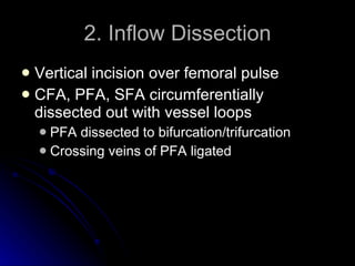
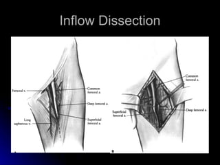
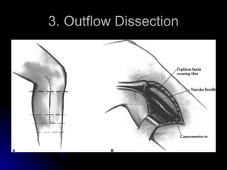
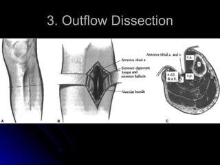
Fem-distal Bypass Josh Wilensky
1. Preop Arteriogram Vein mapping
2. Inflow Dissection Vertical incision over femoral pulse CFA, PFA, SFA circumferentially dissected out with vessel loops PFA dissected to bifurcation/trifurcation Crossing veins of PFA ligated
Inflow Dissection
3. Outflow Dissection
4. Vein Harvest Ipsilateral GSV with skip incisions Distended with heparinized saline Side branches ligated with 4-0 silk Reversed vs Non-reversed Non-reversed – better size match
5. Tunneling - Anatomic location  umbilical tape is left in the tunnel distal end of the vein is tied with a 2–0 silk tie to the tunneling device and passed back through the previously made tunnel
6. Heparin 10,000 units of heparin
7. Anastomosis Proximal: 6-0 running prolene end-to-side Distal: 7-0 interrupted prolene end-to-side
8. Arteriogram Check feet for pulses and signals Completion arteriogram Cannulate CFA with 20-gauge butterfly needle
9. Wound Closure Wounds closed in layers with 3-0 vicryl Skin with staples
10. Post op Staples out in 2 weeks ABIs in 6 weeks Q 6mo ABIs

Fem distal bypass

Editor's Notes

  • #4 Double looped, heiphitz on PFA
  • #6 lower leg medial incision made approximately 1 cm below the medial border of the tibia . the medial head of the gastrocnemius as well as the soleus muscle are identified and retracted posteromedially
  • #7 The anterior tibial artery is exposed through a lateral skin incision made halfway between the lateral edge of the tibia and the fibula. The subcutaneous tissue and fascia are incised and the intramuscular septum between the tibialis anterior muscle medially and the extensor digitorum longus muscle laterally is located and incised
  • #9 A: Medial tunnel from common femoral artery to posterior tibial artery B: Lateral tunnel from common femoral artery to anterior tibial artery.
  • #11 Incorporation of distal branch