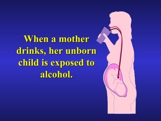
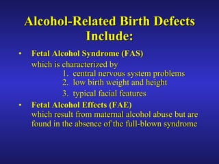
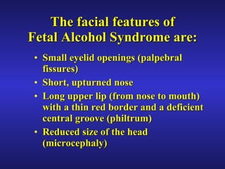
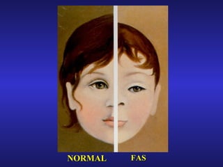
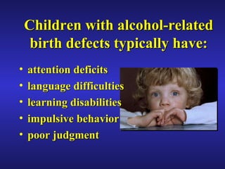
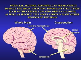
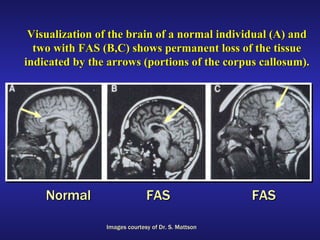
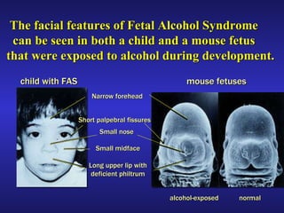
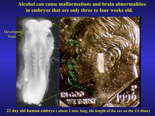
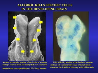
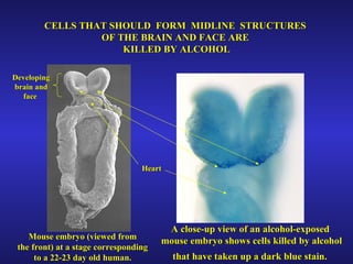
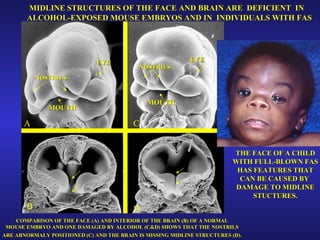
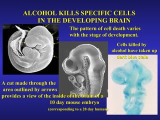
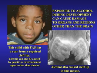
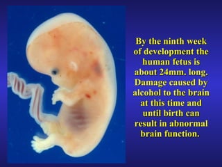
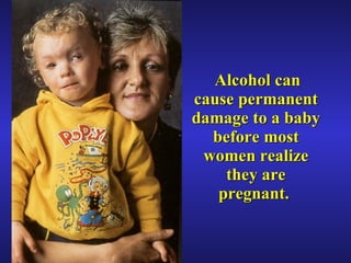
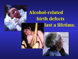
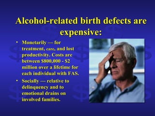
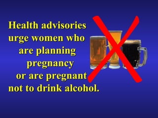
Better Safe Than Sorry: The Biological Basis of  Fetal Alcohol Syndrome  and other Alcohol-Related Birth Defects
When a mother drinks, her unborn child is exposed to alcohol.
Alcohol-Related Birth Defects Include: Fetal Alcohol Syndrome   (FAS)   which is characterized by 1.  central nervous system problems  2.  low birth weight and height 3.  typical facial features Fetal Alcohol Effects   (FAE) which result from maternal alcohol abuse but are found in the absence of the full-blown syndrome
The facial features of Fetal Alcohol Syndrome are: Small eyelid openings (palpebral fissures) Short, upturned nose Long upper lip (from nose to mouth) with a thin red border and a deficient central groove (philtrum)  Reduced size of the head (microcephaly)
FAS NORMAL
Full-blown fetal alcohol syndrome (FAS)    represents only the    “tip of the iceberg”    relative to all alcohol-related birth   defects (ARBDs). FAS ARBDs
MATERNAL ALCOHOL ABUSE IS THE LEADING KNOWN CAUSE OF MENTAL RETARDATION IN THE WESTERN WORLD
Children with alcohol-related birth defects typically have: attention deficits language difficulties learning disabilities impulsive behavior poor judgment
Cross-section Whole brain cerebral hemispheres cerebellum corpus callosum PRENATAL ALCOHOL EXPOSURE CAN PERMANENTLY  DAMAGE THE BRAIN, AFFECTING IMPORTANT STRUCTURES  SUCH AS THE CEREBELLUM AND CORPUS CALLOSUM, AS WELL AS SPECIFIC CELL POPULATIONS IN MANY OTHER  REGIONS OF THE BRAIN
Visualization of the brain of a normal individual (A) and two with FAS (B,C) shows permanent loss of the tissue indicated by the arrows (portions of the corpus callosum). Images courtesy of Dr. S. Mattson Normal FAS FAS
Similarities in mouse and human embryogenesis provide opportunities  to study the effects  of alcohol on development. 5 mm. Mouse (10 days old) Human  (approx. 28 days old) EYE HEART UPPER LIMB ( ARM ) 3 mm.
Narrow forehead Short palpebral fissures Small nose Small midface Long upper lip with deficient philtrum child with FAS normal alcohol-exposed mouse fetuses The facial features of Fetal Alcohol Syndrome  can be seen in both a child and a mouse fetus  that were exposed to alcohol during development.
The amount and timing of maternal alcohol use determine the type and extent of resulting birth defects.
Alcohol can cause malformations and brain abnormalities  in embryos that are only three to four weeks old. 22 day old human embryo  ( about 2 mm. long, the length of the ear on the US dime)   Developing brain
ALCOHOL KILLS SPECIFIC CELLS  IN THE DEVELOPING BRAIN Arrows surround a portion of the brain of a mouse  embryo (viewed from the back) that is at a develop- mental stage corresponding to a 22-23 day human.   Cells killed by alcohol in the brain of a mouse  embryo (at a comparable stage of development  to that on the left) have taken up a dark blue stain.
CELLS THAT SHOULD  FORM  MIDLINE  STRUCTURES  OF THE BRAIN AND FACE ARE  KILLED BY ALCOHOL Developing brain and face  Heart Mouse embryo (viewed from the front) at a stage corresponding  to a 22-23 day old human. A close-up view of an alcohol-exposed  mouse embryo shows cells killed by alcohol that have taken up a dark blue stain.
EYE EYE A C B D MOUTH MOUTH NOSTRILS NOSTRILS MIDLINE STRUCTURES OF THE FACE AND BRAIN ARE  DEFICIENT  IN ALCOHOL-EXPOSED MOUSE EMBRYOS AND IN  INDIVIDUALS WITH FAS THE FACE OF A CHILD WITH FULL-BLOWN FAS HAS FEATURES THAT CAN BE CAUSED BY  DAMAGE TO MIDLINE STUCTURES. COMPARISON OF THE FACE (A) AND INTERIOR OF THE BRAIN (B) OF A NORMAL  MOUSE EMBRYO AND ONE DAMAGED BY ALCOHOL (C&D) SHOWS THAT THE NOSTRILS  ARE ABNORMALY POSITIONED (C) AND THE BRAIN IS MISSING MIDLINE STRUCTURES (D).
ALCOHOL KILLS SPECIFIC CELLS    IN THE DEVELOPING BRAIN A cut made through the area outlined by arrows provides a view of the inside of the brain of a  10 day mouse embryo  (corresponding to a 28 day human) Cells killed by  alcohol   have taken up  dark blue stain   The pattern of cell death varies  with the stage of development.
EXPOSURE TO ALCOHOL  DURING DEVELOPMENT CAN CAUSE DAMAGE  TO ORGANS AND REGIONS  OTHER THAN THE BRAIN This child with FAS has  a scar from a repaired cleft lip. Cleft lip can also be caused by genetic or environmental agents other than alcohol. Alcohol also caused cleft lip  in this mouse.
By the ninth week of development the human fetus is about 24mm. long. Damage caused by alcohol to the brain at this time and until birth can result in abnormal brain function.
Excessive alcohol exposure can cause damage during all stages of prenatal development. Pre-implantation:   first 2 weeks Embryonic :   3-8 weeks after conception Fetal : from week 9 until birth
  Alcohol can cause permanent damage to a baby before most women realize they are pregnant.
Alcohol-related birth defects last a lifetime.
Alcohol-related birth defects are expensive: Monetarily — for treatment,  care , and lost productivity. Costs are between $800,000 - $2 million over a lifetime for each individual with FAS. Socially — relative to delinquency and to emotional drains on involved families. $ $ $ $ $ $ $ $ $ $
??? How much is too much ???
How much alcohol is in a drink? 12 oz beer  =  5 oz wine  =  shot of liquor Each contains the same amount of alcohol in a mixed drink
Some drinks contain more than a “serving” of alcohol WARNING
NO ONE KNOWS WHAT A  “ SAFE” AMOUNT OF ALCOHOL CONSUMPTION  DURING PREGNANCY MAY BE.
Health advisories urge women who    are planning    pregnancy    or are pregnant  not to drink alcohol.
Despite warnings, frequent drinking among pregnant women    appears to be increasing.   Frequent drinking is defined as 7  or more drinks per week or 5 or more  drinks on at least one occasion.
ALCOHOL-RELATED  BIRTH DEFECTS  ARE PREVENTABLE prevention prevention prevention prevention prevention preve prevention prevention prevention

Fas

  • 1.
    Better Safe ThanSorry: The Biological Basis of Fetal Alcohol Syndrome and other Alcohol-Related Birth Defects
  • 2.
    When a motherdrinks, her unborn child is exposed to alcohol.
  • 3.
    Alcohol-Related Birth DefectsInclude: Fetal Alcohol Syndrome (FAS) which is characterized by 1. central nervous system problems 2. low birth weight and height 3. typical facial features Fetal Alcohol Effects (FAE) which result from maternal alcohol abuse but are found in the absence of the full-blown syndrome
  • 4.
    The facial featuresof Fetal Alcohol Syndrome are: Small eyelid openings (palpebral fissures) Short, upturned nose Long upper lip (from nose to mouth) with a thin red border and a deficient central groove (philtrum) Reduced size of the head (microcephaly)
  • 5.
  • 6.
    Full-blown fetal alcoholsyndrome (FAS) represents only the “tip of the iceberg” relative to all alcohol-related birth defects (ARBDs). FAS ARBDs
  • 7.
    MATERNAL ALCOHOL ABUSEIS THE LEADING KNOWN CAUSE OF MENTAL RETARDATION IN THE WESTERN WORLD
  • 8.
    Children with alcohol-relatedbirth defects typically have: attention deficits language difficulties learning disabilities impulsive behavior poor judgment
  • 9.
    Cross-section Whole braincerebral hemispheres cerebellum corpus callosum PRENATAL ALCOHOL EXPOSURE CAN PERMANENTLY DAMAGE THE BRAIN, AFFECTING IMPORTANT STRUCTURES SUCH AS THE CEREBELLUM AND CORPUS CALLOSUM, AS WELL AS SPECIFIC CELL POPULATIONS IN MANY OTHER REGIONS OF THE BRAIN
  • 10.
    Visualization of thebrain of a normal individual (A) and two with FAS (B,C) shows permanent loss of the tissue indicated by the arrows (portions of the corpus callosum). Images courtesy of Dr. S. Mattson Normal FAS FAS
  • 11.
    Similarities in mouseand human embryogenesis provide opportunities to study the effects of alcohol on development. 5 mm. Mouse (10 days old) Human (approx. 28 days old) EYE HEART UPPER LIMB ( ARM ) 3 mm.
  • 12.
    Narrow forehead Shortpalpebral fissures Small nose Small midface Long upper lip with deficient philtrum child with FAS normal alcohol-exposed mouse fetuses The facial features of Fetal Alcohol Syndrome can be seen in both a child and a mouse fetus that were exposed to alcohol during development.
  • 13.
    The amount andtiming of maternal alcohol use determine the type and extent of resulting birth defects.
  • 14.
    Alcohol can causemalformations and brain abnormalities in embryos that are only three to four weeks old. 22 day old human embryo ( about 2 mm. long, the length of the ear on the US dime) Developing brain
  • 15.
    ALCOHOL KILLS SPECIFICCELLS IN THE DEVELOPING BRAIN Arrows surround a portion of the brain of a mouse embryo (viewed from the back) that is at a develop- mental stage corresponding to a 22-23 day human. Cells killed by alcohol in the brain of a mouse embryo (at a comparable stage of development to that on the left) have taken up a dark blue stain.
  • 16.
    CELLS THAT SHOULD FORM MIDLINE STRUCTURES OF THE BRAIN AND FACE ARE KILLED BY ALCOHOL Developing brain and face Heart Mouse embryo (viewed from the front) at a stage corresponding to a 22-23 day old human. A close-up view of an alcohol-exposed mouse embryo shows cells killed by alcohol that have taken up a dark blue stain.
  • 17.
    EYE EYE AC B D MOUTH MOUTH NOSTRILS NOSTRILS MIDLINE STRUCTURES OF THE FACE AND BRAIN ARE DEFICIENT IN ALCOHOL-EXPOSED MOUSE EMBRYOS AND IN INDIVIDUALS WITH FAS THE FACE OF A CHILD WITH FULL-BLOWN FAS HAS FEATURES THAT CAN BE CAUSED BY DAMAGE TO MIDLINE STUCTURES. COMPARISON OF THE FACE (A) AND INTERIOR OF THE BRAIN (B) OF A NORMAL MOUSE EMBRYO AND ONE DAMAGED BY ALCOHOL (C&D) SHOWS THAT THE NOSTRILS ARE ABNORMALY POSITIONED (C) AND THE BRAIN IS MISSING MIDLINE STRUCTURES (D).
  • 18.
    ALCOHOL KILLS SPECIFICCELLS IN THE DEVELOPING BRAIN A cut made through the area outlined by arrows provides a view of the inside of the brain of a 10 day mouse embryo (corresponding to a 28 day human) Cells killed by alcohol have taken up dark blue stain The pattern of cell death varies with the stage of development.
  • 19.
    EXPOSURE TO ALCOHOL DURING DEVELOPMENT CAN CAUSE DAMAGE TO ORGANS AND REGIONS OTHER THAN THE BRAIN This child with FAS has a scar from a repaired cleft lip. Cleft lip can also be caused by genetic or environmental agents other than alcohol. Alcohol also caused cleft lip in this mouse.
  • 20.
    By the ninthweek of development the human fetus is about 24mm. long. Damage caused by alcohol to the brain at this time and until birth can result in abnormal brain function.
  • 21.
    Excessive alcohol exposurecan cause damage during all stages of prenatal development. Pre-implantation: first 2 weeks Embryonic : 3-8 weeks after conception Fetal : from week 9 until birth
  • 22.
    Alcoholcan cause permanent damage to a baby before most women realize they are pregnant.
  • 23.
  • 24.
    Alcohol-related birth defectsare expensive: Monetarily — for treatment, care , and lost productivity. Costs are between $800,000 - $2 million over a lifetime for each individual with FAS. Socially — relative to delinquency and to emotional drains on involved families. $ $ $ $ $ $ $ $ $ $
  • 25.
    ??? How muchis too much ???
  • 26.
    How much alcoholis in a drink? 12 oz beer = 5 oz wine = shot of liquor Each contains the same amount of alcohol in a mixed drink
  • 27.
    Some drinks containmore than a “serving” of alcohol WARNING
  • 28.
    NO ONE KNOWSWHAT A “ SAFE” AMOUNT OF ALCOHOL CONSUMPTION DURING PREGNANCY MAY BE.
  • 29.
    Health advisories urgewomen who are planning pregnancy or are pregnant not to drink alcohol.
  • 30.
    Despite warnings, frequentdrinking among pregnant women appears to be increasing. Frequent drinking is defined as 7 or more drinks per week or 5 or more drinks on at least one occasion.
  • 31.
    ALCOHOL-RELATED BIRTHDEFECTS ARE PREVENTABLE prevention prevention prevention prevention prevention preve prevention prevention prevention