FACIAL NERVE ppt.pptx M56BOOKSTORESHAMA SARANSH D SARAN RAJ SHALINI SANDHIYA M56BOOKSTORE JOIN US
•Download as PPTX, PDF•
0 likes•25 views

FACIAL NERVE & BELLS PALSY PPT ---PRESENTED BY – SHAMA SARANSH D SARAN RAJ SHALINI SANDHIYA M56BOOKSTORE JOIN US

Recommended
More Related Content
Similar to FACIAL NERVE ppt.pptx M56BOOKSTORESHAMA SARANSH D SARAN RAJ SHALINI SANDHIYA M56BOOKSTORE JOIN US
Similar to FACIAL NERVE ppt.pptx M56BOOKSTORESHAMA SARANSH D SARAN RAJ SHALINI SANDHIYA M56BOOKSTORE JOIN US (20)
More from M56BOOKSTORE PRODUCT/SERVICE
More from M56BOOKSTORE PRODUCT/SERVICE (16)
Recently uploaded
Recently uploaded (20)
FACIAL NERVE ppt.pptx M56BOOKSTORESHAMA SARANSH D SARAN RAJ SHALINI SANDHIYA M56BOOKSTORE JOIN US
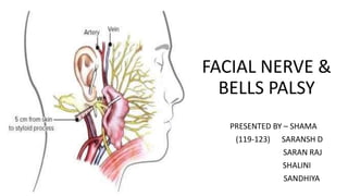
- 1. FACIAL NERVE & BELLS PALSY PRESENTED BY – SHAMA (119-123) SARANSH D SARAN RAJ SHALINI SANDHIYA
- 2. FACIAL NERVE CONTENTS - • Introduction • Formation • Fibers • Functions • Nuclei • Origin & course • Branches & distribution
- 3. Competency based presentation - A 40-year-old male comes to the OPD with the history of inability to close the right eye and dribbling of saliva on the right side of mouth since early morning. He gives history of exposure to cold wind while travelling on the bus. On examination, there is loss of wrinkling on the right side of forehead and angle of the mouth is deviated to the left side.
- 4. INTRODUCTION- Facial nerve is a vital nerve for facial expression. Facial nerve – seventh cranial nerve Facial nerve – nerve of 2nd branchial arch/pharyngeal arch –
- 5. Formation -
- 6. Fibers - • Sensory fibers ( afferent ) • Special sensory fibers • Visceral/autonomic motor ( efferent ) • Somatic motor fibers What are fibers ? A nerve fiber is a threadlike extension of a nerve cell (the neuron) and consists of an axon and myelin sheath.
- 7. • Sensory fibers – Transmitting signals to brain -----from external acoustic meatus as well as skin over mastoid & lateral pinna. • Special sensory fibers – Receving & transmitting taste information from anterior 2/3rd of tongue. • Visceral/autonomic motor fibers – innervating ----- • Lacrimal gland then innervating mucous memb.of • Submandibular gland nasal cavity & hard – soft palate. • Sublingual gland Allowing production of tears & saliva.
- 8. Somatic motor – innervating muscles of facial expression-----Like – • Muscle in scalp • Stapedius muscle of ear • Posterior belly of digastric muscle • Stylohyoid muscle
- 9. Nuclei - • Nerve fibers connected to 4 x nuclei – situated in lower pons • Motor nucleus • Superior salivatory nucleus • Lacrimatory nuclei • Nucleus of tractus solitarius NTS cranial nerves ? Imp – 2mark Q
- 10. COURSE – Brainstem -------middle ear --------parotid gland • Attached to brainstem by ---- 02 roots of motor & sensory • 02 roots of facial nerve ------ attached to lateral part of pons. • 02 roots run laterally & forward with 08th cranial N. ( VBC ) to reach Internal acoustic meatus
- 11. • In meatus – 2 x roots – sensory & motor fuse • 1st part – laterally above vestibule • 2nd part – medial wall above promontory • 3rd part - vertically downward behind promontory. • Facial nerve leave skull by passing through stylomastoid foramen.
- 12. Extra cranial course - • Facial nerve crosses -------lateral sidebase of styloid process. • Enters – posteromedial surface of parotid gland. • Runs forward through the gland -----crossing retromandubular vein &ECA. • Behind the neck of mandible -----divided into 5 x terminal branches emerge • Anterior border of parotid gland.
- 13. Divided into 5 x terminal branches - 1. Temporal branch 2. Zygomatic branch 3. Buccal branch 4. Mandibular branch 5. Cervical branch
- 14. Branches & distributions - Terminal branches – with in parotid gland – Temporal branch Zygomatic branch Buccal branch Mandibular branch Cervical branch With in facial canal – greater petrosal nerve stapedius nerve Chordatympani Exit from stylomastoid foramen – Posterior auricular Post. Belly of digastric Stylohyoid
- 16. • Bell’s palsy – • Sudden paralysis of nerve at stylomastoid foramen----result in asymmetry of corner of mouth • Inability to close to eye • Wrinkling of skin of forehead on the same side.
- 17. How to remember – bell’s palsy - Trick – B – blink reflex abnormal E – Earache L – lacrimation L - loss of tase S – sudden onset P – paralysis of facial nerve
- 18. • Lesions – bell’s palsy –
- 19. • A 40-year-old male comes to the OPD with the history of inability to close the right eye anddribbling of saliva on the right side of mouth since early morning. He gives history ofexposure to cold wind while travelling on the bus. On examination, there is loss of wrinklingon the right side of forehead and angle of the mouth is deviated to the left side. • 1. Identify the condition. • 2. Which cranial nerve is affected? • 3. Is this an upper motor neuron or lower motor neuron type of lesion? • 4. Which part of the nerve is involved in the lesion? • 5.Mention the nucleus, course, branches and distribution of involved nerve. • Answers – 1. The condition is Bell's palsy. 2. The cranial nerve affected is the facial nerve (CN VII) . 3. Bell's palsy is typically a lower motor neuron type of lesion. 4. The lesion is likely affecting the facial nerve as it exits the skull. 5. The facial nerve originates from the facial nucleus in the pons courses through the internal auditory canal branches out to innervate the muscles of facial expression – including the forehead, eyelids, nose, and mouth.