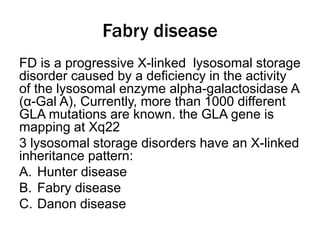
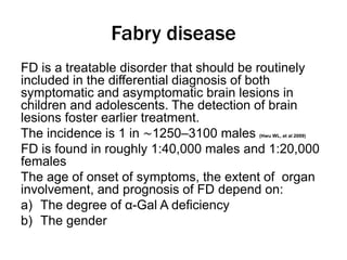
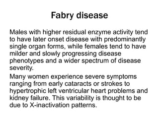
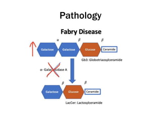
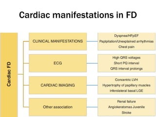
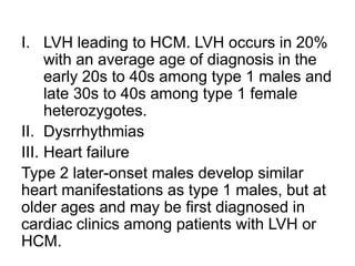
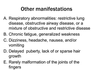
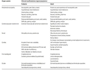
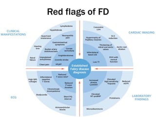
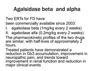
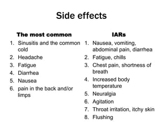
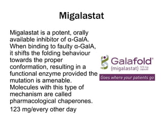
Fabry disease
Alpha-galactosidase A
deficiency
By
DR MOHAMMUD IBRAHEEM
Synonyms
1) Alpha-galactosidase A deficiency
2) Anderson-Fabry disease
3) Angiokeratoma corporis diffusum
4) Angiokeratoma diffuse
5) GLA deficiency
Fabry disease
FD is a progressive X-linked lysosomal storage
disorder caused by a deficiency in the activity
of the lysosomal enzyme alpha-galactosidase A
(α-Gal A), Currently, more than 1000 different
GLA mutations are known. the GLA gene is
mapping at Xq22
3 lysosomal storage disorders have an X-linked
inheritance pattern:
A. Hunter disease
B. Fabry disease
C. Danon disease
Fabry disease
FD is a treatable disorder that should be routinely
included in the differential diagnosis of both
symptomatic and asymptomatic brain lesions in
children and adolescents. The detection of brain
lesions foster earlier treatment.
The incidence is 1 in ∼1250–3100 males (Hwu WL, et al 2009)
FD is found in roughly 1:40,000 males and 1:20,000
females
The age of onset of symptoms, the extent of organ
involvement, and prognosis of FD depend on:
a) The degree of α-Gal A deficiency
b) The gender
Fabry disease
Males with higher residual enzyme activity tend
to have later onset disease with predominantly
single organ forms, while females tend to have
milder and slowly progressing disease
phenotypes and a wider spectrum of disease
severity.
Many women experience severe symptoms
ranging from early cataracts or strokes to
hypertrophic left ventricular heart problems and
kidney failure. This variability is thought to be
due to X-inactivation patterns.
Pathology
Pathology
Deposition of glycosphingolipids, such as
globotriaosylceramide (Gb3) and its deacylated
derivative globotriaosylsphingosine, within
lysosomes in virtually all cell types including
a) Capillary endothelial cells
b) Renal cells (podocytes, tubular cells,
glomerular endothelial, mesangial, and
interstitial cells)
c) Cardiac cells (cardiomyocytes and
fbroblasts)
d) Nerve cells
Pathology
Foam cells with vacuolated
cytoplasm are found in
smooth and striated
muscle, In marrow and
renal glomeruli.
In the CNS, storage is
confined to walls of the
blood vessels and, to a
lesser extent, to the ANS.
Types
I. Type 1 have little or no functional α-Gal A
enzymatic activity (<3% of normal mean
activity), and marked accumulation of Gb3
and related glycolipids in capillaries and
small blood vessels which cause the major
symptoms in childhood or adolescence.
II. Type 2 “later-onset” phenotype (previously
called cardiac or renal variants) have
residual α-Gal A activity, lack Gb3
accumulation in capillaries and small blood
vessels, and do not show the early
manifestations of type 1
Dermatological manifestations
1. Skin abnormalities, in the form of
punctate tiny, painless angiectatic
papules (angiokeratoma corporis
diffusum) commonly found on the
genitalia or umbilicus or the hips, rarely
involve the face.
Ophthalmological manifestations
I. Corneal dystrophy; Abnormal deposits of
glycolipids in the cornea resulting in a
whorl-like opacity seen by slit-lamp.
II. Blood vessels in the eyes may appear
twisted (cork screw-like; contorted)
and/or slightly enlarged (dilated).
III. Cilioretinal artery occlusion and anterior
ischemic optic neuropathy
3. PNS-Related Manifestations
Small fiber neuropathy is a frequent and affect
80%.
There is clearly identified preferential damage to A-
delta fibers and relative sparing of C fibers.
Topographically, this is a classical length-
dependent and symmetric axonopathy.
Patients have both somatic and autonomic
manifestations, including acroparesthesias, cold
intolerance, burning dysesthesias, and sensory
loss that starts in the distal limbs (palms and
soles).
Neuropathic pain in FD:
i. Chronic
Fabry’s crises
It presents as episodes lasting from hours to
several days. patients suffer excruciating pain
starts in the hands and feet and then spreads
centripetally to the proximal limb regions.
Autonomic manifestations occur during these
crises.
Exercise, fever, and stress are triggers for the
episodes. Each crisis have an acute onset and
end, but recurrence is seen in many individuals
over time.
Later in the disease course, sustained pain
improvement occur, this is attributed to extensive
causes of SFN pain
1. autoimmune thyroiditis
2. autoimmune trochleitis
3. Hashimoto’s
encephalopathy
4. Henoch–Schönlein
purpura
5. brachial plexitis
6. type 1 diabetes
7. RhF
8. SLE
7. post-viral arthritis
8. immune
thrombocytopenic
purpura
9. Crohn’s disease
10.Sjögren’s spectrum
disorders
11.RhA
12.Raynaud’s disease
13.growing pains
3. Autonomic neuropathy
a) Cold intolerance, Hypohidrosis and
exercise intolerance are conspicuous trait
in FD.
b) Xerostomia and xerophthalmia
c) Gastrointestinal dysmotility
4. CNS-Related Manifestations
The prevalence of CNS manifestations is 12–31%
among men and 5–18% among women but TIA is
more prevalent in women (16% vs. 11%)
I. Tinnitus, hearing impairment, vertigo
II. Psychiatric disorder; depression is frequent,
with rates that range from 15 to 62%.
Depression is directly related to chronic pain
III. cognitive impairment mainly affects executive
functioning, information processing speed, and
attention
IV. Cerebrovascular events as stroke and TIA.
The mean age of CVS events in men is 28.8–
34 years, while in women is 40.3–50 years.
Cerebrovascular
Cochleovestibular Manifestations
i. cochlear impairment; Tinnitus and
hearing loss. Hearing loss is commonly
bilateral, asymmetric, and sensorineural
ii. vestibular symptoms as vertigo,
dizziness, and instability.
This manifestation to be found in
approximately 70% of patients
Renal manifestations
Progressive proteinuric kidney disease.
i. In type 1, the decline begins with podocyte
involvement causing proteinuria and
decreasing GFR, all leading to RF and the
need for dialysis or transplantation by 35 to
45 years of age.
ii. In type 2 males, kidney involvement occurs
in the 4th decade or later, but some patients
do not develop RF. Kidney involvement in
type 1 female heterozygotes is more
variable. 10-15% of type 1 females develop
kidney failure.
Cardiac manifestations in FD
I. LVH leading to HCM. LVH occurs in 20%
with an average age of diagnosis in the
early 20s to 40s among type 1 males and
late 30s to 40s among type 1 female
heterozygotes.
II. Dysrrhythmias
III. Heart failure
Type 2 later-onset males develop similar
heart manifestations as type 1 males, but at
older ages and may be first diagnosed in
cardiac clinics among patients with LVH or
HCM.
Other manifestations
A. Respiratory abnormalities: restrictive lung
disease, obstructive airway disease, or a
mixture of obstructive and restrictive disease
B. Chronic fatigue, generalized weakness
C. Dizziness, headache, nausea, and/or
vomiting
D. Delayed puberty, lack of or sparse hair
growth
E. Rarely malformation of the joints of the
fingers
Red flags of FD
Diagnosis
Diagnosis is confirmed early by
determination of
alpha-galactosidase in plasma, leukocytes
or fibroblasts in males.
In females, genetic analysis is necessary.
Kidney biopsy may also be suggestive of
Fabry disease if excessive lipid buildup is
noted.
DD
A. Type II Schindler disease (Kanzaki disease)
is the adult-onset form with symptoms
presenting in the 2nd or 3rd decade of life.
The disorder is characterized by
angiokeratoma.
B. Fucosidosis; Symptoms include a skin lesion
similar to FD (angiokeratoma), progressive
deterioration of the brain and spinal cord ,
intellectual disability, and growth retardation.
C. Erythromelalgia is AD rare condition that
primarily affects the feet and, less
commonly, the hands. It is characterized
by intense burning pain of affected
extremities, severe redness, and
increased skin temperature that may be
episodic or almost continuous in nature.
D. Familial Mediterranean Fever (FMF)
E. Juvenile systemic lupus erythematosus
F. Celiac disease
Investigations
A. peripheral neuropathy
I. Specifc tests for SFN as quantitative sensory testing
(QST), a psychophysical test that measures
detection thresholds for warmth and cold in the hands
and feet. These thresholds are extremely elevated in
patients with FD
II. Skin biopsy with intraepidermal nerve fiber density
(IEND) quantifcation is the gold standard test to
diagnose small fiber neuropathy. There is reduced
IEND in FD
III. Quantitative sudomotor axonal refex test (QSART)
assesses the integrity of postganglionic cholinergic
fibers responsible for sweat production. Patients with
FD present blunted QSART responses
a) CNS
i. lesions affect white and gray matter and are
more frequent in the posterior circulation
ii. vessel ectasia frequent in the posterior
circulation
iii. chronic findings might be detected in
magnetic resonance imaging
iv. MRI, characterized by progressive white-
matter lesions, which occur early in the
course of FD
TREATMENT
Agalsidase beta and alpha
Two ERTs for FD have
been commercially available since 2003:
I. agalsidase beta (1mg/kg every 2 weeks)
II. agalsidase alfa (0.2mg/kg every 2 weeks)
The pharmacokinetic profiles of the two drugs
are similar, with half-lives of approximately 2
hours.
Treated patients have demonstrated a
reduction in Gb3 accumulation, improvement in
neuropathic pain, and trends toward
improvement in renal function and reduction in
major clinical events
Pegunigalsidase alfa
Pegunigalsidase alfa is a recombinant human
α-galactosidase-A indicated for long-term ERT
in patients with FD
It is composed of two subunits of AGAL
covalently bound by a chain of polyethylene
glycol, which increases its stability and reduces
its clearance.
Pegunigalsidase alfa is administered every 2
weeks (±3 days) and patients is observed for
24 hours after each of the first 7 infusions
Pegunigalsidase alfa dosing
0.2- and 1.0-mg/kg is diluted with 0.9% NaCl
to a volume of 150 mL, and in the 2.0-mg/kg
group to a final volume of 350 mL.
The mean durations of the initial infusions in
the 0.2-mg/kg, 1.0-mg/kg, and 2.0-mg/kg
groups are 4.0, 5.5, and 6.4 hours, and are
sequentially reduced to mean durations of
1.5, 3.3, and 3.1 hours at 12 months,
respectively.
Pegunigalsidase alfa
Pretreating with antihistamines, antipyretics,
and/or corticosteroids and appropriate medical
support measures, including cardiopulmonary
resuscitation equipment is recomended
Compared with agalsidase alfa and beta, both
of which exhibit a T1/2γ of ≤2 hours and
maintain measurable plasma levels for <24
hours , pegunigalsidase alfa has a T1/2γ of
about 80 hours with
Pegunigalsidase alfa
19% develope treatment-induced ADAs, the
ADA response is transient, with no observed
impact on pharmacodynamics, efficacy, or
safety.
14% experience hypersensitivity reactions.
Anaphylaxis occurre within 5 to 40 minutes of
the start of the initial infusion. Signs and
symptoms included headache, nausea,
vomiting, throat tightness, facial and oral
edema, truncal rash, tachycardia, hypotension,
rigors, urticaria, intense pruritus, moderate
upper airway obstructions, macroglossia, and
mild lip edema.
Side effects
The most common
1. Sinusitis and the common
cold
2. Headache
3. Fatigue
4. Diarrhea
5. Nausea
6. pain in the back and/or
limps
IARs
1. Nausea, vomiting,
abdominal pain, diarrhea
2. Fatigue, chills
3. Chest pain, shortness of
breath
4. Increased body
temperature
5. Neuralgia
6. Agitation
7. Throat irritation, itchy skin
8. Flushing
Migalastat
Migalastat is a potent, orally
available inhibitor of α-GalA.
When binding to faulty α-GalA,
it shifts the folding behaviour
towards the proper
conformation, resulting in a
functional enzyme provided the
mutation is amenable.
Molecules with this type of
mechanism are called
pharmacological chaperones.
123 mg/every other day
Organ-specific treatment
1. Gabapentin, pregabalin, carbamazepine,
and amitriptyline
2. Antihypertensive
3. Antiplatelet
4. Cardiac management
THANK YOU

Fabry disease.pptx

  • 1.
  • 3.
    Synonyms 1) Alpha-galactosidase Adeficiency 2) Anderson-Fabry disease 3) Angiokeratoma corporis diffusum 4) Angiokeratoma diffuse 5) GLA deficiency
  • 4.
    Fabry disease FD isa progressive X-linked lysosomal storage disorder caused by a deficiency in the activity of the lysosomal enzyme alpha-galactosidase A (α-Gal A), Currently, more than 1000 different GLA mutations are known. the GLA gene is mapping at Xq22 3 lysosomal storage disorders have an X-linked inheritance pattern: A. Hunter disease B. Fabry disease C. Danon disease
  • 5.
    Fabry disease FD isa treatable disorder that should be routinely included in the differential diagnosis of both symptomatic and asymptomatic brain lesions in children and adolescents. The detection of brain lesions foster earlier treatment. The incidence is 1 in ∼1250–3100 males (Hwu WL, et al 2009) FD is found in roughly 1:40,000 males and 1:20,000 females The age of onset of symptoms, the extent of organ involvement, and prognosis of FD depend on: a) The degree of α-Gal A deficiency b) The gender
  • 6.
    Fabry disease Males withhigher residual enzyme activity tend to have later onset disease with predominantly single organ forms, while females tend to have milder and slowly progressing disease phenotypes and a wider spectrum of disease severity. Many women experience severe symptoms ranging from early cataracts or strokes to hypertrophic left ventricular heart problems and kidney failure. This variability is thought to be due to X-inactivation patterns.
  • 7.
  • 8.
    Pathology Deposition of glycosphingolipids,such as globotriaosylceramide (Gb3) and its deacylated derivative globotriaosylsphingosine, within lysosomes in virtually all cell types including a) Capillary endothelial cells b) Renal cells (podocytes, tubular cells, glomerular endothelial, mesangial, and interstitial cells) c) Cardiac cells (cardiomyocytes and fbroblasts) d) Nerve cells
  • 9.
    Pathology Foam cells withvacuolated cytoplasm are found in smooth and striated muscle, In marrow and renal glomeruli. In the CNS, storage is confined to walls of the blood vessels and, to a lesser extent, to the ANS.
  • 10.
    Types I. Type 1have little or no functional α-Gal A enzymatic activity (<3% of normal mean activity), and marked accumulation of Gb3 and related glycolipids in capillaries and small blood vessels which cause the major symptoms in childhood or adolescence. II. Type 2 “later-onset” phenotype (previously called cardiac or renal variants) have residual α-Gal A activity, lack Gb3 accumulation in capillaries and small blood vessels, and do not show the early manifestations of type 1
  • 11.
    Dermatological manifestations 1. Skinabnormalities, in the form of punctate tiny, painless angiectatic papules (angiokeratoma corporis diffusum) commonly found on the genitalia or umbilicus or the hips, rarely involve the face.
  • 13.
    Ophthalmological manifestations I. Cornealdystrophy; Abnormal deposits of glycolipids in the cornea resulting in a whorl-like opacity seen by slit-lamp. II. Blood vessels in the eyes may appear twisted (cork screw-like; contorted) and/or slightly enlarged (dilated). III. Cilioretinal artery occlusion and anterior ischemic optic neuropathy
  • 16.
    3. PNS-Related Manifestations Smallfiber neuropathy is a frequent and affect 80%. There is clearly identified preferential damage to A- delta fibers and relative sparing of C fibers. Topographically, this is a classical length- dependent and symmetric axonopathy. Patients have both somatic and autonomic manifestations, including acroparesthesias, cold intolerance, burning dysesthesias, and sensory loss that starts in the distal limbs (palms and soles). Neuropathic pain in FD: i. Chronic
  • 17.
    Fabry’s crises It presentsas episodes lasting from hours to several days. patients suffer excruciating pain starts in the hands and feet and then spreads centripetally to the proximal limb regions. Autonomic manifestations occur during these crises. Exercise, fever, and stress are triggers for the episodes. Each crisis have an acute onset and end, but recurrence is seen in many individuals over time. Later in the disease course, sustained pain improvement occur, this is attributed to extensive
  • 18.
    causes of SFNpain 1. autoimmune thyroiditis 2. autoimmune trochleitis 3. Hashimoto’s encephalopathy 4. Henoch–Schönlein purpura 5. brachial plexitis 6. type 1 diabetes 7. RhF 8. SLE 7. post-viral arthritis 8. immune thrombocytopenic purpura 9. Crohn’s disease 10.Sjögren’s spectrum disorders 11.RhA 12.Raynaud’s disease 13.growing pains
  • 19.
    3. Autonomic neuropathy a)Cold intolerance, Hypohidrosis and exercise intolerance are conspicuous trait in FD. b) Xerostomia and xerophthalmia c) Gastrointestinal dysmotility
  • 20.
    4. CNS-Related Manifestations Theprevalence of CNS manifestations is 12–31% among men and 5–18% among women but TIA is more prevalent in women (16% vs. 11%) I. Tinnitus, hearing impairment, vertigo II. Psychiatric disorder; depression is frequent, with rates that range from 15 to 62%. Depression is directly related to chronic pain III. cognitive impairment mainly affects executive functioning, information processing speed, and attention IV. Cerebrovascular events as stroke and TIA. The mean age of CVS events in men is 28.8– 34 years, while in women is 40.3–50 years. Cerebrovascular
  • 21.
    Cochleovestibular Manifestations i. cochlearimpairment; Tinnitus and hearing loss. Hearing loss is commonly bilateral, asymmetric, and sensorineural ii. vestibular symptoms as vertigo, dizziness, and instability. This manifestation to be found in approximately 70% of patients
  • 22.
    Renal manifestations Progressive proteinurickidney disease. i. In type 1, the decline begins with podocyte involvement causing proteinuria and decreasing GFR, all leading to RF and the need for dialysis or transplantation by 35 to 45 years of age. ii. In type 2 males, kidney involvement occurs in the 4th decade or later, but some patients do not develop RF. Kidney involvement in type 1 female heterozygotes is more variable. 10-15% of type 1 females develop kidney failure.
  • 23.
  • 24.
    I. LVH leadingto HCM. LVH occurs in 20% with an average age of diagnosis in the early 20s to 40s among type 1 males and late 30s to 40s among type 1 female heterozygotes. II. Dysrrhythmias III. Heart failure Type 2 later-onset males develop similar heart manifestations as type 1 males, but at older ages and may be first diagnosed in cardiac clinics among patients with LVH or HCM.
  • 25.
    Other manifestations A. Respiratoryabnormalities: restrictive lung disease, obstructive airway disease, or a mixture of obstructive and restrictive disease B. Chronic fatigue, generalized weakness C. Dizziness, headache, nausea, and/or vomiting D. Delayed puberty, lack of or sparse hair growth E. Rarely malformation of the joints of the fingers
  • 27.
  • 28.
    Diagnosis Diagnosis is confirmedearly by determination of alpha-galactosidase in plasma, leukocytes or fibroblasts in males. In females, genetic analysis is necessary. Kidney biopsy may also be suggestive of Fabry disease if excessive lipid buildup is noted.
  • 29.
    DD A. Type IISchindler disease (Kanzaki disease) is the adult-onset form with symptoms presenting in the 2nd or 3rd decade of life. The disorder is characterized by angiokeratoma. B. Fucosidosis; Symptoms include a skin lesion similar to FD (angiokeratoma), progressive deterioration of the brain and spinal cord , intellectual disability, and growth retardation.
  • 30.
    C. Erythromelalgia isAD rare condition that primarily affects the feet and, less commonly, the hands. It is characterized by intense burning pain of affected extremities, severe redness, and increased skin temperature that may be episodic or almost continuous in nature. D. Familial Mediterranean Fever (FMF) E. Juvenile systemic lupus erythematosus F. Celiac disease
  • 31.
    Investigations A. peripheral neuropathy I.Specifc tests for SFN as quantitative sensory testing (QST), a psychophysical test that measures detection thresholds for warmth and cold in the hands and feet. These thresholds are extremely elevated in patients with FD II. Skin biopsy with intraepidermal nerve fiber density (IEND) quantifcation is the gold standard test to diagnose small fiber neuropathy. There is reduced IEND in FD III. Quantitative sudomotor axonal refex test (QSART) assesses the integrity of postganglionic cholinergic fibers responsible for sweat production. Patients with FD present blunted QSART responses
  • 32.
    a) CNS i. lesionsaffect white and gray matter and are more frequent in the posterior circulation ii. vessel ectasia frequent in the posterior circulation iii. chronic findings might be detected in magnetic resonance imaging iv. MRI, characterized by progressive white- matter lesions, which occur early in the course of FD
  • 34.
  • 35.
    Agalsidase beta andalpha Two ERTs for FD have been commercially available since 2003: I. agalsidase beta (1mg/kg every 2 weeks) II. agalsidase alfa (0.2mg/kg every 2 weeks) The pharmacokinetic profiles of the two drugs are similar, with half-lives of approximately 2 hours. Treated patients have demonstrated a reduction in Gb3 accumulation, improvement in neuropathic pain, and trends toward improvement in renal function and reduction in major clinical events
  • 36.
    Pegunigalsidase alfa Pegunigalsidase alfais a recombinant human α-galactosidase-A indicated for long-term ERT in patients with FD It is composed of two subunits of AGAL covalently bound by a chain of polyethylene glycol, which increases its stability and reduces its clearance. Pegunigalsidase alfa is administered every 2 weeks (±3 days) and patients is observed for 24 hours after each of the first 7 infusions
  • 37.
    Pegunigalsidase alfa dosing 0.2-and 1.0-mg/kg is diluted with 0.9% NaCl to a volume of 150 mL, and in the 2.0-mg/kg group to a final volume of 350 mL. The mean durations of the initial infusions in the 0.2-mg/kg, 1.0-mg/kg, and 2.0-mg/kg groups are 4.0, 5.5, and 6.4 hours, and are sequentially reduced to mean durations of 1.5, 3.3, and 3.1 hours at 12 months, respectively.
  • 38.
    Pegunigalsidase alfa Pretreating withantihistamines, antipyretics, and/or corticosteroids and appropriate medical support measures, including cardiopulmonary resuscitation equipment is recomended Compared with agalsidase alfa and beta, both of which exhibit a T1/2γ of ≤2 hours and maintain measurable plasma levels for <24 hours , pegunigalsidase alfa has a T1/2γ of about 80 hours with
  • 39.
    Pegunigalsidase alfa 19% developetreatment-induced ADAs, the ADA response is transient, with no observed impact on pharmacodynamics, efficacy, or safety. 14% experience hypersensitivity reactions. Anaphylaxis occurre within 5 to 40 minutes of the start of the initial infusion. Signs and symptoms included headache, nausea, vomiting, throat tightness, facial and oral edema, truncal rash, tachycardia, hypotension, rigors, urticaria, intense pruritus, moderate upper airway obstructions, macroglossia, and mild lip edema.
  • 40.
    Side effects The mostcommon 1. Sinusitis and the common cold 2. Headache 3. Fatigue 4. Diarrhea 5. Nausea 6. pain in the back and/or limps IARs 1. Nausea, vomiting, abdominal pain, diarrhea 2. Fatigue, chills 3. Chest pain, shortness of breath 4. Increased body temperature 5. Neuralgia 6. Agitation 7. Throat irritation, itchy skin 8. Flushing
  • 41.
    Migalastat Migalastat is apotent, orally available inhibitor of α-GalA. When binding to faulty α-GalA, it shifts the folding behaviour towards the proper conformation, resulting in a functional enzyme provided the mutation is amenable. Molecules with this type of mechanism are called pharmacological chaperones. 123 mg/every other day
  • 42.
    Organ-specific treatment 1. Gabapentin,pregabalin, carbamazepine, and amitriptyline 2. Antihypertensive 3. Antiplatelet 4. Cardiac management
  • 43.