HARAMAYA UNIVERSITY COLLEGEOF SPORT SCIENCE
ACADEMY
DEPARTMENT OF SPORT SCIENCE
COURSE: EXERCISE PHYSIOLOGY
COURSE TITLE: INTRODUCTION TO THE STRUCTURE AND
FUNCTION OF MUSCLES WITH A PARTICULAR EMPHASIS
TO PLASTICITY AND ADAPTIVE RESPONSE TO LOAD AND
INTENSITY
AND MEAUREMENT ANALYSIS OF PHYSIOLOGICAL
FUNCTION OF SPORTMEN
Student's name:Tamene Deksisa
IDNO:PGMR0212/17
Submitted to : Dr.Desita Enyew
2.
Contents of title
-Introduction
- Types of muscle and muscle contraction
-structure and function of muscle in exercise
- adaptation to exercise
- factor that influence muscle plasticity
- measurment analysis of physiological function of
sportmen
3.
INTRODUCTION
Muscles are madeup of muscle fibers that are
responsible for movement and other functions in the
body:
Structure
Muscles are made up of thousands of elastic fibers that
are bundled together and wrapped in a thin membrane
called a perimysium. Each fiber is made up of myofibrils,
which contain proteins and molecules that provide
energy and oxygen for muscle contraction.
Function
Muscles generate force, create movement, and provide
shape and form to the body. They also help with
posture, joint stability, and heat production.
4.
Types Of Muscles
Thereare three types of muscles : skeletal, cardiac, and
smooth:
Skeletal muscles: These muscles are voluntary and work
with bones, tendons, and ligaments to support weight
and move the body.
Cardiac muscles: These muscles propel blood and help
maintain oxygenation in the body.
Smooth muscles: These muscles are involuntary and are
found in organs like the stomach, bladder, and blood
vessels. They use contractile force to propel contents
across the lumen of organ systems.
5.
When exercising, musclescan grow through
hypertrophy, which is the growth of existing muscle
fibers. This can happen when stress is placed on
sarcomeres during physical activity.
When a muscle is not exercise the miscle is
athrophy.Causes of AtrophyAtrophy is due to one or
more number of causes such as:Poor
nourishmentDecreased blood supply Lack of workload
or exerciseLoss of control by nerves or
hormonesIntrinsic disease of the tissue or organ.
(Www.ncbi.nlm.nih.gov) , Rechel E.Noto (may 1,2023)
Muscles, attached to bones or internal organs and
blood vessels, are responsible for movement. Nearly all
movement in the body is the result of muscle
6.
Types of MuscleContractions
1.Isotonic contraction:Any contraction that creates force
(tension) & moves a load Change in length of the muscle
but tension constant (tension > load)
2. Isometric contractionContraction that create force
(tension) with out moving a load.The muscle as a whole
does not change length The tension produced never
exceeds the resistance (load). (tension < load)
7.
Muscle Fatigue: definedas a reversible, exercise-induced
reduction in the ability of a muscle to generate
force. .Causes of Muscle Fatigue:
Failure of nerve impulses in the motor neuron to release
enoughMuscle Fatigue: defined as a reversible, exercise-
induced reduction in the ability of a muscle to generate
force. .Causes of Muscle Fatigue:
Failure of nerve impulses in the motor neuron to release
enough Acetylcholine (↓Neural stimulation) ↓ Release of
Ca ions from the sarcoplasmic reticulum Depletion of
Nutrients (creatine phosphate & glycogen)Accumulation of
Lactic acid in the blood stream. This interfere with
excitation-contraction coupling.
Acetylcholine (↓Neural stimulation) ↓ Release of Ca ions
from the sarcoplasmic reticulum Depletion of Nutrients
8.
Structure and functionof muscle in exercise
Muscle tissue plays a vital role in exercise, facilitating
movement, maintaining posture, and generating heat.
Understanding the structure and function of muscle can
help elucidate how muscles respond to various forms of
exercise. Here’s a comprehensive overview:
Structure of Muscle
Muscle tissue is primarily categorized into three types:
skeletal, cardiac, and smooth muscle. However, skeletal
muscle is the primary focus in the context of exercise.
9.
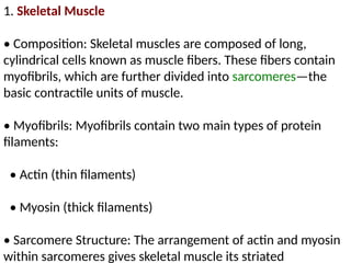
1. Skeletal Muscle
•Composition: Skeletal muscles are composed of long,
cylindrical cells known as muscle fibers. These fibers contain
myofibrils, which are further divided into sarcomeres—the
basic contractile units of muscle.
• Myofibrils: Myofibrils contain two main types of protein
filaments:
• Actin (thin filaments)
• Myosin (thick filaments)
• Sarcomere Structure: The arrangement of actin and myosin
within sarcomeres gives skeletal muscle its striated
10.
striated appearance. Thesliding filament theory explains
how muscle contraction occurs through the sliding of these
filaments past each other.
• Connective Tissue: Skeletal muscle is surrounded by
connective tissues:
• Epimysium: Outer layer surrounding the entire muscle.
• Perimysium: Surrounds bundles of muscle fibers
(fascicles).
• Endomysium: Surrounds individual muscle fibers.
11.
2. Cardiac Muscle
•Location: Found only in the heart.
• Structure: Striated like skeletal muscle but involuntary
and interconnected through intercalated discs, allowing
coordinated contractions.
12.
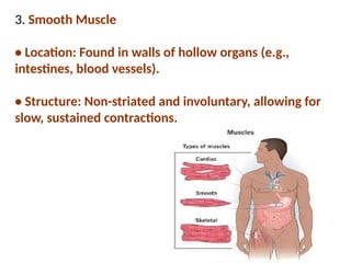
3. Smooth Muscle
•Location: Found in walls of hollow organs (e.g.,
intestines, blood vessels).
• Structure: Non-striated and involuntary, allowing for
slow, sustained contractions.
13.
Function of SkeletalMuscle in Exercise
Skeletal muscle serves several key functions during exercise:
▎1. Contraction and Movement
• Voluntary Control: Skeletal muscles are under voluntary
control, allowing for precise movements.
14.
Types of Contractions:
•Isometric: Muscle generates force without changing
length (e.g., holding a weight).
• Isotonic: Muscle changes length while generating force
(e.g., lifting a weight).
• Concentric: Muscle shortens during contraction (e.g.,
lifting).
• Eccentric: Muscle lengthens under tension (e.g.,
lowering
15.
2. Energy Production
•ATP Generation: Muscle contractions require
adenosine triphosphate (ATP). Muscles can generate ATP
through:
• Aerobic Metabolism: Uses oxygen to produce ATP
from carbohydrates and fats; predominant during
prolonged, moderate-intensity exercise.
• Anaerobic Metabolism: Produces ATP without
oxygen; dominant during short bursts of high-intensity
exercise (e.g., sprinting).
16.
3. Heat Production
•During exercise, muscle contractions generate heat as a
byproduct, helping to maintain body temperature.
▎4. Posture and Stability
• Skeletal muscles play a crucial role in maintaining posture
and stabilizing joints during movement
17.
5. Muscle FiberTypes
• Different muscle fiber types contribute to various
functional capacities:
• Type I (Slow-Twitch): More endurance-oriented, fatigue-
resistant, and primarily use aerobic metabolism.
• Type II (Fast-Twitch): Better for short bursts of power and
speed; can be further divided into:
• Type IIa: Intermediate fibers that can use both aerobic
and anaerobic metabolism.
• Type IIb: Primarily anaerobic and suited for explosive
movements.
18.
Adaptations to Exercise
Withregular training, skeletal muscle undergoes several
adaptations:
• Hypertrophy: Increase in muscle fiber size due to
resistance training.
• Increased Mitochondrial Density: Enhances aerobic
capacity.
• Improved Neural Efficiency: Better motor unit
recruitment and coordination.
• Enhanced Glycogen Storage: Increases energy availability
during exercise.
19.
Muscle plasticity isthe ability of muscle to change its
structure and function in response to environmental stimuli,
such as exercise( Anne Bruton,July 2002 ).The response of
muscle to exercise depends on the type of exercise, the
intensity, and the duration:
Low-intensity endurance exercise. type exercise leads to
qualitative changes of muscle tissue characterized mainly by
an increase in structures supporting oxygen delivery and
consumption (Hans Hoppeler 2011 Jul).
High-load strength-type exercise: Muscle fibers grow by
increasing contractile proteins.
Eccentric contractions: Associated with exercise-induced
injuries, these contractions elicit varied muscle adaptation
and regenerative responses.
20.
Factors that influencemuscle plasticity include:
Exercise type
Different types of exercise, such as resistance training and
endurance training, can cause different responses in muscles.
Physical activity and exercise can induce muscle plasticity.
During exercise, muscle fibers release myokines that can have
anabolic and catabolic effects on muscle fibers and satellite
cells.
21.
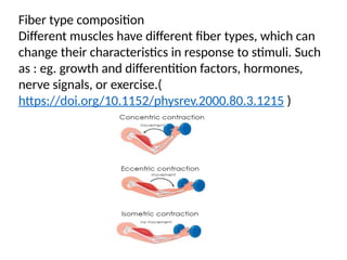
Fiber type composition
Differentmuscles have different fiber types, which can
change their characteristics in response to stimuli. Such
as : eg. growth and differentition factors, hormones,
nerve signals, or exercise.(
https://doi.org/10.1152/physrev.2000.80.3.1215 )
22.
Training
Different types oftraining can lead to different
adaptations in muscle:
Strength training :Can increase muscle fiber cross-
sectional area. Thus an increase in strength will enable
the muscle to resist an increased load. In normal
subjects, muscle strength can easily be increased by
training, provided that the training loads used exceed
those of normal daily activities: Can increase muscle
fiber cross-sectional area(Komi and Hakkinen, 1991 ).
Endurance training: Can increase mitochondrial
volume.
23.
Muscle adaptive RESPONSEto load and intensity
Muscle adaptive responses to load and intensity are
critical components of how the body adapts to resistance
training and physical activity. These adaptations occur at
various levels, including cellular, molecular, and systemic,
and are influenced by factors such as the type of
exercise, intensity, frequency, and duration. Here’s an
overview of the key adaptive responses:
1. Muscle Hypertrophy
• Definition: An increase in the size of muscle fibers,
24.
• Mechanism: Musclefibers undergo micro-tears during
intense exercise, leading to inflammation and repair
processes that stimulate muscle growth. Satellite cells
(muscle stem cells) play a crucial role in this repair and
growth process.
▎2. Muscle Strength
• Definition: The ability of a muscle or group of muscles to
exert force.
• Mechanism: Strength gains occur through neural
adaptations (improved motor unit recruitment and firing
rates) and muscle hypertrophy. Initially, most strength
gains are due to neural adaptations rather than increases
in muscle size.
25.
3. Metabolic Adaptations
•Increased Enzyme Activity: Resistance training can
enhance the activity of enzymes involved in energy
production (e.g., creatine kinase, phosphofructokinase).
• Glycogen Storage: Muscles adapt to store more glycogen,
improving endurance and performance during high-intensity
activities.
▎4. Changes in Muscle Fiber Composition
• Fiber Type Shifts: Regular training can lead to shifts in
muscle fiber types. For example, resistance training may
increase the proportion of fast-twitch fibers (Type II), which
are more suited for explosive movements, while endurance
26.
5. Tendon andLigament Strength
• Adaptation: Tendons and ligaments also adapt to
increased loads by becoming thicker and stronger,
which helps prevent injuries and improves overall
joint stability.
▎6. Neuromuscular Adaptations
• Improved Coordination: Training enhances the
coordination between different muscle groups and
improves the efficiency of movement patterns.
27.
Motor Unit Recruitment:Increased ability to recruit more
motor units during high-intensity efforts.
▎7. Hormonal Responses
• Anabolic Hormones: Resistance training stimulates the
release of hormones like testosterone and growth
hormone, which play roles in muscle repair and growth.
• Cortisol: Intense exercise can increase cortisol levels,
which is involved in energy metabolism but can also lead
to muscle breakdown if chronically elevated.
28.
8. Endurance andAerobic Capacity
• Although primarily associated with cardiovascular
training, resistance training can also improve muscular
endurance, allowing muscles to perform repeated
contractions over time without fatigue.
▎9. Recovery Adaptations
• Improved Recovery: With consistent training,
muscles adapt to recover more efficiently from bouts
of exercise, reducing soreness and recovery time.
29.
10. Specificity ofTraining
• The principle of specificity states that muscles adapt
specifically to the type of load and intensity they are
exposed to. For example, heavy weights with low
repetitions promote strength, while lighter weights with
higher repetitions enhance muscular endurance
To optimize muscle adaptive responses, it is essential to
incorporate a well-rounded training program that includes
variations in load, intensity, volume, and exercise
selection. Additionally, adequate nutrition, rest, and
recovery are crucial for supporting these adaptations
effectively.
30.
Measurement analysis ofphysiological function of
sportmen
Measurement and Analysis of Physiological Function in
Athletes
The physiological function of athletes is critical for
understanding their performance capabilities, training
adaptations, and overall health. Various measurement
techniques are used to assess different physiological
parameters, helping coaches and sports scientists tailor
training programs and monitor athlete progress. This
overview covers key physiological functions and the methods
used to analyze them.
31.
Key Physiological Parameters
1.Cardiovascular Function
• Heart Rate (HR): Measures the number of heartbeats
per minute. It provides insights into cardiovascular fitness
and recovery.
• Stroke Volume (SV): The amount of blood pumped by
the heart with each beat. It indicates the heart's efficiency.
• Cardiac Output (CO): The total volume of blood the
heart pumps per minute (CO = HR x SV). It reflects overall
cardiovascular performance.
32.
• Blood Pressure(BP): Measures the force of blood
against arterial walls, important for assessing
cardiovascular health.
2. Respiratory Function
• Ventilation Rate (VE): The volume of air inhaled or
exhaled per minute. It indicates respiratory efficiency
during exercise.
• Oxygen Consumption (VO2): The amount of oxygen
the body uses during physical activity. VO2 max is a key
indicator of aerobic fitness and endurance capacity
33.
• Respiratory ExchangeRatio (RER): The ratio of carbon
dioxide produced to oxygen consumed, providing insight
into metabolic processes during exercise.
3. Muscular Function
• Muscle Strength: Measured through tests like one-
repetition maximum (1RM) or isokinetic dynamometry.
• Muscle Endurance: Assessed through repeated
contractions over time, often measured with exercises
like push-ups or sit-ups.
34.
• Power gometeror jump mats to measure explosive
strength.
4. Metabolic Function
• Lactate Threshold: The exercise intensity at which
lactate begins to accumulate in the blood, indicating a
shift from aerobic to anaerobic metabolism.
• Substrate Utilization: Analyzed through indirect
calorimetry to determine the proportion of carbohydrates
and fats used as fuel during exercise
35.
5. Body Composition
•Body Fat Percentage: Measured using methods such as
skinfold calipers, bioelectrical impedance analysis (BIA), or
dual-energy X-ray absorptiometry (DEXA).
• Lean Muscle Mass: Assessed alongside body fat to
evaluate overall body composition.
6. Thermoregulation
• Core Temperature: Monitored using ingestible
temperature sensors or skin thermometers to assess an
athlete's ability to regulate body temperature during
exercise.
36.
Measurement Techniques
1. LaboratoryTests
• Maximal Exercise Testing: Conducted on a treadmill or
cycle ergometer to assess VO2 max, lactate threshold, and
other cardiovascular and metabolic responses.
• Metabolic Cart: Measures gas exchange during
exercise, providing data on VO2, VCO2, and RER.
• Electromyography (EMG): Assesses muscle activation
patterns during movement.
37.
2. Field Tests
•Cooper Test: A 12-minute run to estimate VO2 max
based on distance covered.
• Yo-Yo Intermittent Recovery Test: Measures an
athlete's ability to perform repeated high-intensity efforts
with short recovery periods.
• Vertical Jump Test: Assesses lower body power and
explosiveness.
38.
3. Wearable Technology
•Heart Rate Monitors: Provide real-time HR data during
training sessions.
• GPS Devices: Track distance, speed, and movement
patterns in team sports.
• Accelerometers: Measure physical activity levels and
intensity.
4. Blood Sampling
• Used to assess biochemical markers such as lactate
levels, hormones (e.g., cortisol), and electrolytes to evaluate
39.
5.Hydration assisment
Urine specificgravity or osmolality tests can provide insights
into hydration status, which is critical for performance.
▎Data Analysis and Interpretation
1. Statistical Analysis:
• Use software tools (e.g., SPSS, R) to analyze data from test
and experiments, determining significance and correlations
between variables.
2. Performance Monitoring:
40.
2. Performance Monitoring:
•Regular assessments can track an athlete's progress over
time, identifying trends in fitness improvements or potential
overtraining.
3. Training Load Monitoring:
• Training Impulse (TRIMP) or Session Rating of Perceived
Exertion (RPE) can quantify training load and help prevent
injuries.
4. Individualization of Training Programs:
• Data collected from physiological assessments allows for
tailored training regimens that address individual strengths
41.
individual strengths andweaknesses.
Conclusion
The measurement and analysis of physiological functions
in athletes provide invaluable insights into their
performance capabilities and overall health. By utilizing a
combination of laboratory tests, field assessments,
wearable technology, and data analysis, coaches and
sports scientists can optimize training programs, enhance
performance, and promote athlete well-being. Regular
monitoring helps ensure athletes are progressing
appropriately while minimizing the risk of injury and
burnout