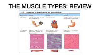
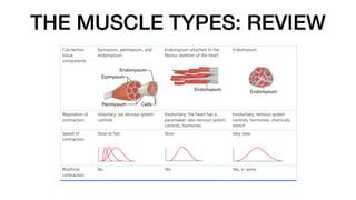
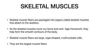
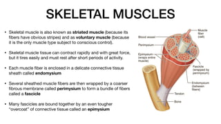
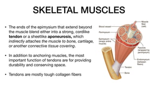
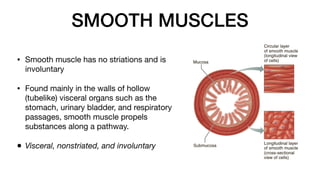
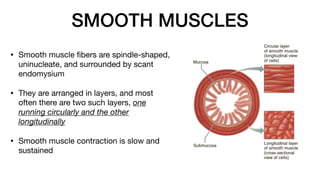
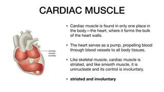
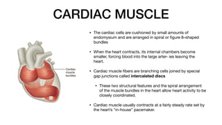
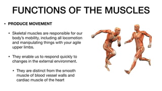
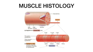
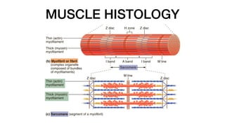
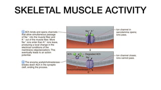
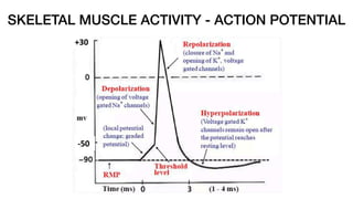
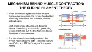
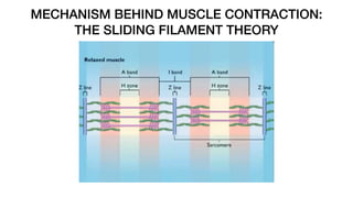
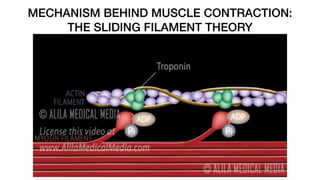
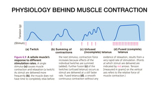
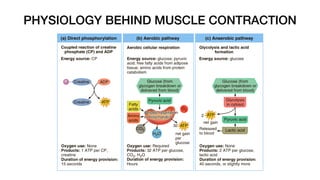
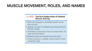
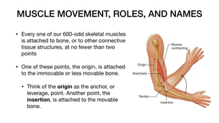
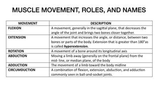
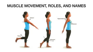
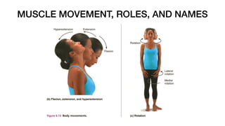
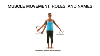
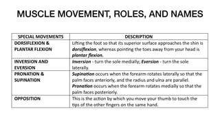
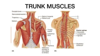
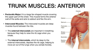
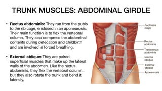
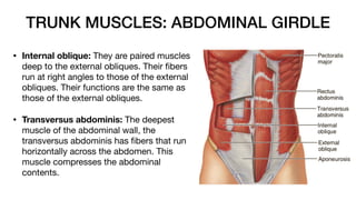
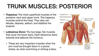
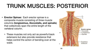
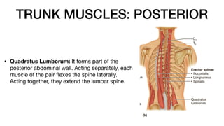
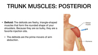
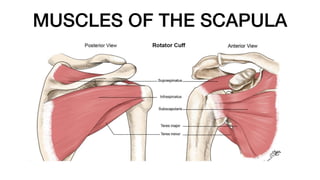
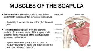
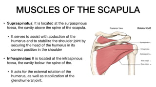
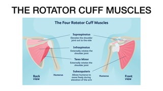
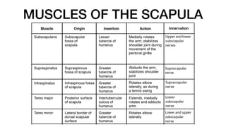
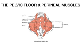
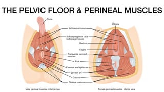
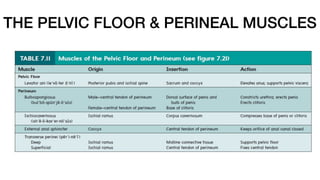
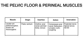
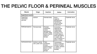
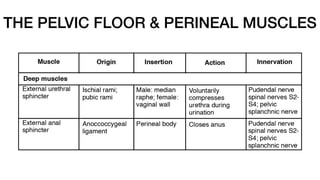
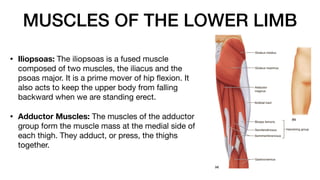
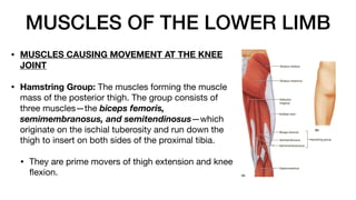
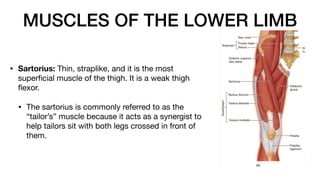
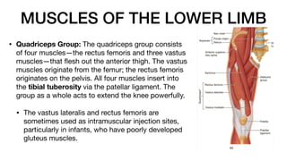
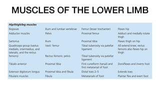
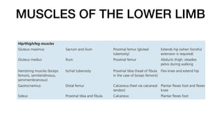
THE MUSCULAR SYSTEM
Prepared by: Renz Victor T. Guangco, M.D.
THE MUSCLE TYPES: REVIEW
SKELETAL MUSCLES
• Skeletal muscle
fi
bers are packaged into organs called skeletal muscles
that attach to the skeleton.
• As the skeletal muscles cover our bone and carti- lage framework, they
help form the smooth contours of the body.
• Skeletal muscle
fi
bers are large, cigar-shaped, multinucleate cells.
• They are the largest muscle
fi
bers
SKELETAL MUSCLES
• Skeletal muscle is also known as striated muscle (because its
fi
bers have obvious stripes) and as voluntary muscle (because
it is the only muscle type subject to conscious control).
• Skeletal muscle tissue can contract rapidly and with great force,
but it tires easily and must rest after short periods of activity.
• Each muscle
fi
ber is enclosed in a delicate connective tissue
sheath called endomysium
• Several sheathed muscle
fi
bers are then wrapped by a coarser
fi
brous membrane called perimysium to form a bundle of
fi
bers
called a fascicle
• Many fascicles are bound together by an even tougher
“overcoat” of connective tissue called an epimysium
SKELETAL MUSCLES
• The ends of the epimysium that extend beyond
the muscle blend either into a strong, cordlike
tendon or a sheetlike aponeurosis, which
indirectly attaches the muscle to bone, cartilage,
or another connective tissue covering.
• In addition to anchoring muscles, the most
important function of tendons are for providing
durability and conserving space.
• Tendons are mostly tough collagen
fi
bers
SMOOTH MUSCLES
• Smooth muscle has no striations and is
involuntary
• Found mainly in the walls of hollow
(tubelike) visceral organs such as the
stomach, urinary bladder, and respiratory
passages, smooth muscle propels
substances along a pathway.
• Visceral, nonstriated, and involuntary
SMOOTH MUSCLES
• Smooth muscle
fi
bers are spindle-shaped,
uninucleate, and surrounded by scant
endomysium
• They are arranged in layers, and most
often there are two such layers, one
running circularly and the other
longitudinally
• Smooth muscle contraction is slow and
sustained
CARDIAC MUSCLE
• Cardiac muscle is found in only one place in
the body—the heart, where it forms the bulk
of the heart walls.
• The heart serves as a pump, propelling blood
through blood vessels to all body tissues.
• Like skeletal muscle, cardiac muscle is
striated, and like smooth muscle, it is
uninucleate and its control is involuntary.
• striated and involuntary
CARDIAC MUSCLE
• The cardiac cells are cushioned by small amounts of
endomysium and are arranged in spiral or
fi
gure 8–shaped
bundles
• When the heart contracts, its internal chambers become
smaller, forcing blood into the large arter- ies leaving the
heart.
• Cardiac muscle
fi
bers are branching cells joined by special
gap junctions called intercalated discs
• These two structural features and the spiral arrangement
of the muscle bundles in the heart allow heart activity to be
closely coordinated.
• Cardiac muscle usually contracts at a fairly steady rate set by
the heart’s “in-house” pacemaker.
FUNCTIONS OF THE MUSCLES
• PRODUCE MOVEMENT
• Skeletal muscles are responsible for our
body’s mobility, including all locomotion
and manipulating things with your agile
upper limbs.
• They enable us to respond quickly to
changes in the external environment.
• They are distinct from the smooth
muscle of blood vessel walls and
cardiac muscle of the heart
FUNCTION OF THE MUSCLES
• MAINTAIN POSTURE AND BODY
POSITION
• they function almost continuously, making
one tiny adjustment after another so that
we maintain an erect or seated posture,
• even when we slouch, despite the never-
ending downward pull of gravity.
FUNCTION OF THE MUSCLES
• STABILIZE JOINTS
• Muscles and tendons are extremely important in
reinforcing and stabilizing joints that have poorly
articulating surfaces, such as the shoulder and knee
joints.
• GENERATE HEAT
• Muscle activity generates body heat as a byproduct.
• As ATP is used to power muscle contraction, nearly
three-quarters of its energy escapes as heat.
• This heat is vital in maintaining normal body
temperature
FUNCTION OF THE MUSCLES
• OTHER FUNCTIONS
• Muscles perform other important functions as well. Smooth muscles form
valves that regulate the passage of substances through internal body
openings, dilate and constrict the pupils of our eyes, and make up the
arrector pili muscles that cause our hairs to stand on end.
• Skeletal muscles form valves that are under voluntary control, and they
enclose and protect fragile internal organs.
MUSCLE HISTOLOGY
MUSCLE HISTOLOGY
SKELETAL MUSCLE ACTIVITY
• Muscle
fi
bers have several special functional properties that enable them
to perform their duties.
• IRRITABILITY, also termed responsiveness, which is the ability to receive
and respond to a stimulus.
• CONTRACTILITY, is the ability to forcibly shorten when adequately
stimulated. This property sets muscle apart from all other tissue types.
• EXTENSIBILITY is the ability of muscle
fi
bers to stretch, whereas
ELASTICITY is their ability to recoil and resume their resting length after
being stretched.
SKELETAL MUSCLE ACTIVITY
SKELETAL MUSCLE ACTIVITY - ACTION POTENTIAL
MECHANISM BEHIND MUSCLE CONTRACTION:
THE SLIDING FILAMENT THEORY
• When the nervous system activates muscle
fi
bers as just described, the myosin heads attach
to binding sites on the thin
fi
laments, and the
sliding begins.
• Each cross bridge attaches and detaches
several times during a contraction, generating
tension that helps pull the thin
fi
laments toward
the center of the sarcomere.
• The formation of cross bridges—when the
myosin heads attach to actin—requires calcium
ions (Ca2+) and ATP (to “energize” the myosin
heads)
MECHANISM BEHIND MUSCLE CONTRACTION:
THE SLIDING FILAMENT THEORY
MECHANISM BEHIND MUSCLE CONTRACTION:
THE SLIDING FILAMENT THEORY
PHYSIOLOGY BEHIND MUSCLE CONTRACTION
• GRADED RESPONSES
• In skeletal muscles, the “all-or-none” law of muscle physiology applies to the
muscle
fi
ber, not to the whole muscle.
• It states that a muscle
fi
ber will contract to its fullest extent when it is
stimulated adequately; it never partially contracts.
• However, the whole muscle reacts to stimuli with graded responses, or di
ff
erent
degrees of shortening, which generate di
ff
erent amounts of force.
• In general, graded muscle contractions can be produced two ways: (1) by
changing the frequency of muscle stimulation and (2) by changing the number of
muscle
fi
bers being stimulated at one time
PHYSIOLOGY BEHIND MUSCLE CONTRACTION
• Muscle Response to Increasingly Rapid Stimulation
• In most types of muscle activity, nerve impulses are delivered to the muscle
at a very rapid rate—so rapid that the muscle does not get a chance to relax
completely between stimuli:
• The e
ff
ects of the successive contractions are “summed” (added)
together, and the contractions of the muscle get stronger and smoother.
The muscle exhibits unfused tetanus or incomplete tetanus.
• When the muscle is stimulated so rapidly that no evidence of relaxation is
seen and the contractions are com- pletely smooth and sustained, the
muscle is in fused or complete tetanus.
PHYSIOLOGY BEHIND MUSCLE CONTRACTION
• Muscle Response to Stronger Stimuli
• Tetanus produces stronger (more forceful) muscle contractions, but its
primary role is to produce smooth and prolonged muscle contractions.
• How forcefully a muscle contracts depends to a large extent on how many of
its cells are stimulated.
• When only a few cells are stimulated, the muscle as a whole contracts
only slightly.
• When all the motor units are active and all the muscle
fi
bers are
stimulated, the muscle contraction is as strong as it can get.
PHYSIOLOGY BEHIND MUSCLE CONTRACTION
MUSCLE FATIGUE AND OXYGEN DEFICIT
• If we exercise our muscles strenuously for a long time, muscle fatigue
occurs. A muscle is fatigued when it is unable to contract even though
it is still being stimulated.
• Without rest, a working muscle begins to tire and contracts more weakly
until it
fi
nally ceases reacting and stops contracting.
• Factors that contribute to muscle fatigue are not fully known. Suspected
causes are imbalances in ions (Ca2+, K+) and problems at the
neuromuscular junction.
• However, many agree that the major factor is the oxygen de
fi
cit that
occurs during prolonged muscle activity.
MUSCLE FATIGUE AND OXYGEN DEFICIT
• When muscles lack su
ffi
cient oxygen for aerobic respiration, lactic acid begins to
accumulate in the muscle via the anaerobic pathway.
• the muscle’s ATP supply starts to run low, and ionic imbalance tends to occur.
• Together these factors cause the muscle to contract less and less e
ff
ectively
and
fi
nally to stop contracting altogether.
• During the recovery period after activity, the individual breathes rapidly and deeply.
• This continues until the muscles have received the amount of oxygen needed to
get rid of the accumulated lactic acid and replenish ATP and creatine phosphate
reserves.
TYPES OF MUSCLE CONTRACTIONS
• Isotonic contractions: the myo
fi
laments are successful in their sliding
movements, the muscle shortens, and movement occurs.
• Isometric contractions: Contractions in which the muscles do not
shorten
MUSCLE TONE
• One aspect of skeletal muscle activity cannot be consciously controlled.
• Even when a muscle is voluntarily relaxed, some of its
fi
bers are con-
tracting—
fi
rst one group and then another.
• This state of continuous partial contractions is called muscle tone.
• Muscle tone is the result of di
ff
erent motor units, which are scattered
through the muscle, being stimulated by the nervous system in a
systematic way.
EFFECTS OF EXERCISE IN MUSCLES
• regular exercise increases muscle size, strength, and endurance
• Aerobic exercise, or endurance exercise, results in stronger, more
fl
exible muscles with greater resistance to fatigue.
• These changes come about, at least partly, because the blood supply to
the muscles increases, and the individual muscle
fi
bers form more
mitochondria and store more oxygen.
• Aerobic exercise helps us reach a steady rate of ATP production and
improves the e
ffi
ciency of aerobic respiration.
EFFECTS OF EXERCISE IN MUSCLES
• regular exercise increases muscle size, strength, and endurance
• Aerobic exercise, or endurance exercise, results in stronger, more
fl
exible
muscles with greater resistance to fatigue.
• These changes come about, at least partly, because the blood supply to the
muscles increases, and the individual muscle
fi
bers form more mitochondria and
store more oxygen.
• Aerobic exercise helps us reach a steady rate of ATP production and improves
the e
ffi
ciency of aerobic respiration.
• Resistance exercise, or isometric exercise which pit the muscles against an
immovable object
EFFECTS OF EXERCISE IN MUSCLES
• exercise, or isometric exercise (Figure 6.11b), which pit the muscles
against an immovable
MUSCLE MOVEMENT, ROLES, AND NAMES
• Every one of our 600-odd skeletal muscles
is attached to bone, or to other connective
tissue structures, at no fewer than two
points
• One of these points, the origin, is attached
to the immovable or less movable bone.
• Think of the origin as the anchor, or
leverage, point. Another point, the
insertion, is attached to the movable
bone.
MUSCLE MOVEMENT, ROLES, AND NAMES
INTERACTION OF SKELETAL MUSCLES
IN THE BODY
• Muscles can’t push—they can only pull as they contract—so most often
body movements result from two or more muscles acting together or
against each other.
• Muscles are arranged so that whatever one muscle (or group of
muscles) can do, other muscles can reverse.
INTERACTION OF SKELETAL MUSCLES
IN THE BODY
• The muscle that has the major responsibility for causing a particular
movement is called the prime mover.
• Muscles that oppose or reverse a movement are antagonists.
• Synergists help prime movers by producing the same movement or by
reducing undesirable movements.
• Fixators are specialized synergists. They hold a bone still or stabilize the
origin of a prime mover so all the tension can be used to move the
insertion bone.
NAMING SKELETAL MUSCLES
ARRANGEMENT OF FASICLES
HEAD AND NECK MUSCLES
• The frontalis, which covers the frontal bone, runs
from the cranial aponeurosis to the skin of the
eyebrows, where it inserts. This muscle allows
you to raise your eyebrows, as in surprise, and to
wrinkle your forehead.
• At the posterior end of the cranial aponeurosis is
the small occipitalis muscle, which covers the
posterior aspect of the muscle, which covers the
posterior aspect of the skull and pulls the scalp
posteriorly.
HEAD AND NECK MUSCLES
• Orbicularis Oculi: The
fi
bers of the orbicularis oculi
run in circles around the eyes. It allows you to close
your eyes, squint, blink, and wink.
• Orbicularis Oris: The circular muscle of the lips.
Often called the “kissing” muscle, it closes the mouth
and protrudes the lips.
• Buccinator: It runs horizontally across the cheek and
inserts into the orbicularis oris. It
fl
attens the cheek
(as in whistling or blowing a trumpet). It is also listed
as a chewing muscle because it compresses the
cheek to hold food between the teeth during chewing.
HEAD AND NECK MUSCLES
• Zygomaticus: It extends from the corner of the mouth
to the cheekbone. It is often referred to as the “smiling”
muscle because it raises the corners of the mouth.
• Masseter: As it runs from the zygomatic process of the
temporal bone to the mandible, the masseter covers
the angle of the lower jaw. This muscle closes the jaw
by elevating the mandible.
• Temporalis: The temporalis is a fan-shaped muscle
overlying the temporal bone. It inserts into the
mandible and acts as a synergist of the masseter in
closing the jaw.
HEAD AND NECK MUSCLES
• Platysma: It is a single sheetlike muscle that covers the
anterolateral neck. It originates from the connective tissue
covering of the chest muscles and inserts into the area around
the mouth. Its action is to pull the corners of the mouth
inferiorly, producing a downward sag of the mouth.
• Sternocleidomastoid: Two-headed muscles, one found on
each side of the neck. Of the two heads of each muscle, one
arises from the sternum, and the other arises from the clavicle.
The heads fuse before inserting into the mastoid process of the
temporal bone.
• When both sternocleidomastoid muscles contract together,
they
fl
ex your neck. If just one muscle contracts, the face is
rotated toward the shoulder on the opposite side and tilts the
head to its own side.
HEAD AND NECK MUSCLES
TRUNK MUSCLES
TRUNK MUSCLES: ANTERIOR
• Pectoralis Major: It is a large fan-shaped muscle covering
the upper part of the chest. This muscle forms the anterior
wall of the axilla and acts to adduct and
fl
ex the arm.
• Intercostal Muscles: The intercostal muscles are deep
muscles found between the ribs.
• The external intercostals are important in breathing
because they help to raise the rib cage when you
inhale.
• The internal intercostals, which lie deep to the
external intercostals, depress the rib cage, helping to
move air out of the lungs when you exhale forcibly.
TRUNK MUSCLES: ABDOMINAL GIRDLE
• Rectus abdominis: They run from the pubis
to the rib cage, enclosed in an aponeurosis.
Their main function is to
fl
ex the vertebral
column. They also compress the abdominal
contents during defecation and childbirth
and are involved in forced breathing.
• External oblique: They are paired
super
fi
cial muscles that make up the lateral
walls of the abdomen. Like the rectus
abdominis, they
fl
ex the vertebral column,
but they also rotate the trunk and bend it
laterally.
TRUNK MUSCLES: ABDOMINAL GIRDLE
• Internal oblique: They are paired muscles
deep to the external obliques. Their
fi
bers
run at right angles to those of the external
obliques. Their functions are the same as
those of the external obliques.
• Transversus abdominis: The deepest
muscle of the abdominal wall, the
transversus abdominis has
fi
bers that run
horizontally across the abdomen. This
muscle compresses the abdominal
contents.
TRUNK MUSCLES: POSTERIOR
• Trapezius: The most super
fi
cial muscles of the
posterior neck and upper trunk. The trapezius
muscles extend the head. They also can
elevate, depress, adduct, and stabilize the
scapula.
• Latissimus Dorsi: The two large,
fl
at muscles
that cover the lower back. Each latissimus dorsi
extends and adducts the humerus.
• These are very important muscles when the
arm must be brought down in a power
stroke, as when swimming or striking a blow.
TRUNK MUSCLES: POSTERIOR
• Erector Spinae: Each erector spinae is a
composite muscle consisting of three muscle
columns (longissimus, iliocostalis, and spinalis)
that collectively span the entire length of the
vertebral column.
• These muscles not only act as powerful back
extensors but also provide resistance that
helps control the action of bending over at the
waist.
TRUNK MUSCLES: POSTERIOR
• Quadratus Lumborum: It forms part of the
posterior abdominal wall. Acting separately, each
muscle of the pair
fl
exes the spine laterally.
Acting together, they extend the lumbar spine.
TRUNK MUSCLES: POSTERIOR
• Deltoid: The deltoids are
fl
eshy, triangle-shaped
muscles that form the rounded shape of your
shoulders. Because they are so bulky, they are a
favorite injection site.
• The deltoids are the prime movers of arm
abduction.
TRUNK MUSCLES
MUSCLES OF THE UPPER LIMB
• Biceps Brachii: It originates by two heads from
the shoulder girdle and inserts into the radial
tuberosity. This muscle is the powerful prime
mover for
fl
exion of the forearm and acts to
supinate the forearm.
• Brachialis: The brachialis lies deep to the biceps
brachii and, like the biceps, is a prime mover in
elbow
fl
exion. The brachialis lifts the ulna as the
biceps lifts the radius.
MUSCLES OF THE UPPER LIMB
• Brachioradialis The brachioradialis is a fairly weak
muscle that arises on the humerus and inserts into
the distal forearm.
• Triceps Brachii: The triceps brachii is the only
muscle
fl
eshing out the posterior humerus. Its
three heads arise from the shoulder girdle and
proximal humerus, and it inserts into the olecranon
process of the ulna.
• Being the powerful prime mover of elbow
extension, it is the antagonist of the biceps
brachii and brachialis. This muscle straightens
the arm
MUSCLES OF THE UPPER LIMB
MUSCLES OF THE SCAPULA
• Subscapularis: The subscapularis muscle lies
underneath the anterior
fl
at surface of the scapula.
• It medially It rotates the arm at the glenohumeral
joint.
• Teres Major: It originates from the posterior
surface of the inferior angle of the scapula and it
attaches to the medial lip of the intertubercular
groove of the humerus.
• It pulls the anterior surface of the humerus
medially towards the trunk and it can extend the
arm from the
fl
exed position
MUSCLES OF THE SCAPULA
• Teres Minor: It originates from the posterior
surface of the scapula, adjacent to its lateral
border. It attaches to the greater tubercle of
the humerus
• It stabilizes the ball-and-socket
glenohumeral joint by helping hold the
humeral head (ball) into the shallow glenoid
• It also laterally or externally rotates the arm
at the shoulder joint.
MUSCLES OF THE SCAPULA
• Supraspinatus: It is located at the surpaspinous
fossa, the cavity above the spine of the scapula.
• It serves to assist with abduction of the
humerus and to stabilize the shoulder joint by
securing the head of the humerus in its
correct position in the shoulder.
• Infraspinatus: It is located at the infraspinous
fossa, the cavity below the spine of the.
• It acts for the external rotation of the
humerus, as well as stabilization of the
glenohumeral joint.
THE ROTATOR CUFF MUSCLES
MUSCLES OF THE SCAPULA
THE PELVIC FLOOR & PERINEAL MUSCLES
MUSCLES OF THE LOWER LIMB
• MUSCLES CAUSING MOVEMENT AT THE HIP JOINT
• Gluteus Maximus: The gluteus maximus is a super
fi
cial
muscle of the hip that forms most of the
fl
esh of the
buttock. It is a powerful hip extensor that acts to bring
the thigh in a straight line with the pelvis.
• Gluteus Medius: The gluteus medius runs from the ilium
to the femur, beneath the gluteus maximus for most of
its length. The gluteus medius is a hip abductor and is
important in steadying the pelvis during walking.
• It is also an important site for giving intramuscular
injections, particularly when administering more than 5
ml.
MUSCLES OF THE LOWER LIMB
• Iliopsoas: The iliopsoas is a fused muscle
composed of two muscles, the iliacus and the
psoas major. It is a prime mover of hip
fl
exion. It
also acts to keep the upper body from falling
backward when we are standing erect.
• Adductor Muscles: The muscles of the adductor
group form the muscle mass at the medial side of
each thigh. They adduct, or press, the thighs
together.
MUSCLES OF THE LOWER LIMB
• MUSCLES CAUSING MOVEMENT AT THE KNEE
JOINT
• Hamstring Group: The muscles forming the muscle
mass of the posterior thigh. The group consists of
three muscles—the biceps femoris,
semimembranosus, and semitendinosus—which
originate on the ischial tuberosity and run down the
thigh to insert on both sides of the proximal tibia.
• They are prime movers of thigh extension and knee
fl
exion.
MUSCLES OF THE LOWER LIMB
• Sartorius: Thin, straplike, and it is the most
super
fi
cial muscle of the thigh. It is a weak thigh
fl
exor.
• The sartorius is commonly referred to as the
“tailor’s” muscle because it acts as a synergist to
help tailors sit with both legs crossed in front of
them.
MUSCLES OF THE LOWER LIMB
• Quadriceps Group: The quadriceps group consists
of four muscles—the rectus femoris and three vastus
muscles—that
fl
esh out the anterior thigh. The vastus
muscles originate from the femur; the rectus femoris
originates on the pelvis. All four muscles insert into
the tibial tuberosity via the patellar ligament. The
group as a whole acts to extend the knee powerfully.
• The vastus lateralis and rectus femoris are
sometimes used as intramuscular injection sites,
particularly in infants, who have poorly developed
gluteus muscles.
MUSCLES OF THE LOWER LIMB
• MUSCLES CAUSING MOVEMENT AT THE ANKLES
AND FOOT
• Tibialis Anterior: The tibialis anterior is a super
fi
cial
muscle on the anterior leg. It acts to dorsi
fl
ex and invert
the foot.
• Extensor Digitorum Longus: Lateral to the tibialis
anterior. It is a prime mover of toe extension.
MUSCLES OF THE LOWER LIMB
• Fibularis Muscles: The three
fi
bularis muscles—
longus, brevis, and tertius—are found on the lateral
part of the leg. The group as a whole plantar
fl
exes and
everts the foot, which is antagonistic to the tibialis
anterior.
MUSCLES OF THE LOWER LIMB
• Gastrocnemius: It is a two-bellied muscle that forms the
curved calf of the posterior leg. It arises by two heads,
one from each side of the distal femur, and inserts
through the large calcaneal (Achilles) tendon into the heel
of the foot.
• It is a prime mover for plantar
fl
exion of the foot; for
this reason it is often called the “toe dancer’s” muscle.
• If the calcaneal tendon is severely damaged or cut,
walking is very di
ffi
cult. The foot drags because it is
not able to “push o
ff
” the toe (raise the heel).
MUSCLES OF THE LOWER LIMB
• Soleus: Deep to the gastrocnemius is the
fl
eshy soleus
muscle. Because it arises on the tibia and
fi
bula (rather
than the femur), it does not a
ff
ect knee movement
• but like the gastrocnemius, it inserts into the calcaneal
tendon and is a strong plantar
fl
exor of the foot.
MUSCLES OF THE LOWER LIMB
• END OF DISCUSSION
• Source: Essentials of Anatomy and Physiology, 13th Edition by Marieb
and Keller (2019)
•

05 - The Muscular System (2024 Revised).pdf

  • 1.
    THE MUSCULAR SYSTEM Preparedby: Renz Victor T. Guangco, M.D.
  • 2.
  • 3.
  • 4.
    SKELETAL MUSCLES • Skeletalmuscle fi bers are packaged into organs called skeletal muscles that attach to the skeleton. • As the skeletal muscles cover our bone and carti- lage framework, they help form the smooth contours of the body. • Skeletal muscle fi bers are large, cigar-shaped, multinucleate cells. • They are the largest muscle fi bers
  • 5.
    SKELETAL MUSCLES • Skeletalmuscle is also known as striated muscle (because its fi bers have obvious stripes) and as voluntary muscle (because it is the only muscle type subject to conscious control). • Skeletal muscle tissue can contract rapidly and with great force, but it tires easily and must rest after short periods of activity. • Each muscle fi ber is enclosed in a delicate connective tissue sheath called endomysium • Several sheathed muscle fi bers are then wrapped by a coarser fi brous membrane called perimysium to form a bundle of fi bers called a fascicle • Many fascicles are bound together by an even tougher “overcoat” of connective tissue called an epimysium
  • 6.
    SKELETAL MUSCLES • Theends of the epimysium that extend beyond the muscle blend either into a strong, cordlike tendon or a sheetlike aponeurosis, which indirectly attaches the muscle to bone, cartilage, or another connective tissue covering. • In addition to anchoring muscles, the most important function of tendons are for providing durability and conserving space. • Tendons are mostly tough collagen fi bers
  • 7.
    SMOOTH MUSCLES • Smoothmuscle has no striations and is involuntary • Found mainly in the walls of hollow (tubelike) visceral organs such as the stomach, urinary bladder, and respiratory passages, smooth muscle propels substances along a pathway. • Visceral, nonstriated, and involuntary
  • 8.
    SMOOTH MUSCLES • Smoothmuscle fi bers are spindle-shaped, uninucleate, and surrounded by scant endomysium • They are arranged in layers, and most often there are two such layers, one running circularly and the other longitudinally • Smooth muscle contraction is slow and sustained
  • 9.
    CARDIAC MUSCLE • Cardiacmuscle is found in only one place in the body—the heart, where it forms the bulk of the heart walls. • The heart serves as a pump, propelling blood through blood vessels to all body tissues. • Like skeletal muscle, cardiac muscle is striated, and like smooth muscle, it is uninucleate and its control is involuntary. • striated and involuntary
  • 10.
    CARDIAC MUSCLE • Thecardiac cells are cushioned by small amounts of endomysium and are arranged in spiral or fi gure 8–shaped bundles • When the heart contracts, its internal chambers become smaller, forcing blood into the large arter- ies leaving the heart. • Cardiac muscle fi bers are branching cells joined by special gap junctions called intercalated discs • These two structural features and the spiral arrangement of the muscle bundles in the heart allow heart activity to be closely coordinated. • Cardiac muscle usually contracts at a fairly steady rate set by the heart’s “in-house” pacemaker.
  • 11.
    FUNCTIONS OF THEMUSCLES • PRODUCE MOVEMENT • Skeletal muscles are responsible for our body’s mobility, including all locomotion and manipulating things with your agile upper limbs. • They enable us to respond quickly to changes in the external environment. • They are distinct from the smooth muscle of blood vessel walls and cardiac muscle of the heart
  • 12.
    FUNCTION OF THEMUSCLES • MAINTAIN POSTURE AND BODY POSITION • they function almost continuously, making one tiny adjustment after another so that we maintain an erect or seated posture, • even when we slouch, despite the never- ending downward pull of gravity.
  • 13.
    FUNCTION OF THEMUSCLES • STABILIZE JOINTS • Muscles and tendons are extremely important in reinforcing and stabilizing joints that have poorly articulating surfaces, such as the shoulder and knee joints. • GENERATE HEAT • Muscle activity generates body heat as a byproduct. • As ATP is used to power muscle contraction, nearly three-quarters of its energy escapes as heat. • This heat is vital in maintaining normal body temperature
  • 14.
    FUNCTION OF THEMUSCLES • OTHER FUNCTIONS • Muscles perform other important functions as well. Smooth muscles form valves that regulate the passage of substances through internal body openings, dilate and constrict the pupils of our eyes, and make up the arrector pili muscles that cause our hairs to stand on end. • Skeletal muscles form valves that are under voluntary control, and they enclose and protect fragile internal organs.
  • 15.
  • 16.
  • 17.
    SKELETAL MUSCLE ACTIVITY •Muscle fi bers have several special functional properties that enable them to perform their duties. • IRRITABILITY, also termed responsiveness, which is the ability to receive and respond to a stimulus. • CONTRACTILITY, is the ability to forcibly shorten when adequately stimulated. This property sets muscle apart from all other tissue types. • EXTENSIBILITY is the ability of muscle fi bers to stretch, whereas ELASTICITY is their ability to recoil and resume their resting length after being stretched.
  • 18.
  • 19.
  • 20.
  • 21.
    SKELETAL MUSCLE ACTIVITY- ACTION POTENTIAL
  • 22.
    MECHANISM BEHIND MUSCLECONTRACTION: THE SLIDING FILAMENT THEORY • When the nervous system activates muscle fi bers as just described, the myosin heads attach to binding sites on the thin fi laments, and the sliding begins. • Each cross bridge attaches and detaches several times during a contraction, generating tension that helps pull the thin fi laments toward the center of the sarcomere. • The formation of cross bridges—when the myosin heads attach to actin—requires calcium ions (Ca2+) and ATP (to “energize” the myosin heads)
  • 23.
    MECHANISM BEHIND MUSCLECONTRACTION: THE SLIDING FILAMENT THEORY
  • 24.
    MECHANISM BEHIND MUSCLECONTRACTION: THE SLIDING FILAMENT THEORY
  • 25.
    PHYSIOLOGY BEHIND MUSCLECONTRACTION • GRADED RESPONSES • In skeletal muscles, the “all-or-none” law of muscle physiology applies to the muscle fi ber, not to the whole muscle. • It states that a muscle fi ber will contract to its fullest extent when it is stimulated adequately; it never partially contracts. • However, the whole muscle reacts to stimuli with graded responses, or di ff erent degrees of shortening, which generate di ff erent amounts of force. • In general, graded muscle contractions can be produced two ways: (1) by changing the frequency of muscle stimulation and (2) by changing the number of muscle fi bers being stimulated at one time
  • 26.
    PHYSIOLOGY BEHIND MUSCLECONTRACTION • Muscle Response to Increasingly Rapid Stimulation • In most types of muscle activity, nerve impulses are delivered to the muscle at a very rapid rate—so rapid that the muscle does not get a chance to relax completely between stimuli: • The e ff ects of the successive contractions are “summed” (added) together, and the contractions of the muscle get stronger and smoother. The muscle exhibits unfused tetanus or incomplete tetanus. • When the muscle is stimulated so rapidly that no evidence of relaxation is seen and the contractions are com- pletely smooth and sustained, the muscle is in fused or complete tetanus.
  • 27.
    PHYSIOLOGY BEHIND MUSCLECONTRACTION • Muscle Response to Stronger Stimuli • Tetanus produces stronger (more forceful) muscle contractions, but its primary role is to produce smooth and prolonged muscle contractions. • How forcefully a muscle contracts depends to a large extent on how many of its cells are stimulated. • When only a few cells are stimulated, the muscle as a whole contracts only slightly. • When all the motor units are active and all the muscle fi bers are stimulated, the muscle contraction is as strong as it can get.
  • 28.
  • 29.
  • 30.
    MUSCLE FATIGUE ANDOXYGEN DEFICIT • If we exercise our muscles strenuously for a long time, muscle fatigue occurs. A muscle is fatigued when it is unable to contract even though it is still being stimulated. • Without rest, a working muscle begins to tire and contracts more weakly until it fi nally ceases reacting and stops contracting. • Factors that contribute to muscle fatigue are not fully known. Suspected causes are imbalances in ions (Ca2+, K+) and problems at the neuromuscular junction. • However, many agree that the major factor is the oxygen de fi cit that occurs during prolonged muscle activity.
  • 31.
    MUSCLE FATIGUE ANDOXYGEN DEFICIT • When muscles lack su ffi cient oxygen for aerobic respiration, lactic acid begins to accumulate in the muscle via the anaerobic pathway. • the muscle’s ATP supply starts to run low, and ionic imbalance tends to occur. • Together these factors cause the muscle to contract less and less e ff ectively and fi nally to stop contracting altogether. • During the recovery period after activity, the individual breathes rapidly and deeply. • This continues until the muscles have received the amount of oxygen needed to get rid of the accumulated lactic acid and replenish ATP and creatine phosphate reserves.
  • 32.
    TYPES OF MUSCLECONTRACTIONS • Isotonic contractions: the myo fi laments are successful in their sliding movements, the muscle shortens, and movement occurs. • Isometric contractions: Contractions in which the muscles do not shorten
  • 33.
    MUSCLE TONE • Oneaspect of skeletal muscle activity cannot be consciously controlled. • Even when a muscle is voluntarily relaxed, some of its fi bers are con- tracting— fi rst one group and then another. • This state of continuous partial contractions is called muscle tone. • Muscle tone is the result of di ff erent motor units, which are scattered through the muscle, being stimulated by the nervous system in a systematic way.
  • 34.
    EFFECTS OF EXERCISEIN MUSCLES • regular exercise increases muscle size, strength, and endurance • Aerobic exercise, or endurance exercise, results in stronger, more fl exible muscles with greater resistance to fatigue. • These changes come about, at least partly, because the blood supply to the muscles increases, and the individual muscle fi bers form more mitochondria and store more oxygen. • Aerobic exercise helps us reach a steady rate of ATP production and improves the e ffi ciency of aerobic respiration.
  • 35.
    EFFECTS OF EXERCISEIN MUSCLES • regular exercise increases muscle size, strength, and endurance • Aerobic exercise, or endurance exercise, results in stronger, more fl exible muscles with greater resistance to fatigue. • These changes come about, at least partly, because the blood supply to the muscles increases, and the individual muscle fi bers form more mitochondria and store more oxygen. • Aerobic exercise helps us reach a steady rate of ATP production and improves the e ffi ciency of aerobic respiration. • Resistance exercise, or isometric exercise which pit the muscles against an immovable object
  • 36.
    EFFECTS OF EXERCISEIN MUSCLES • exercise, or isometric exercise (Figure 6.11b), which pit the muscles against an immovable
  • 37.
  • 38.
    MUSCLE MOVEMENT, ROLES,AND NAMES • Every one of our 600-odd skeletal muscles is attached to bone, or to other connective tissue structures, at no fewer than two points • One of these points, the origin, is attached to the immovable or less movable bone. • Think of the origin as the anchor, or leverage, point. Another point, the insertion, is attached to the movable bone.
  • 39.
  • 40.
  • 41.
  • 42.
  • 43.
  • 44.
  • 45.
    INTERACTION OF SKELETALMUSCLES IN THE BODY • Muscles can’t push—they can only pull as they contract—so most often body movements result from two or more muscles acting together or against each other. • Muscles are arranged so that whatever one muscle (or group of muscles) can do, other muscles can reverse.
  • 46.
    INTERACTION OF SKELETALMUSCLES IN THE BODY • The muscle that has the major responsibility for causing a particular movement is called the prime mover. • Muscles that oppose or reverse a movement are antagonists. • Synergists help prime movers by producing the same movement or by reducing undesirable movements. • Fixators are specialized synergists. They hold a bone still or stabilize the origin of a prime mover so all the tension can be used to move the insertion bone.
  • 47.
  • 48.
  • 49.
  • 50.
  • 51.
    HEAD AND NECKMUSCLES • The frontalis, which covers the frontal bone, runs from the cranial aponeurosis to the skin of the eyebrows, where it inserts. This muscle allows you to raise your eyebrows, as in surprise, and to wrinkle your forehead. • At the posterior end of the cranial aponeurosis is the small occipitalis muscle, which covers the posterior aspect of the muscle, which covers the posterior aspect of the skull and pulls the scalp posteriorly.
  • 52.
    HEAD AND NECKMUSCLES • Orbicularis Oculi: The fi bers of the orbicularis oculi run in circles around the eyes. It allows you to close your eyes, squint, blink, and wink. • Orbicularis Oris: The circular muscle of the lips. Often called the “kissing” muscle, it closes the mouth and protrudes the lips. • Buccinator: It runs horizontally across the cheek and inserts into the orbicularis oris. It fl attens the cheek (as in whistling or blowing a trumpet). It is also listed as a chewing muscle because it compresses the cheek to hold food between the teeth during chewing.
  • 53.
    HEAD AND NECKMUSCLES • Zygomaticus: It extends from the corner of the mouth to the cheekbone. It is often referred to as the “smiling” muscle because it raises the corners of the mouth. • Masseter: As it runs from the zygomatic process of the temporal bone to the mandible, the masseter covers the angle of the lower jaw. This muscle closes the jaw by elevating the mandible. • Temporalis: The temporalis is a fan-shaped muscle overlying the temporal bone. It inserts into the mandible and acts as a synergist of the masseter in closing the jaw.
  • 54.
    HEAD AND NECKMUSCLES • Platysma: It is a single sheetlike muscle that covers the anterolateral neck. It originates from the connective tissue covering of the chest muscles and inserts into the area around the mouth. Its action is to pull the corners of the mouth inferiorly, producing a downward sag of the mouth. • Sternocleidomastoid: Two-headed muscles, one found on each side of the neck. Of the two heads of each muscle, one arises from the sternum, and the other arises from the clavicle. The heads fuse before inserting into the mastoid process of the temporal bone. • When both sternocleidomastoid muscles contract together, they fl ex your neck. If just one muscle contracts, the face is rotated toward the shoulder on the opposite side and tilts the head to its own side.
  • 55.
  • 56.
  • 57.
    TRUNK MUSCLES: ANTERIOR •Pectoralis Major: It is a large fan-shaped muscle covering the upper part of the chest. This muscle forms the anterior wall of the axilla and acts to adduct and fl ex the arm. • Intercostal Muscles: The intercostal muscles are deep muscles found between the ribs. • The external intercostals are important in breathing because they help to raise the rib cage when you inhale. • The internal intercostals, which lie deep to the external intercostals, depress the rib cage, helping to move air out of the lungs when you exhale forcibly.
  • 58.
    TRUNK MUSCLES: ABDOMINALGIRDLE • Rectus abdominis: They run from the pubis to the rib cage, enclosed in an aponeurosis. Their main function is to fl ex the vertebral column. They also compress the abdominal contents during defecation and childbirth and are involved in forced breathing. • External oblique: They are paired super fi cial muscles that make up the lateral walls of the abdomen. Like the rectus abdominis, they fl ex the vertebral column, but they also rotate the trunk and bend it laterally.
  • 59.
    TRUNK MUSCLES: ABDOMINALGIRDLE • Internal oblique: They are paired muscles deep to the external obliques. Their fi bers run at right angles to those of the external obliques. Their functions are the same as those of the external obliques. • Transversus abdominis: The deepest muscle of the abdominal wall, the transversus abdominis has fi bers that run horizontally across the abdomen. This muscle compresses the abdominal contents.
  • 60.
    TRUNK MUSCLES: POSTERIOR •Trapezius: The most super fi cial muscles of the posterior neck and upper trunk. The trapezius muscles extend the head. They also can elevate, depress, adduct, and stabilize the scapula. • Latissimus Dorsi: The two large, fl at muscles that cover the lower back. Each latissimus dorsi extends and adducts the humerus. • These are very important muscles when the arm must be brought down in a power stroke, as when swimming or striking a blow.
  • 61.
    TRUNK MUSCLES: POSTERIOR •Erector Spinae: Each erector spinae is a composite muscle consisting of three muscle columns (longissimus, iliocostalis, and spinalis) that collectively span the entire length of the vertebral column. • These muscles not only act as powerful back extensors but also provide resistance that helps control the action of bending over at the waist.
  • 62.
    TRUNK MUSCLES: POSTERIOR •Quadratus Lumborum: It forms part of the posterior abdominal wall. Acting separately, each muscle of the pair fl exes the spine laterally. Acting together, they extend the lumbar spine.
  • 63.
    TRUNK MUSCLES: POSTERIOR •Deltoid: The deltoids are fl eshy, triangle-shaped muscles that form the rounded shape of your shoulders. Because they are so bulky, they are a favorite injection site. • The deltoids are the prime movers of arm abduction.
  • 64.
  • 65.
    MUSCLES OF THEUPPER LIMB • Biceps Brachii: It originates by two heads from the shoulder girdle and inserts into the radial tuberosity. This muscle is the powerful prime mover for fl exion of the forearm and acts to supinate the forearm. • Brachialis: The brachialis lies deep to the biceps brachii and, like the biceps, is a prime mover in elbow fl exion. The brachialis lifts the ulna as the biceps lifts the radius.
  • 66.
    MUSCLES OF THEUPPER LIMB • Brachioradialis The brachioradialis is a fairly weak muscle that arises on the humerus and inserts into the distal forearm. • Triceps Brachii: The triceps brachii is the only muscle fl eshing out the posterior humerus. Its three heads arise from the shoulder girdle and proximal humerus, and it inserts into the olecranon process of the ulna. • Being the powerful prime mover of elbow extension, it is the antagonist of the biceps brachii and brachialis. This muscle straightens the arm
  • 67.
    MUSCLES OF THEUPPER LIMB
  • 68.
    MUSCLES OF THEUPPER LIMB
  • 69.
  • 70.
    MUSCLES OF THESCAPULA • Subscapularis: The subscapularis muscle lies underneath the anterior fl at surface of the scapula. • It medially It rotates the arm at the glenohumeral joint. • Teres Major: It originates from the posterior surface of the inferior angle of the scapula and it attaches to the medial lip of the intertubercular groove of the humerus. • It pulls the anterior surface of the humerus medially towards the trunk and it can extend the arm from the fl exed position
  • 71.
    MUSCLES OF THESCAPULA • Teres Minor: It originates from the posterior surface of the scapula, adjacent to its lateral border. It attaches to the greater tubercle of the humerus • It stabilizes the ball-and-socket glenohumeral joint by helping hold the humeral head (ball) into the shallow glenoid • It also laterally or externally rotates the arm at the shoulder joint.
  • 72.
    MUSCLES OF THESCAPULA • Supraspinatus: It is located at the surpaspinous fossa, the cavity above the spine of the scapula. • It serves to assist with abduction of the humerus and to stabilize the shoulder joint by securing the head of the humerus in its correct position in the shoulder. • Infraspinatus: It is located at the infraspinous fossa, the cavity below the spine of the. • It acts for the external rotation of the humerus, as well as stabilization of the glenohumeral joint.
  • 73.
  • 74.
  • 75.
    THE PELVIC FLOOR& PERINEAL MUSCLES
  • 76.
    THE PELVIC FLOOR& PERINEAL MUSCLES
  • 77.
    THE PELVIC FLOOR& PERINEAL MUSCLES
  • 78.
    THE PELVIC FLOOR& PERINEAL MUSCLES
  • 79.
    THE PELVIC FLOOR& PERINEAL MUSCLES
  • 80.
    THE PELVIC FLOOR& PERINEAL MUSCLES
  • 81.
    MUSCLES OF THELOWER LIMB • MUSCLES CAUSING MOVEMENT AT THE HIP JOINT • Gluteus Maximus: The gluteus maximus is a super fi cial muscle of the hip that forms most of the fl esh of the buttock. It is a powerful hip extensor that acts to bring the thigh in a straight line with the pelvis. • Gluteus Medius: The gluteus medius runs from the ilium to the femur, beneath the gluteus maximus for most of its length. The gluteus medius is a hip abductor and is important in steadying the pelvis during walking. • It is also an important site for giving intramuscular injections, particularly when administering more than 5 ml.
  • 82.
    MUSCLES OF THELOWER LIMB • Iliopsoas: The iliopsoas is a fused muscle composed of two muscles, the iliacus and the psoas major. It is a prime mover of hip fl exion. It also acts to keep the upper body from falling backward when we are standing erect. • Adductor Muscles: The muscles of the adductor group form the muscle mass at the medial side of each thigh. They adduct, or press, the thighs together.
  • 83.
    MUSCLES OF THELOWER LIMB • MUSCLES CAUSING MOVEMENT AT THE KNEE JOINT • Hamstring Group: The muscles forming the muscle mass of the posterior thigh. The group consists of three muscles—the biceps femoris, semimembranosus, and semitendinosus—which originate on the ischial tuberosity and run down the thigh to insert on both sides of the proximal tibia. • They are prime movers of thigh extension and knee fl exion.
  • 84.
    MUSCLES OF THELOWER LIMB • Sartorius: Thin, straplike, and it is the most super fi cial muscle of the thigh. It is a weak thigh fl exor. • The sartorius is commonly referred to as the “tailor’s” muscle because it acts as a synergist to help tailors sit with both legs crossed in front of them.
  • 85.
    MUSCLES OF THELOWER LIMB • Quadriceps Group: The quadriceps group consists of four muscles—the rectus femoris and three vastus muscles—that fl esh out the anterior thigh. The vastus muscles originate from the femur; the rectus femoris originates on the pelvis. All four muscles insert into the tibial tuberosity via the patellar ligament. The group as a whole acts to extend the knee powerfully. • The vastus lateralis and rectus femoris are sometimes used as intramuscular injection sites, particularly in infants, who have poorly developed gluteus muscles.
  • 86.
    MUSCLES OF THELOWER LIMB • MUSCLES CAUSING MOVEMENT AT THE ANKLES AND FOOT • Tibialis Anterior: The tibialis anterior is a super fi cial muscle on the anterior leg. It acts to dorsi fl ex and invert the foot. • Extensor Digitorum Longus: Lateral to the tibialis anterior. It is a prime mover of toe extension.
  • 87.
    MUSCLES OF THELOWER LIMB • Fibularis Muscles: The three fi bularis muscles— longus, brevis, and tertius—are found on the lateral part of the leg. The group as a whole plantar fl exes and everts the foot, which is antagonistic to the tibialis anterior.
  • 88.
    MUSCLES OF THELOWER LIMB • Gastrocnemius: It is a two-bellied muscle that forms the curved calf of the posterior leg. It arises by two heads, one from each side of the distal femur, and inserts through the large calcaneal (Achilles) tendon into the heel of the foot. • It is a prime mover for plantar fl exion of the foot; for this reason it is often called the “toe dancer’s” muscle. • If the calcaneal tendon is severely damaged or cut, walking is very di ffi cult. The foot drags because it is not able to “push o ff ” the toe (raise the heel).
  • 89.
    MUSCLES OF THELOWER LIMB • Soleus: Deep to the gastrocnemius is the fl eshy soleus muscle. Because it arises on the tibia and fi bula (rather than the femur), it does not a ff ect knee movement • but like the gastrocnemius, it inserts into the calcaneal tendon and is a strong plantar fl exor of the foot.
  • 90.
    MUSCLES OF THELOWER LIMB
  • 91.
    MUSCLES OF THELOWER LIMB
  • 92.
    • END OFDISCUSSION • Source: Essentials of Anatomy and Physiology, 13th Edition by Marieb and Keller (2019) •