SlideShare a Scribd company logo
1 of 8
Esophageal Conditions: A
Radiological Perspective
This comprehensive list outlines the radiological findings associated with
various esophageal conditions, as observed through barium
esophagograms. From mild esophagitis to complex structural
abnormalities, these diagnostic insights can aid clinicians in accurately
identifying and managing a wide range of esophageal disorders.
Na by Nagasai Pelala
Inflammatory Conditions
Mild Esophagitis
Characterized by fold
thickening, this condition
represents the mildest form
of esophageal
inflammation. The barium
esophagogram may reveal
subtle changes in the
esophageal mucosal
pattern, indicating the
presence of this condition.
Moderate Esophagitis
Superficial erosions,
appearing as lines and
punctate defects, are the
hallmark of moderate
esophagitis on the barium
esophagogram. These
findings indicate a more
advanced stage of
esophageal inflammation.
Severe Esophagitis
In severe esophagitis, the
barium esophagogram may
reveal a few short and fixed
transverse folds, reflecting
the more significant
mucosal damage and
scarring associated with
this condition.
Anatomical Variations
1 Feline Folds
(Physiologic)
These numerous, fine,
delicate, symmetric,
and transient folds are
a normal anatomical
variant and do not
indicate any
underlying pathology.
2 Chronic Reflux
Induced Strictures
Chronic
gastroesophageal
reflux can lead to the
development of short
segment strictures
above the
gastroesophageal
junction, often
accompanied by a
short type esophageal
hernia.
3 Intramural
Esophageal
Pseudodiverticulo
sis
Diffuse tiny
outpouchings, most
commonly due to
chronic reflux
esophagitis, can be
observed on the
barium
esophagogram.
Metaplastic and Neoplastic Conditions
Barrett's Esophagus
This condition is
characterized by a mid-
esophagus stricture, often
accompanied by a hiatal
hernia and possible
reticular changes, as seen
on the barium
esophagogram. These
findings are indicative of the
metaplastic changes
associated with Barrett's
esophagus.
Medication-Induced
Esophagitis
Certain medications can
cause esophageal irritation
and inflammation, leading
to the formation of strictures
at anatomical narrowings,
as observed on the barium
esophagogram.
Eosinophilic
Esophagitis
Luminal narrowing and
furrowing in the upper
esophagus are
characteristic radiological
findings associated with
eosinophilic esophagitis, a
condition driven by an
eosinophilic immune
response.
Infectious and Inflammatory
Conditions
Candidiasis
Multiple plaque-like filling defects on
the barium esophagogram are
indicative of esophageal candidiasis, a
fungal infection that can occur in
immunocompromised individuals.
Herpes Esophagitis
The barium esophagogram may reveal
multiple discrete ulcerations on an
otherwise normal esophageal mucosal
background, characteristic of herpes
esophagitis.
Cytomegalovirus (CMV) Infection
A large solitary discrete ulcer, especially in HIV-positive patients, is a typical radiological
finding associated with CMV esophagitis.
Motility Disorders
Achalasia
The barium esophagogram
in achalasia typically
shows a dilated
esophagus with a standing
column of contrast and a
beak-like stricture near the
gastroesophageal junction,
reflecting the impaired
esophageal motility and
incomplete relaxation of
the lower esophageal
sphincter.
Pseudoachalasia /
Carcinoma
In cases of
pseudoachalasia or
esophageal carcinoma, the
barium esophagogram
may reveal a fixed rigid
stricture with a soft tissue
mass evident on CT,
distinguishing it from the
classic achalasia findings.
Diffuse Esophageal
Spasm (DES)
The characteristic
corkscrew appearance on
the barium esophagogram
is a hallmark of diffuse
esophageal spasm, a
motility disorder
characterized by
uncoordinated, non-
peristaltic contractions of
the esophageal smooth
muscle.
Structural Abnormalities
Leiomyoma
A submucosal, circumscribed
mass with a smooth
impression on the
esophageal contour is
characteristic of an
esophageal leiomyoma, a
benign smooth muscle
tumor.
Esophageal Varices
Serpentine filling defects with
a scalloped contour in the
distal esophagus are
indicative of esophageal
varices, dilated veins that
can develop due to portal
hypertension.
Zenker's Diverticulum
A diverticulum in the
posterior wall of the cervical
esophagus, known as a
Zenker's diverticulum, can be
visualized on the barium
esophagogram.
Miscellaneous Conditions
Caustic Strictures Long segment narrowing with dysmotility,
irregular margins, edema, and plaques of
sloughing mucosa.
Glycogenic Acanthosis Nodular filling defect resembling reticular
changes.
Foreign Bodies Hard objects typically lodged in the upper
esophagus.
Meat Impaction Commonly found at the gastroesophageal
junction.

More Related Content

Similar to Esophageal-Conditions-A-Radiological-Perspective.pptx

Presentation1.pptx, radiological imaging of large bowel diseases
Presentation1.pptx, radiological imaging of large bowel diseasesPresentation1.pptx, radiological imaging of large bowel diseases
Presentation1.pptx, radiological imaging of large bowel diseases
Abdellah Nazeer
 

Similar to Esophageal-Conditions-A-Radiological-Perspective.pptx (20)

Abdominal Tuberculosis by Dr Sabu Augustine
Abdominal Tuberculosis by Dr Sabu AugustineAbdominal Tuberculosis by Dr Sabu Augustine
Abdominal Tuberculosis by Dr Sabu Augustine
 
Imaging of Acute Diverticulitis
Imaging of Acute Diverticulitis  Imaging of Acute Diverticulitis
Imaging of Acute Diverticulitis
 
Presentation1.pptx, radiological imaging of large bowel diseases
Presentation1.pptx, radiological imaging of large bowel diseasesPresentation1.pptx, radiological imaging of large bowel diseases
Presentation1.pptx, radiological imaging of large bowel diseases
 
Stomach and duodenum_-_benign_lesions[2]
Stomach and duodenum_-_benign_lesions[2]Stomach and duodenum_-_benign_lesions[2]
Stomach and duodenum_-_benign_lesions[2]
 
X ray signs of abdomen
X ray signs of abdomenX ray signs of abdomen
X ray signs of abdomen
 
Pediatric High and Low intestinal Obstruction.pptx
Pediatric High and Low intestinal Obstruction.pptxPediatric High and Low intestinal Obstruction.pptx
Pediatric High and Low intestinal Obstruction.pptx
 
Lect.3.diseases of esophagus
Lect.3.diseases of  esophagusLect.3.diseases of  esophagus
Lect.3.diseases of esophagus
 
Bowel obstruction
Bowel obstruction Bowel obstruction
Bowel obstruction
 
Git oral manifestation
Git oral manifestationGit oral manifestation
Git oral manifestation
 
Alimentary Tract Duplications
Alimentary Tract DuplicationsAlimentary Tract Duplications
Alimentary Tract Duplications
 
Git 4th 3rd.
Git 4th 3rd.Git 4th 3rd.
Git 4th 3rd.
 
Lecture diseases of the git
Lecture diseases of the gitLecture diseases of the git
Lecture diseases of the git
 
PATHOLOGY OF GASTROINTESTINAL TRACT( Oesophagus).pptx
PATHOLOGY OF GASTROINTESTINAL TRACT( Oesophagus).pptxPATHOLOGY OF GASTROINTESTINAL TRACT( Oesophagus).pptx
PATHOLOGY OF GASTROINTESTINAL TRACT( Oesophagus).pptx
 
Lect.3.diseases of esophagus
Lect.3.diseases of  esophagusLect.3.diseases of  esophagus
Lect.3.diseases of esophagus
 
USG in gall bladder pathologies
USG in gall bladder pathologiesUSG in gall bladder pathologies
USG in gall bladder pathologies
 
Presentation main surgery 123456nhnhnhnahko
Presentation main surgery 123456nhnhnhnahkoPresentation main surgery 123456nhnhnhnahko
Presentation main surgery 123456nhnhnhnahko
 
GASTROINTESTINAL PATHOLOGY
GASTROINTESTINAL PATHOLOGY GASTROINTESTINAL PATHOLOGY
GASTROINTESTINAL PATHOLOGY
 
Diagnositc Imaging of the Esophagus
Diagnositc Imaging of the EsophagusDiagnositc Imaging of the Esophagus
Diagnositc Imaging of the Esophagus
 
Imaging of Acute Abdomen
Imaging of Acute Abdomen Imaging of Acute Abdomen
Imaging of Acute Abdomen
 
Prenatal imaging of git & Liver
Prenatal imaging of git &  LiverPrenatal imaging of git &  Liver
Prenatal imaging of git & Liver
 

More from Nagasai Pelala

PPT on SDG Priority Indicators 130223 (1).pptx
PPT on SDG  Priority Indicators  130223 (1).pptxPPT on SDG  Priority Indicators  130223 (1).pptx
PPT on SDG Priority Indicators 130223 (1).pptx
Nagasai Pelala
 

More from Nagasai Pelala (16)

PPT on SDG Priority Indicators 130223 (1).pptx
PPT on SDG  Priority Indicators  130223 (1).pptxPPT on SDG  Priority Indicators  130223 (1).pptx
PPT on SDG Priority Indicators 130223 (1).pptx
 
Chest signs spotters in xray andbct.pptx
Chest signs spotters in xray andbct.pptxChest signs spotters in xray andbct.pptx
Chest signs spotters in xray andbct.pptx
 
Mediastinal and lung masses xrays images radiology.pdf
Mediastinal and lung masses xrays images radiology.pdfMediastinal and lung masses xrays images radiology.pdf
Mediastinal and lung masses xrays images radiology.pdf
 
RADIOLOGICAL ANATOMY OF THE MEDIASTINUM.PDF
RADIOLOGICAL ANATOMY OF THE MEDIASTINUM.PDFRADIOLOGICAL ANATOMY OF THE MEDIASTINUM.PDF
RADIOLOGICAL ANATOMY OF THE MEDIASTINUM.PDF
 
Tomosynthesis vs digital mammography efficacy comparison using birads.pptx
Tomosynthesis vs digital mammography efficacy comparison using birads.pptxTomosynthesis vs digital mammography efficacy comparison using birads.pptx
Tomosynthesis vs digital mammography efficacy comparison using birads.pptx
 
Management of low flow vascularmalformations.pptx
Management of low flow vascularmalformations.pptxManagement of low flow vascularmalformations.pptx
Management of low flow vascularmalformations.pptx
 
High Resolution Computer TomographyTEMPORAL BONE Pathology.pptx
High Resolution Computer TomographyTEMPORAL BONE Pathology.pptxHigh Resolution Computer TomographyTEMPORAL BONE Pathology.pptx
High Resolution Computer TomographyTEMPORAL BONE Pathology.pptx
 
atrial fibrillation anticoagulants early vs late doac.pptx
atrial fibrillation anticoagulants early vs late doac.pptxatrial fibrillation anticoagulants early vs late doac.pptx
atrial fibrillation anticoagulants early vs late doac.pptx
 
ROLE OF HIGH-RESOLUTION COMPUTER TOMOGRAPHY IN SPUTUM POSITIVE.pptx
ROLE OF HIGH-RESOLUTION COMPUTER TOMOGRAPHY IN SPUTUM POSITIVE.pptxROLE OF HIGH-RESOLUTION COMPUTER TOMOGRAPHY IN SPUTUM POSITIVE.pptx
ROLE OF HIGH-RESOLUTION COMPUTER TOMOGRAPHY IN SPUTUM POSITIVE.pptx
 
PANCREATIC TUMOURS radiology imaging.pptx
PANCREATIC TUMOURS radiology imaging.pptxPANCREATIC TUMOURS radiology imaging.pptx
PANCREATIC TUMOURS radiology imaging.pptx
 
LUNG ULTRASOUND IN NEONATAL SPECIFIC LUNG DISEASES.pptx
LUNG ULTRASOUND IN NEONATAL SPECIFIC LUNG DISEASES.pptxLUNG ULTRASOUND IN NEONATAL SPECIFIC LUNG DISEASES.pptx
LUNG ULTRASOUND IN NEONATAL SPECIFIC LUNG DISEASES.pptx
 
MRS magnetic resonance spectroscopy IN CNS.pptx
MRS magnetic resonance spectroscopy IN CNS.pptxMRS magnetic resonance spectroscopy IN CNS.pptx
MRS magnetic resonance spectroscopy IN CNS.pptx
 
Xray filters beam restrictors and Grids Radiodiagnosis.ppt
Xray filters beam restrictors and Grids Radiodiagnosis.pptXray filters beam restrictors and Grids Radiodiagnosis.ppt
Xray filters beam restrictors and Grids Radiodiagnosis.ppt
 
PPT on SDG Priority Indicators 130223 (1).pptx
PPT on SDG  Priority Indicators  130223 (1).pptxPPT on SDG  Priority Indicators  130223 (1).pptx
PPT on SDG Priority Indicators 130223 (1).pptx
 
13698601.ppt
13698601.ppt13698601.ppt
13698601.ppt
 
Review with DMHOs 3.8.pptx
Review with DMHOs 3.8.pptxReview with DMHOs 3.8.pptx
Review with DMHOs 3.8.pptx
 

Recently uploaded

SURVEY I created for uni project research
SURVEY I created for uni project researchSURVEY I created for uni project research
SURVEY I created for uni project research
CaitlinCummins3
 
會考英文會考英文會考英文會考英文會考英文會考英文會考英文會考英文會考英文會考英文會考英文
會考英文會考英文會考英文會考英文會考英文會考英文會考英文會考英文會考英文會考英文會考英文會考英文會考英文會考英文會考英文會考英文會考英文會考英文會考英文會考英文會考英文會考英文
會考英文會考英文會考英文會考英文會考英文會考英文會考英文會考英文會考英文會考英文會考英文
中 央社
 
Personalisation of Education by AI and Big Data - Lourdes Guàrdia
Personalisation of Education by AI and Big Data - Lourdes GuàrdiaPersonalisation of Education by AI and Big Data - Lourdes Guàrdia
Personalisation of Education by AI and Big Data - Lourdes Guàrdia
EADTU
 

Recently uploaded (20)

Spring gala 2024 photo slideshow - Celebrating School-Community Partnerships
Spring gala 2024 photo slideshow - Celebrating School-Community PartnershipsSpring gala 2024 photo slideshow - Celebrating School-Community Partnerships
Spring gala 2024 photo slideshow - Celebrating School-Community Partnerships
 
SURVEY I created for uni project research
SURVEY I created for uni project researchSURVEY I created for uni project research
SURVEY I created for uni project research
 
e-Sealing at EADTU by Kamakshi Rajagopal
e-Sealing at EADTU by Kamakshi Rajagopale-Sealing at EADTU by Kamakshi Rajagopal
e-Sealing at EADTU by Kamakshi Rajagopal
 
PSYPACT- Practicing Over State Lines May 2024.pptx
PSYPACT- Practicing Over State Lines May 2024.pptxPSYPACT- Practicing Over State Lines May 2024.pptx
PSYPACT- Practicing Over State Lines May 2024.pptx
 
Mattingly "AI & Prompt Design: Named Entity Recognition"
Mattingly "AI & Prompt Design: Named Entity Recognition"Mattingly "AI & Prompt Design: Named Entity Recognition"
Mattingly "AI & Prompt Design: Named Entity Recognition"
 
ĐỀ THAM KHẢO KÌ THI TUYỂN SINH VÀO LỚP 10 MÔN TIẾNG ANH FORM 50 CÂU TRẮC NGHI...
ĐỀ THAM KHẢO KÌ THI TUYỂN SINH VÀO LỚP 10 MÔN TIẾNG ANH FORM 50 CÂU TRẮC NGHI...ĐỀ THAM KHẢO KÌ THI TUYỂN SINH VÀO LỚP 10 MÔN TIẾNG ANH FORM 50 CÂU TRẮC NGHI...
ĐỀ THAM KHẢO KÌ THI TUYỂN SINH VÀO LỚP 10 MÔN TIẾNG ANH FORM 50 CÂU TRẮC NGHI...
 
會考英文會考英文會考英文會考英文會考英文會考英文會考英文會考英文會考英文會考英文會考英文
會考英文會考英文會考英文會考英文會考英文會考英文會考英文會考英文會考英文會考英文會考英文會考英文會考英文會考英文會考英文會考英文會考英文會考英文會考英文會考英文會考英文會考英文
會考英文會考英文會考英文會考英文會考英文會考英文會考英文會考英文會考英文會考英文會考英文
 
OS-operating systems- ch05 (CPU Scheduling) ...
OS-operating systems- ch05 (CPU Scheduling) ...OS-operating systems- ch05 (CPU Scheduling) ...
OS-operating systems- ch05 (CPU Scheduling) ...
 
Mattingly "AI and Prompt Design: LLMs with NER"
Mattingly "AI and Prompt Design: LLMs with NER"Mattingly "AI and Prompt Design: LLMs with NER"
Mattingly "AI and Prompt Design: LLMs with NER"
 
24 ĐỀ THAM KHẢO KÌ THI TUYỂN SINH VÀO LỚP 10 MÔN TIẾNG ANH SỞ GIÁO DỤC HẢI DƯ...
24 ĐỀ THAM KHẢO KÌ THI TUYỂN SINH VÀO LỚP 10 MÔN TIẾNG ANH SỞ GIÁO DỤC HẢI DƯ...24 ĐỀ THAM KHẢO KÌ THI TUYỂN SINH VÀO LỚP 10 MÔN TIẾNG ANH SỞ GIÁO DỤC HẢI DƯ...
24 ĐỀ THAM KHẢO KÌ THI TUYỂN SINH VÀO LỚP 10 MÔN TIẾNG ANH SỞ GIÁO DỤC HẢI DƯ...
 
Observing-Correct-Grammar-in-Making-Definitions.pptx
Observing-Correct-Grammar-in-Making-Definitions.pptxObserving-Correct-Grammar-in-Making-Definitions.pptx
Observing-Correct-Grammar-in-Making-Definitions.pptx
 
Sternal Fractures & Dislocations - EMGuidewire Radiology Reading Room
Sternal Fractures & Dislocations - EMGuidewire Radiology Reading RoomSternal Fractures & Dislocations - EMGuidewire Radiology Reading Room
Sternal Fractures & Dislocations - EMGuidewire Radiology Reading Room
 
Andreas Schleicher presents at the launch of What does child empowerment mean...
Andreas Schleicher presents at the launch of What does child empowerment mean...Andreas Schleicher presents at the launch of What does child empowerment mean...
Andreas Schleicher presents at the launch of What does child empowerment mean...
 
Major project report on Tata Motors and its marketing strategies
Major project report on Tata Motors and its marketing strategiesMajor project report on Tata Motors and its marketing strategies
Major project report on Tata Motors and its marketing strategies
 
How To Create Editable Tree View in Odoo 17
How To Create Editable Tree View in Odoo 17How To Create Editable Tree View in Odoo 17
How To Create Editable Tree View in Odoo 17
 
Stl Algorithms in C++ jjjjjjjjjjjjjjjjjj
Stl Algorithms in C++ jjjjjjjjjjjjjjjjjjStl Algorithms in C++ jjjjjjjjjjjjjjjjjj
Stl Algorithms in C++ jjjjjjjjjjjjjjjjjj
 
FICTIONAL SALESMAN/SALESMAN SNSW 2024.pdf
FICTIONAL SALESMAN/SALESMAN SNSW 2024.pdfFICTIONAL SALESMAN/SALESMAN SNSW 2024.pdf
FICTIONAL SALESMAN/SALESMAN SNSW 2024.pdf
 
Graduate Outcomes Presentation Slides - English (v3).pptx
Graduate Outcomes Presentation Slides - English (v3).pptxGraduate Outcomes Presentation Slides - English (v3).pptx
Graduate Outcomes Presentation Slides - English (v3).pptx
 
How to Manage Website in Odoo 17 Studio App.pptx
How to Manage Website in Odoo 17 Studio App.pptxHow to Manage Website in Odoo 17 Studio App.pptx
How to Manage Website in Odoo 17 Studio App.pptx
 
Personalisation of Education by AI and Big Data - Lourdes Guàrdia
Personalisation of Education by AI and Big Data - Lourdes GuàrdiaPersonalisation of Education by AI and Big Data - Lourdes Guàrdia
Personalisation of Education by AI and Big Data - Lourdes Guàrdia
 

Esophageal-Conditions-A-Radiological-Perspective.pptx

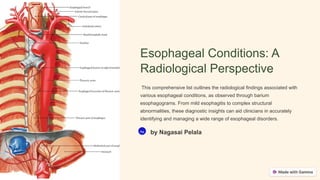
  • 1. Esophageal Conditions: A Radiological Perspective This comprehensive list outlines the radiological findings associated with various esophageal conditions, as observed through barium esophagograms. From mild esophagitis to complex structural abnormalities, these diagnostic insights can aid clinicians in accurately identifying and managing a wide range of esophageal disorders. Na by Nagasai Pelala
  • 2. Inflammatory Conditions Mild Esophagitis Characterized by fold thickening, this condition represents the mildest form of esophageal inflammation. The barium esophagogram may reveal subtle changes in the esophageal mucosal pattern, indicating the presence of this condition. Moderate Esophagitis Superficial erosions, appearing as lines and punctate defects, are the hallmark of moderate esophagitis on the barium esophagogram. These findings indicate a more advanced stage of esophageal inflammation. Severe Esophagitis In severe esophagitis, the barium esophagogram may reveal a few short and fixed transverse folds, reflecting the more significant mucosal damage and scarring associated with this condition.
  • 3. Anatomical Variations 1 Feline Folds (Physiologic) These numerous, fine, delicate, symmetric, and transient folds are a normal anatomical variant and do not indicate any underlying pathology. 2 Chronic Reflux Induced Strictures Chronic gastroesophageal reflux can lead to the development of short segment strictures above the gastroesophageal junction, often accompanied by a short type esophageal hernia. 3 Intramural Esophageal Pseudodiverticulo sis Diffuse tiny outpouchings, most commonly due to chronic reflux esophagitis, can be observed on the barium esophagogram.
  • 4. Metaplastic and Neoplastic Conditions Barrett's Esophagus This condition is characterized by a mid- esophagus stricture, often accompanied by a hiatal hernia and possible reticular changes, as seen on the barium esophagogram. These findings are indicative of the metaplastic changes associated with Barrett's esophagus. Medication-Induced Esophagitis Certain medications can cause esophageal irritation and inflammation, leading to the formation of strictures at anatomical narrowings, as observed on the barium esophagogram. Eosinophilic Esophagitis Luminal narrowing and furrowing in the upper esophagus are characteristic radiological findings associated with eosinophilic esophagitis, a condition driven by an eosinophilic immune response.
  • 5. Infectious and Inflammatory Conditions Candidiasis Multiple plaque-like filling defects on the barium esophagogram are indicative of esophageal candidiasis, a fungal infection that can occur in immunocompromised individuals. Herpes Esophagitis The barium esophagogram may reveal multiple discrete ulcerations on an otherwise normal esophageal mucosal background, characteristic of herpes esophagitis. Cytomegalovirus (CMV) Infection A large solitary discrete ulcer, especially in HIV-positive patients, is a typical radiological finding associated with CMV esophagitis.
  • 6. Motility Disorders Achalasia The barium esophagogram in achalasia typically shows a dilated esophagus with a standing column of contrast and a beak-like stricture near the gastroesophageal junction, reflecting the impaired esophageal motility and incomplete relaxation of the lower esophageal sphincter. Pseudoachalasia / Carcinoma In cases of pseudoachalasia or esophageal carcinoma, the barium esophagogram may reveal a fixed rigid stricture with a soft tissue mass evident on CT, distinguishing it from the classic achalasia findings. Diffuse Esophageal Spasm (DES) The characteristic corkscrew appearance on the barium esophagogram is a hallmark of diffuse esophageal spasm, a motility disorder characterized by uncoordinated, non- peristaltic contractions of the esophageal smooth muscle.
  • 7. Structural Abnormalities Leiomyoma A submucosal, circumscribed mass with a smooth impression on the esophageal contour is characteristic of an esophageal leiomyoma, a benign smooth muscle tumor. Esophageal Varices Serpentine filling defects with a scalloped contour in the distal esophagus are indicative of esophageal varices, dilated veins that can develop due to portal hypertension. Zenker's Diverticulum A diverticulum in the posterior wall of the cervical esophagus, known as a Zenker's diverticulum, can be visualized on the barium esophagogram.
  • 8. Miscellaneous Conditions Caustic Strictures Long segment narrowing with dysmotility, irregular margins, edema, and plaques of sloughing mucosa. Glycogenic Acanthosis Nodular filling defect resembling reticular changes. Foreign Bodies Hard objects typically lodged in the upper esophagus. Meat Impaction Commonly found at the gastroesophageal junction.