
Recommended
Recommended
More Related Content
Similar to ENIGMA_Cortical_QC_2.0.pdf
Similar to ENIGMA_Cortical_QC_2.0.pdf (20)
Recently uploaded
Recently uploaded (20)
ENIGMA_Cortical_QC_2.0.pdf
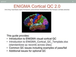
- 1. 1 ENIGMA Cortical QC 2.0 This guide provides: • Introduction to ENIGMA visual cortical QC • Introduction to ENIGMA_Cortical_QC_Template.xlsx (standardizes qc records across sites) • Common QC issues including examples of pass/fail • Additional issues for optional QC Chris Ching, Faisal Rashid, Sophia Thomopoulos, Lianne Schmaal, Premika Boedhoe, Paul Thompson, and Neda Jahanshad 4/11/17
- 2. FreeSurfer Cortical Quality Check • If you’re new to the cortical QC, we highly recommend spending some time viewing some subjects in Freeview to get familiarized with the FreeSurfer output (see next slide for directions). • Use the ENIGMA Internal QC method for checking cortical segmentation quality which is good for spotting under/ overestimations. • Use the ENIGMA External QC for checking cortical labels, anatomical boundaries, and confirming errors spotted on internal QC. • Make sure to QC all subjects, not just those flagged in the outlier.log file. • Use the ENIGMA_Cortical_QC_Template.xlsx to record your QC ratings. 2
- 3. View some subjects in Freeview (optional) Use the following example commands to view subjects in Freeview, especially if you’re new to cortical QC: • Replace yellow highlighted portions with your own paths • Replace ${SUBJECT}with a subject ID from your sample • Copy and paste into command line to load subject 3 ####Configure FreeSurfer#### export FREESURFER_HOME="/usr/local/freesurfer-5.3.0_64bit" export LIBGL_ALWAYS_INDIRECT=1 source $FREESURFER_HOME/SetUpFreeSurfer.sh export SUBJECTS_DIR=/path/to/freesurfer/output cd ${SUBJECTS_DIR} ####To view internal QC#### freeview -v ${SUBJECT}/mri/orig.mgz $SUBJECT/mri/aparc+aseg.mgz:colormap=lut:opacity=0.4 ####To view external QC:#### freeview -f ${SUBJECT}/surf/lh.pial:annot=aparc.annot:name=pial_aparc:visible=0 ${SUBJECT}/surf/rh.pial:annot=aparc.annot:name=pial_aparc:visible=0 --viewport 3d https://surfer.nmr.mgh.harvard.edu/fswiki/FreeviewGuide
- 5. Use the “ENIGMA_Cortical_QC_Template.xlsx” to record your QC First Tab: QC Subject: Subject ID’s Internal/External_QC Columns: • Pass – no issues with internal/external QC • Moderate – fail particular regions (indicate R=right, L=Left or both=R/L) • Fail – severe pathology, image artifacts, registration problems causing severe mislabeling (list R/L for all regions) Second Tab: QC_Code_Key • Provides standardized codes for the “QC_Code” column on first tab to keep track of common errors • Examples of these common errors are provided in this guide 5 Regions color-coded based on FS labels Download the template here: http://enigma.ini.usc.edu/protocols/imaging-protocols/
- 6. Good examples Labels generally correspond to known anatomical boundaries Grey matter properly segmented (no under/overestimations) 6
- 8. Internal QC Fail Fail entire subject: cerebellum misclassified and other severe global underestimations 8
- 9. Internal QC Fail Complete fail (pathology) 9 Complete fail (motion causing failed segmentation)
- 10. Internal QC: Moderate • Certain regions are under/overestimated but rest of segmentation looks good. • Can use External QC to confirm affected regions (examples to follow). • List regions misclassified in QC spreadsheet (L, R, or R/L) so they can be withheld from analysis. 10
- 11. 11 Internal QC: Moderate Temporal Pole Underestimation • FreeSurfer sometimes (<10% cases) underestimates the anterior temporal and frontal poles. • List underestimated regions in QC spreadsheet (L, R, or R/L) so they can be withheld from analysis.
- 12. External QC Ideal external segmentation (generally correspond to known anatomical boundaries) 12
- 13. External QC: Fail FAIL (Processing Error) Fail (pathology) 13
- 14. Moderate: Banks of superior temporal sulcus overestimation In about 20-30% of subjects, the BanksSTS appears on gyral surface. In some cases (≈15%) the size of the mislabeled BanksSTS may influence the surrounding ROIs (e.g. superior temporal/ middle temporal gyri). BanksSTS QC Steps: 1. Load index.html into browser. 2. Press: “ Command and + ” which zooms in once – match the size of your QC images to the size of image above (**Tip: view the PDF version of the guide at 100% and your QC images should be about the same size as the brain above**) 3. IF the mislabeled BanksSTS is larger than hand/cursor, THEN fail the BanksSTS + surrounding affected regions (e.g. superior temporal gyrus and/or middle frontal gyrus – see examples on next slides.) 14
- 15. OK examples (do not fail BanksSTS) Pass Pass Pass Pass 15
- 16. Bad examples BanksSTS (List affected regions in QC sheet) Fail BanksSTS + STG Fail BanksSTS + STG + MTG Fail BanksSTS + STG + MTG Fail BanksSTS + MTG 16 STG: superior temporal gyrus; MTG: middle temporal gyrus
- 17. Pre/Postcentral Gyrus Issues (<15% cases) 17 Notice flat regions Indicates overestimation of meninges (confirmed on internal QC) Fail Pre/Postcentral gyri and superior parietal Overestimation of the postcentral gyrus Fail pre/postcentral gyri, superior parietal, and supramarginal gyrus (may confirm on internal QC) Meninges overestimations Moderate overestimations of meninges Fail Pre/Postcentral gyri
- 18. 18 Overestimation of postcentral gyrus Fail postcentral, superior parietal, and supramarginal gyri Pre/Postcentral Gyrus Issues (<15% cases) Misclassifications Misclassification (overlap) of pre/postcentral gyri Fail Pre/Postcentral gyri, superior parietal, superior frontal, and caudal middle frontal
- 19. Pericalcarine Overestimations (<5% cases) • Segmentation overestimates pericalcarine region • Note failed regions (above: pericalcarine, lingual, and cuneus regions) 19 GOOD BAD • Segmentation confined to calcarine sulcus
- 20. Additional Issues The following includes regions that have been noted as possibly problematic but to a lesser extent than the previous examples. Choosing to record instances of the following issues may make it easier to perform follow- up analyses. 20
- 21. Parahippocampal (green) and entorhinal cortex (red) underestimations detected in 70-80% of cases • We tend to be less stringent with regard to the segmentation of the entorhinal cortex and parahippocampal gyrus because this region is commonly underestimated • **Possible findings in these regions should be interpreted with caution and it may be useful to track how often these issues to perform follow-up analysis** A B 21
- 22. Other issues: cingulate cortex 22 ● A paracingulate sulcus (PCS) is present in 30–60% of cases and is more frequently found in the left hemisphere (A and B). This can cause segmentation problems in the cingulate and surrounding regions. ● In subjects with prominent paracingulate sulcus (example D, E), portions of the cingulate may be underestimated while superior frontal regions may be overestimated. ● **We tend to be less stringent with the QC in this region because of the variability of the anatomy/segmentation. Findings in this region should be interpreted with caution and it may be useful to track this issue for follow-up analysis.** Wei et al. 2017 D E
- 23. Other issues: Insula (yellow) 23 • This Freesurfer atlas does not include a subgenual ACC region and sometimes assigns this to the insula (yellow) or medial OFC (red/pink) instead – Example A. Example B is probably more anatomically correct, but judging the accuracy of the insula or medial OFC boundaries becomes difficult. • Example C shows other somewhat common issues regarding Insula overestimation into the temporal lobes and midline. • **We tend to be less stringent with the QC of the insula because of the variability of the anatomy/ segmentation and the difficulty with establishing consistent pass/fail criteria. However, it may be useful for some groups to note how often you observe these issues for follow-up analysis.** A. B. C.
- 24. Other issues: Superior Parietal Overestimation • Segmentation overestimates superior parietal region and impacts precuneus/cuneus regions 24 Normal Overestimation • Segmentation obeys known anatomical boundaries and does not overestimate superior parietal region
- 25. Other issues: middle/inferior temporal gyrus • When the middle temporal gyrus looks as if it covers the inferior temporal gyrus, this is usually due to the rotation angle of the brain and is probably Okie Dokie (Pass). • It is considered normal when the middle and inferior temporal gyri are somewhat overlapping on each other as in the above examples (see examples of non-continuous inferior temporal sulcus). 25
- 26. Supramarginal gyrus overestimation: Extends into superior temporal gyrus 26 • In some cases the supramarginal gyrus (green) may appear to invade adjacent regions (in this example the superior temporal gyrus). • We tend to be less stringent with the QC in this region because there is quite a bit of anatomical variability and the exact boundaries dividing the supramarginal gyrus from surrounding regions can be difficult to assess.