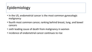
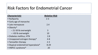
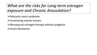
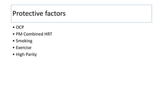
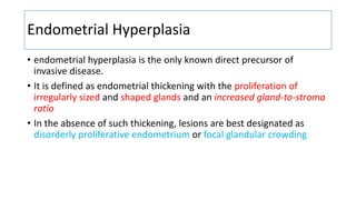
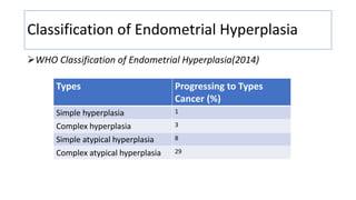
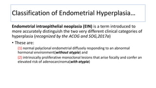
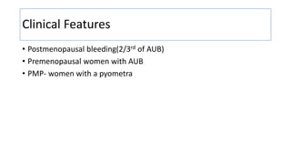
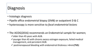
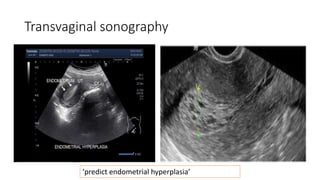
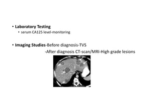
Endometrial Cancer
Girma Hirpasa ( MD, Assistant professor Obstetrics and Gynecology)
Outline
• Epidemiology and risk factors
• Endometrial hyperplasia
• Classification
• Clinical features and diagnosis
• Treatment
• Endometrial cancer
• Pathogenesis
• Prevention
• Diagnosis
• Pathology
• Treatment
Epidemiology
• In the US, endometrial cancer is the most common gynecologic
malignancy
• fourth most common cancer, ranking behind breast, lung, and bowel
cancers
• sixth leading cause of death from malignancy in women
• incidence of endometrial cancer continues to rise
Risk Factors for Endometrial Cancer
Characteristic Relative Risk
• Nulliparity 2-3
 Early age of menarche
• Late menopause 2.4
• Obesity*
– 21-50 lb overweight 3
– >50 lb overweight 10
• Diabetes mellitus, HTN 2.8
• Unopposed estrogen therapy* 4-8
• Tamoxifen therapy 2-3
• Atypical endometrial hyperplasia* 8-29
• HNPCC syndrome* 20
What are the risks for Long-term estrogen
exposure and Chronic Anovulation?
Polycystic ovary syndrome
Functioning ovarian tumors
Menopausal estrogen therapy without progestin
Insulin Resistance
Protective factors
• OCP
• PM Combined HRT
• Smoking
• Exercise
• High Parity
Endometrial Hyperplasia
• endometrial hyperplasia is the only known direct precursor of
invasive disease.
• It is defined as endometrial thickening with the proliferation of
irregularly sized and shaped glands and an increased gland-to-stroma
ratio
• In the absence of such thickening, lesions are best designated as
disorderly proliferative endometrium or focal glandular crowding
Classification of Endometrial Hyperplasia
WHO Classification of Endometrial Hyperplasia(2014)
Types Progressing to Types
Cancer (%)
Simple hyperplasia 1
Complex hyperplasia 3
Simple atypical hyperplasia 8
Complex atypical hyperplasia 29
Classification of Endometrial Hyperplasia…
Endometrial intraepithelial neoplasia (EIN) is a term introduced to
more accurately distinguish the two very different clinical categories of
hyperplasia (recognized by the ACOG and SOG,2017a)
• These are:
(1) normal polyclonal endometrial diffusely responding to an abnormal
hormonal environment(without atypia) and
(2) intrinsically proliferative monoclonal lesions that arise focally and confer an
elevated risk of adenocarcinoma(with atypia)
Clinical Features
• Postmenopausal bleeding(2/3rd of AUB)
• Premenopausal women with AUB
• PMP- women with a pyometra
Diagnosis
• histologic diagnosis
• Pipelle office endometrial biopsy (EMB) or outpatient D & C
• hysteroscopy is more sensitive to focal endometrial lesions
• The ACOG(2016) recommends an Endometrial sample for women;
older than 45 years with AUB.
younger than 45 with chronic excess estrogen exposure, failed medical
management, and persistent AUB.
 postmenopausal bleeding with endometrial thickness >4mm(TVS)
Transvaginal sonography
‘predict endometrial hyperplasia’
Treatment
 mainly depends on
• patient's age,
• comorbid risks for surgery,
• desire for fertility, and
• specific histologic features such as cytologic atypia.
• Hysterectomy is the most definitive treatment
• Hormonal therapy is another option
Treatment…
1.Nonatypical Endometrial Hyperplasia
• Premenopausal Women-may spontaneous regress without therapy
-progestins
• Postmenopausal Women-progestin
• The overall clinical and pathologic regression rates to progestin
therapy range from 70 to 80%
Treatment…
2.Atypical Endometrial Hyperplasia
• Hysterectomy is the preferred treatment
-progression to cancer over time approximates 29%
ENDOMETRIAL CANCER
Pathogenesis
characterized by a dualistic model
1.Type I endometrioid adenocarcinomas
• estrogen-dependent, low grade, and derived from atypical endometrial
hyperplasia
2.Type II cancers are serous, carcinosarcoma, or clear cell histology
• have no precursor lesion; and portend a more aggressive clinical course
ENDOMETRIAL CANCER…cont’d
Type I and II Endometrial Carcinoma Distinguishing Features
Feature Type I Type II
oChronic estrogen present absent
oMenopause status pre-/peri post-
oHyperplasia present absent
oRace white black
oGrade low high
oInvasion Minimal deep
oBehavior stable aggressive
oSubtypes endometroid serous, clear cell
Prevention
• Education
• Counseling
• routine screening of hyperplasia or endometrial cancer??
• Recommended screening(EMB) is for the woman with Lynch
syndrome
every 1 to 2 years beginning at age 30 to 35 years(ACOG,2019)
Diagnosis
Signs and Symptoms
• Irregular vaginal bleeding-In premenopausal women
• Postmenopausal bleeding
• Abnormal vaginal discharge-in older women
• pelvic pain or pressure, bloating, early satiety, and increasing
abdominal girth—advanced disease
• Papanicolaou Test-benign endometrial cells(>45years)
-atypical glandular cells
• Endometrial Sampling-Office Pipelle biopsy or D and C
-MVA*
-hysteroscopy
• Laboratory Testing
• serum CA125 level-monitoring
• Imaging Studies-Before diagnosis-TVS
-After diagnosis CT-scan/MRI-High grade lesions
Pathology
The spectrum of aggressiveness within the histopathologic types of
endometrial cancer is broad
Classification of Endometrial Carcinoma
Endometrioid adenocarcinoma-80%
o Variant with squamous differentiation
o Villoglandular variant
o Secretory variant
o Ciliated cell variant
Mucinous carcinoma-1-2%
Serous carcinoma—5-10%
Clear cell carcinoma
 Mixed cell carcinoma—10%
 Neuroendocrine tumor
 Undifferentiated carcinoma
 Squamous cell carcinoma
 Carcinosarcoma
 Others
Histologic grade
-Histologic grading is primarily determined by the tumor's
architectural growth pattern
Grade Definition
1 5% of a nonsquamous or nonmorular solid growth pattern
2 6-50% of a nonsquamous or nonmorular solid growth pattern
3 >50% of a nonsquamous or nonmorular solid growth pattern
Patterns of Spread
• Type I endometrioid tumors and their variants most commonly
spread in order of frequency, by:
• (I) direct extension, (2) lymphatic metastasis,(3) Hematogenous
dissemination, (4) intraperitoneal exfoliation.
• Type II serous and clear cell carcinoma and carcinosarcoma have a
particular propensity for extrauterine disease, in a pattern that closely
resembles epithelial ovarian cancer
Patterns of Spread…
• Invasion of endometrial stroma and exophytic expansion
• Lymphatic channel invasion haphazard pattern
• Hematogenous dissemination-- most commonly results in metastases
to the lung to the liver, brain, bone, and other sites
• Retrograde transtubal transport
• Port-site metastasis-laparoscopic staging
Treatment
• Surgical treatment
• Chemotraphy
• Radiotherapy
FIGO STAGING,2019
References
• Williams Gynecology 4th edition, 2020.
• Berek & Novak’s Gynecology 16th edition,2020.
• Current obstetrics and gynecology diagnosis and treatment, 11th
edition.
Thank you

Endometrial Cancer...................pptx

  • 1.
    Endometrial Cancer Girma Hirpasa( MD, Assistant professor Obstetrics and Gynecology)
  • 2.
    Outline • Epidemiology andrisk factors • Endometrial hyperplasia • Classification • Clinical features and diagnosis • Treatment • Endometrial cancer • Pathogenesis • Prevention • Diagnosis • Pathology • Treatment
  • 3.
    Epidemiology • In theUS, endometrial cancer is the most common gynecologic malignancy • fourth most common cancer, ranking behind breast, lung, and bowel cancers • sixth leading cause of death from malignancy in women • incidence of endometrial cancer continues to rise
  • 4.
    Risk Factors forEndometrial Cancer Characteristic Relative Risk • Nulliparity 2-3  Early age of menarche • Late menopause 2.4 • Obesity* – 21-50 lb overweight 3 – >50 lb overweight 10 • Diabetes mellitus, HTN 2.8 • Unopposed estrogen therapy* 4-8 • Tamoxifen therapy 2-3 • Atypical endometrial hyperplasia* 8-29 • HNPCC syndrome* 20
  • 5.
    What are therisks for Long-term estrogen exposure and Chronic Anovulation? Polycystic ovary syndrome Functioning ovarian tumors Menopausal estrogen therapy without progestin Insulin Resistance
  • 6.
    Protective factors • OCP •PM Combined HRT • Smoking • Exercise • High Parity
  • 7.
    Endometrial Hyperplasia • endometrialhyperplasia is the only known direct precursor of invasive disease. • It is defined as endometrial thickening with the proliferation of irregularly sized and shaped glands and an increased gland-to-stroma ratio • In the absence of such thickening, lesions are best designated as disorderly proliferative endometrium or focal glandular crowding
  • 8.
    Classification of EndometrialHyperplasia WHO Classification of Endometrial Hyperplasia(2014) Types Progressing to Types Cancer (%) Simple hyperplasia 1 Complex hyperplasia 3 Simple atypical hyperplasia 8 Complex atypical hyperplasia 29
  • 9.
    Classification of EndometrialHyperplasia… Endometrial intraepithelial neoplasia (EIN) is a term introduced to more accurately distinguish the two very different clinical categories of hyperplasia (recognized by the ACOG and SOG,2017a) • These are: (1) normal polyclonal endometrial diffusely responding to an abnormal hormonal environment(without atypia) and (2) intrinsically proliferative monoclonal lesions that arise focally and confer an elevated risk of adenocarcinoma(with atypia)
  • 10.
    Clinical Features • Postmenopausalbleeding(2/3rd of AUB) • Premenopausal women with AUB • PMP- women with a pyometra
  • 11.
    Diagnosis • histologic diagnosis •Pipelle office endometrial biopsy (EMB) or outpatient D & C • hysteroscopy is more sensitive to focal endometrial lesions • The ACOG(2016) recommends an Endometrial sample for women; older than 45 years with AUB. younger than 45 with chronic excess estrogen exposure, failed medical management, and persistent AUB.  postmenopausal bleeding with endometrial thickness >4mm(TVS)
  • 12.
  • 13.
    Treatment  mainly dependson • patient's age, • comorbid risks for surgery, • desire for fertility, and • specific histologic features such as cytologic atypia. • Hysterectomy is the most definitive treatment • Hormonal therapy is another option
  • 14.
    Treatment… 1.Nonatypical Endometrial Hyperplasia •Premenopausal Women-may spontaneous regress without therapy -progestins • Postmenopausal Women-progestin • The overall clinical and pathologic regression rates to progestin therapy range from 70 to 80%
  • 15.
    Treatment… 2.Atypical Endometrial Hyperplasia •Hysterectomy is the preferred treatment -progression to cancer over time approximates 29%
  • 16.
    ENDOMETRIAL CANCER Pathogenesis characterized bya dualistic model 1.Type I endometrioid adenocarcinomas • estrogen-dependent, low grade, and derived from atypical endometrial hyperplasia 2.Type II cancers are serous, carcinosarcoma, or clear cell histology • have no precursor lesion; and portend a more aggressive clinical course
  • 17.
    ENDOMETRIAL CANCER…cont’d Type Iand II Endometrial Carcinoma Distinguishing Features Feature Type I Type II oChronic estrogen present absent oMenopause status pre-/peri post- oHyperplasia present absent oRace white black oGrade low high oInvasion Minimal deep oBehavior stable aggressive oSubtypes endometroid serous, clear cell
  • 18.
    Prevention • Education • Counseling •routine screening of hyperplasia or endometrial cancer?? • Recommended screening(EMB) is for the woman with Lynch syndrome every 1 to 2 years beginning at age 30 to 35 years(ACOG,2019)
  • 19.
    Diagnosis Signs and Symptoms •Irregular vaginal bleeding-In premenopausal women • Postmenopausal bleeding • Abnormal vaginal discharge-in older women • pelvic pain or pressure, bloating, early satiety, and increasing abdominal girth—advanced disease
  • 20.
    • Papanicolaou Test-benignendometrial cells(>45years) -atypical glandular cells • Endometrial Sampling-Office Pipelle biopsy or D and C -MVA* -hysteroscopy
  • 21.
    • Laboratory Testing •serum CA125 level-monitoring • Imaging Studies-Before diagnosis-TVS -After diagnosis CT-scan/MRI-High grade lesions
  • 22.
    Pathology The spectrum ofaggressiveness within the histopathologic types of endometrial cancer is broad Classification of Endometrial Carcinoma Endometrioid adenocarcinoma-80% o Variant with squamous differentiation o Villoglandular variant o Secretory variant o Ciliated cell variant Mucinous carcinoma-1-2% Serous carcinoma—5-10% Clear cell carcinoma  Mixed cell carcinoma—10%  Neuroendocrine tumor  Undifferentiated carcinoma  Squamous cell carcinoma  Carcinosarcoma  Others
  • 23.
    Histologic grade -Histologic gradingis primarily determined by the tumor's architectural growth pattern Grade Definition 1 5% of a nonsquamous or nonmorular solid growth pattern 2 6-50% of a nonsquamous or nonmorular solid growth pattern 3 >50% of a nonsquamous or nonmorular solid growth pattern
  • 24.
    Patterns of Spread •Type I endometrioid tumors and their variants most commonly spread in order of frequency, by: • (I) direct extension, (2) lymphatic metastasis,(3) Hematogenous dissemination, (4) intraperitoneal exfoliation. • Type II serous and clear cell carcinoma and carcinosarcoma have a particular propensity for extrauterine disease, in a pattern that closely resembles epithelial ovarian cancer
  • 25.
    Patterns of Spread… •Invasion of endometrial stroma and exophytic expansion • Lymphatic channel invasion haphazard pattern • Hematogenous dissemination-- most commonly results in metastases to the lung to the liver, brain, bone, and other sites • Retrograde transtubal transport • Port-site metastasis-laparoscopic staging
  • 26.
    Treatment • Surgical treatment •Chemotraphy • Radiotherapy
  • 27.
  • 28.
    References • Williams Gynecology4th edition, 2020. • Berek & Novak’s Gynecology 16th edition,2020. • Current obstetrics and gynecology diagnosis and treatment, 11th edition.
  • 29.