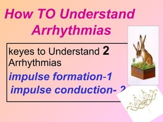
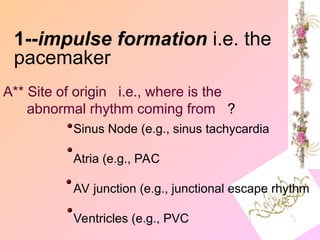
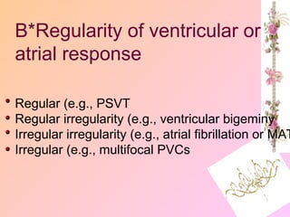
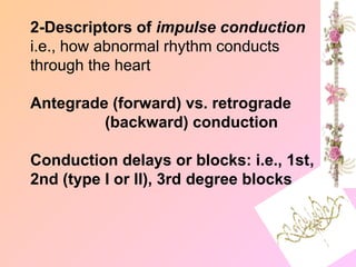
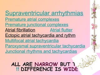
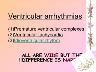
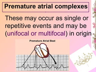
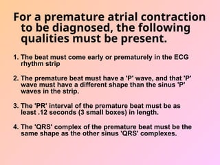
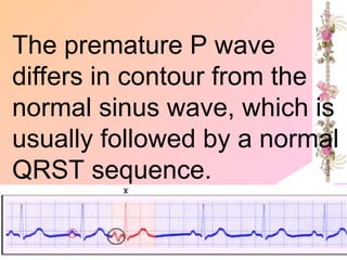
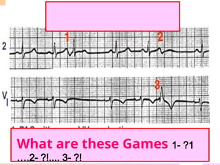
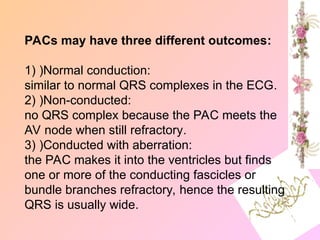
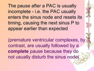
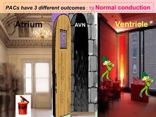
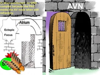
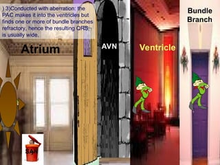
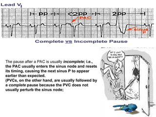
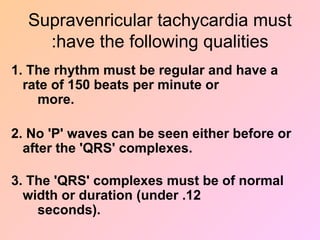
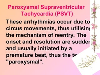
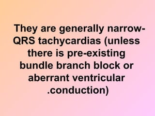
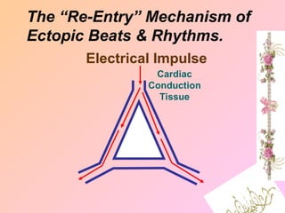
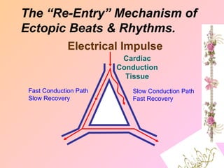
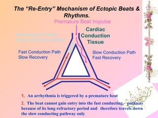
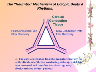
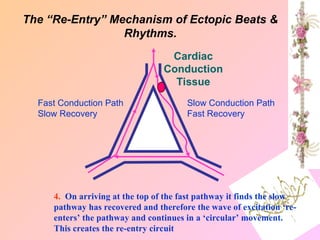
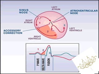
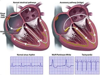
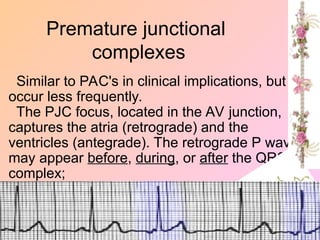
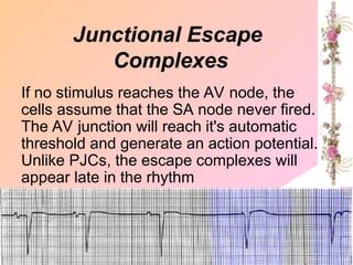
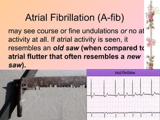
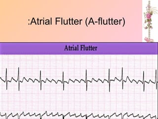
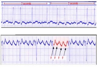
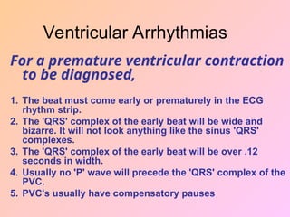
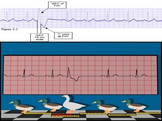
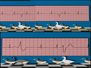
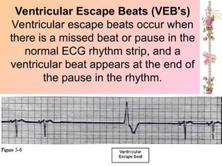
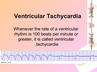
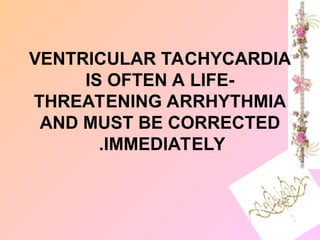
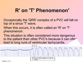
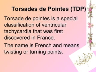
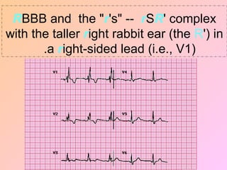
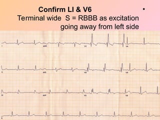
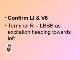
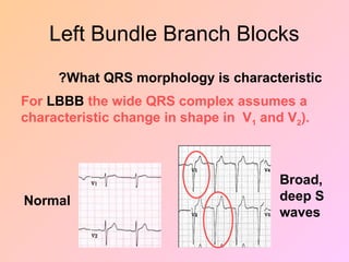
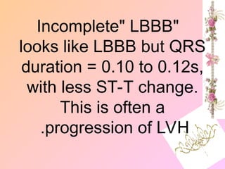
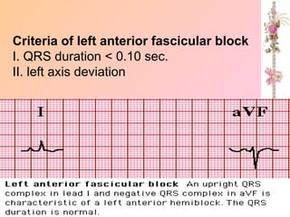
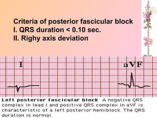
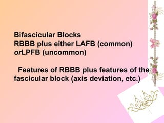
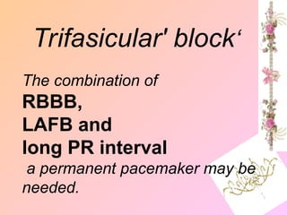
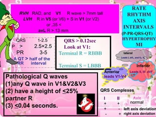
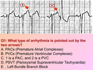
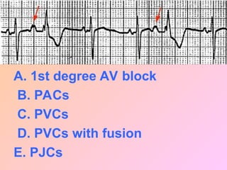
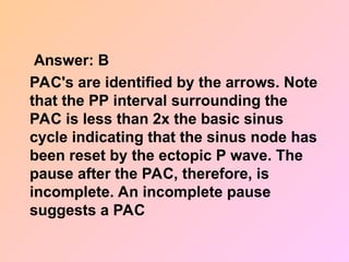
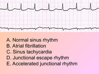
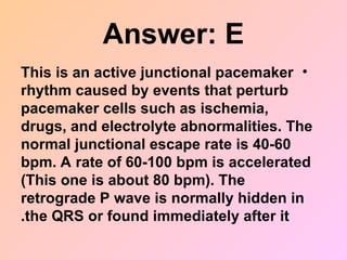
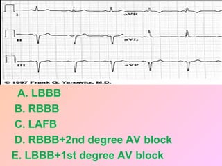
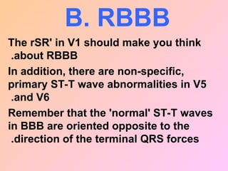
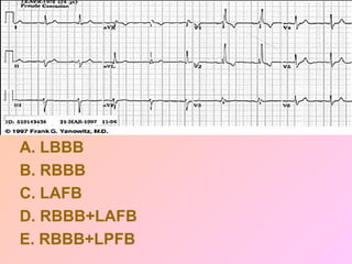
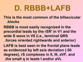
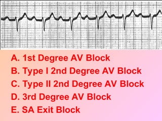
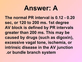
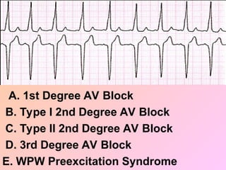
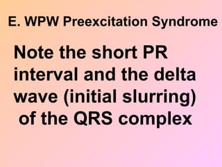
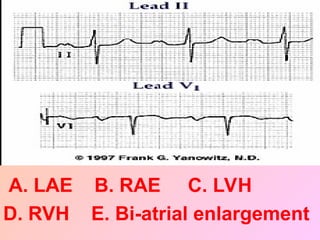
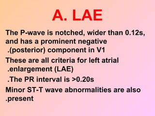
Pathological Q waves
(1)any Q wave in V1&V2&V3
(2) have a height of <25%
partner R
(3) <0.04 seconds.
RVH RAD, and V1 R wave > 7mm tall
LVH R in V5 (or V6) + S in V1 (or V2)
>
35
,
or
avL R > 13 mm
QRS 1-2.5
P > 2.5×2.5
PR 3-5
A QT > half of the
RR interval
QRS > 0.12sec
Look at V1:
Terminal R = RBBB
Terminal S = LBBB
QRS Complexes
I II Axis
+ + normal
+ - left axis deviation
- + right axis deviation
RATE
RHYTHM
AXIS
INTERVALS
(P-PR-QRS-QT)
HYPERTROPHY
MI
lateral
Leads I, aVL, and V5- V6
.
Anterior
leads V1-V4
Inferior
Leads II, III aVF
2
keyes to Understand
Arrhythmias
1
-
impulse formation
2
-
impulse conduction
How TO Understand
Arrhythmias
1--impulse formation i.e. the
pacemaker
A** Site of origin i.e., where is the
abnormal rhythm coming from ?
Sinus Node (e.g., sinus tachycardia
Atria (e.g., PAC
AV junction (e.g., junctional escape rhythm
Ventricles (e.g., PVC
B*Regularity of ventricular or
atrial response
Regular (e.g., PSVT
Regular irregularity (e.g., ventricular bigeminy
Irregular irregularity (e.g., atrial fibrillation or MAT
Irregular (e.g., multifocal PVCs
2-Descriptors of impulse conduction
i.e., how abnormal rhythm conducts
through the heart
Antegrade (forward) vs. retrograde
(backward) conduction
Conduction delays or blocks: i.e., 1st,
2nd (type I or II), 3rd degree blocks
ALL ARE NARROW BUT THE
DIFFERENCE IS WIDE
!!
Supraventricular arrhythmias
Premature atrial complexes
Premature junctional complexes
Atrial fibrillation Atrial flutter
Ectopic atrial tachycardia and rythm
Multifocal atrial tachycardia
Paroxysmal supraventricular tachycardia
Junctional rhythms and tachycardias
IT IS TOO
WIDE
HOW TO
NARROW
??
IT IS TOO WIDE
HOW TO NARROW
??
ALL ARE WIDE BUT THE
DIFFERENCE IS NARROW
!!
Ventricular arrhythmias
(1)Premature ventricular complexes
(2)Ventricular tachycardia
(3)Idioventricular rhythm
These may occur as single or
repetitive events and may be
(unifocal or multifocal) in origin
Premature atrial complexes
Premature atrial contractions, occur
whenever an ectopic site in the
heart's atria produces an electrical
impulse early or prematurely in the
ECG rhythm strip.
Premature contractions are also
know as Premature Beats,
Ectopics, Premature Systoles and
Extra Systoles.
For a premature atrial contraction
to be diagnosed, the following
qualities must be present.
1. The beat must come early or prematurely in the ECG
rhythm strip
2. The premature beat must have a 'P' wave, and that 'P'
wave must have a different shape than the sinus 'P'
waves in the strip.
3. The 'PR' interval of the premature beat must be as
least .12 seconds (3 small boxes) in length.
4. The 'QRS' complex of the premature beat must be the
same shape as the other sinus 'QRS' complexes.
The premature P wave
differs in contour from the
normal sinus wave, which is
usually followed by a normal
QRST sequence.
•Whenever a gap occurs in
a rhythm strip, always
check the beat at the
front of the gap to see if it
is different from the other
beats in the rhythm strip
What are these Games 1- ?1
….2- ?!.... 3- ?!
PACs may have three different outcomes:
1) )Normal conduction:
similar to normal QRS complexes in the ECG.
2) )Non-conducted:
no QRS complex because the PAC meets the
AV node when still refractory.
3) )Conducted with aberration:
the PAC makes it into the ventricles but finds
one or more of the conducting fascicles or
bundle branches refractory, hence the resulting
QRS is usually wide.
The pause after a PAC is usually
incomplete - i.e. the PAC usually
enters the sinus node and resets its
timing, causing the next sinus P to
appear earlier than expected
(premature ventricular complexes, by
contrast, are usually followed by a
complete pause because they do
not usually disturb the sinus node(
Atrium AVN Ventricle
SAN
PACs have 3 different outcomes : 1)) Normal conduction
SAN
SAN
Ectopic
Focus
AVN
2) )Non-conducted: no QRS
complex because the PAC
meets the AV node when still
refractory.
Atrium AVN Ventricle
Bundle
Branch
SAN
) 3)Conducted with aberration: the
PAC makes it into the ventricles but
finds one or more of bundle branches
refractory, hence the resulting QRS
is usually wide.
The pause after a PAC is usually incomplete; i.e.,
the PAC usually enters the sinus node and resets
its timing, causing the next sinus P to appear
earlier than expected.
(PVCs, on the other hand, are usually followed by
a complete pause because the PVC does not
usually perturb the sinus node;
Pathological Q waves
(1)any Q wave in V1&V2&V3
(2) have a height of <25%
partner R
(3) <0.04 seconds.
RVH RAD, and V1 R wave > 7mm tall
LVH R in V5 (or V6) + S in V1 (or V2)
>
35
,
or
avL R > 13 mm
QRS 1-2.5
P > 2.5×2.5
PR 3-5
A QT > half of the
RR interval
QRS > 0.12sec
Look at V1:
Terminal R = RBBB
Terminal S = LBBB
QRS Complexes
I II Axis
+ + normal
+ - left axis deviation
- + right axis deviation
RATE
RHYTHM
AXIS
INTERVALS
(P-PR-QRS-QT)
HYPERTROPHY
MI
lateral
Leads I, aVL, and V5- V6
.
Anterior
leads V1-V4
Inferior
Leads II, III aVF
Enter the goose
(1)PACs Conducted
with aberration &
(2)PVC ?!!!
How to Differentiate
Between:
NO YES (VT)
Comment: likelihood of VT is so high in
patients with previous myocardial infarction (or
substantial organic heart disease) that one
should manage as for ventricular tachycardia
without wasting further time evaluating the
ECG! )
Step 1: Is any one of the following
present: ischaemic heart disease, or
heart failure, or past heart surgery, or
cardiac enlargement?
Comment:. Absence of any RS
complex in the precordial leads
V1..V6 is 100% specific for VT,
but is insensitive (26%)
.
Step 2: Are there any RS
complexes in the V leads?
NO (VT)
YES
Aduration of over 100ms
almost excludes SVT
(98% specific for VT).
Step 3: Is the interval from
start of R to nadir of S
> 100ms?
NO YES (VT)
evidence of AV
dissociation is 98%
specific for VT.
Step 4: Is there AV
dissociation,
NO YES (VT)
Whenever one of these two 'QRS'
shapes appeared as a single beat or
as a run of beats, they were found to
be most likely produced from
aberrant ventricular conduction
A Word of Warning
When there is any doubt whether beats
are ventricular or aberrant, it is best to
always consider them as ventricular
to be on the safe side. Ventricular
rhythms are life-threatening, whereas
aberrant ones are not.
Supraventricular
Tachycardia (SVT)
Supraventricular tachycardia
describes any cardiac
rhythm where the rate is 150
beats per minute or greater,
and there is no evidence of
any 'P' waves being present.
When cardiac rates reach 150 beats per
minute, the 'P' waves are often lost or
buried in the 'T' waves.
This makes it impossible to determine
whether the rhythms are produced by
an
ectopic site in the atria, creating an atrial
tachycardia,
or in the AV node, creating a junctional
tachycardia
Supravenricular tachycardia must
have the following qualities
:
1. The rhythm must be regular and have a
rate of 150 beats per minute or
more.
2. No 'P' waves can be seen either before or
after the 'QRS' complexes.
3. The 'QRS' complexes must be of normal
width or duration (under .12
seconds).
Atrial Tachycardia
• Atrial tachycardia occurs
when a single ectopic site
in one of the atria takes
over pacing the heart
instead of the SA node
An Atrial Tachycardia rhythm
will have the following
1. The rate will be regular.
2. A 'P' wave will precede each
'QRS' complex.
3. The 'PR' intervals will all be the
same and over .12 seconds in
length.
Multifocal Atrial Tachycardia
(MAT)
Discrete, multifocal P' waves occurring at
rates of 100-250/min and with varying P'R
intervals (should see at least 3 different P
wave morphologies in a given lead).
.
Ventricular response is
irregularly irregular (i.e.,
often confused with A-fib).
May be intermittent,
alternating with periods of
normal sinus rhythm
• Multifocal atrial tachycardia will have the
following qualities:
1. It will be irregular.
2. It will have a rate of 150 beats per
minute or greater.
3. It will have different shape 'P' waves all
mixed in with the 'T' wave
Paroxysmal Supraventricular
Tachycardia (PSVT)
When a short burst of SVT
occurs in a rhythm strip, it is
called a run of Paroxysmal
Supraventricular
Tachycardia PSVT).
Pathological Q waves
(1)any Q wave in V1&V2&V3
(2) have a height of <25%
partner R
(3) <0.04 seconds.
RVH RAD, and V1 R wave > 7mm tall
LVH R in V5 (or V6) + S in V1 (or V2)
>
35
,
or
avL R > 13 mm
QRS 1-2.5
P > 2.5×2.5
PR 3-5
A QT > half of the
RR interval
QRS > 0.12sec
Look at V1:
Terminal R = RBBB
Terminal S = LBBB
QRS Complexes
I II Axis
+ + normal
+ - left axis deviation
- + right axis deviation
RATE
RHYTHM
AXIS
INTERVALS
(P-PR-QRS-QT)
HYPERTROPHY
MI
lateral
Leads I, aVL, and V5- V6
.
Anterior
leads V1-V4
Inferior
Leads II, III aVF
These arrhythmias occur due to
circus movements, thus utilising
the mechanism of reentry. The
onset and resolution are sudden
and usually initiated by a
premature beat, thus the term
"paroxysmal".
Paroxysmal Supraventricular
Tachycardia (PSVT)
They are generally narrow-
QRS tachycardias (unless
there is pre-existing
bundle branch block or
aberrant ventricular
conduction)
.
The “Re-Entry” Mechanism of
Ectopic Beats & Rhythms.
Electrical Impulse
Cardiac
Conduction
Tissue
Fast Conduction Path
Slow Recovery
Slow Conduction Path
Fast Recovery
The “Re-Entry” Mechanism of
Ectopic Beats & Rhythms.
Electrical Impulse
Cardiac
Conduction
Tissue
Fast Conduction Path
Slow Recovery
Slow Conduction Path
Fast Recovery
Premature Beat Impulse
Cardiac
Conduction
Tissue
1. An arrhythmia is triggered by a premature beat
2. The beat cannot gain entry into the fast conducting pathway
because of its long refractory period and therefore travels down
the slow conducting pathway only
Repolarizing Tissue
(long refractory period)
The “Re-Entry” Mechanism of Ectopic Beats &
Rhythms.
3. The wave of excitation from the premature beat arrives
at the distal end of the fast conducting pathway, which has
now recovered and therefore travels retrogradely
(backwards) up the fast pathway
Fast Conduction Path
Slow Recovery
Slow Conduction Path
Fast Recovery
Cardiac
Conduction
Tissue
The “Re-Entry” Mechanism of Ectopic Beats &
Rhythms.
4. On arriving at the top of the fast pathway it finds the slow
pathway has recovered and therefore the wave of excitation ‘re-
enters’ the pathway and continues in a ‘circular’ movement.
This creates the re-entry circuit
Fast Conduction Path
Slow Recovery
Slow Conduction Path
Fast Recovery
Cardiac
Conduction
Tissue
The “Re-Entry” Mechanism of Ectopic Beats &
Rhythms.
There are several types
of PSVT, depending
on the location of the
reentry circuit
AV nodal reentrant
tachycardia (AVNRT)
This is the most common form of
PSVT accounting for approximately
50% of all symptomatic PSVTs.
It is the most common cause of SVT in
patients with structurally normal
hearts.
It is accompanied by the following features:
1) ECG shows normal regular QRS complexes with a
rate of 140-240 bpm.
2) QRS complexes may sometimes show typical bundle
branch block/aberration.
3) P waves are either not visible or are seen
immediately before or after the QRS complex.
4) An attack may be spontaneously triggered but
exertion, tea, coffee or alcohol may trigger an AVNRT.
5) Vagal manoeuvres may terminate an acute attack.
6) Beta-blockers or calcium channel blockers may help
to suppress recurrent attacks.
Wolff-Parkinson-White
Syndrome
This is the second most common form of PSVT
and is seen in patients with Wolff-Parkinson-
White (WPW) syndrome. The WPW ECG, seen
in the diagram, shows a short PR, delta wave and
a somewhat widened QRS
.
Similar to PAC's in clinical implications, but
occur less frequently.
The PJC focus, located in the AV junction,
captures the atria (retrograde) and the
ventricles (antegrade). The retrograde P wave
may appear before, during, or after the QRS
complex;
Premature junctional
complexes
If no stimulus reaches the AV node, the
cells assume that the SA node never fired.
The AV junction will reach it's automatic
threshold and generate an action potential.
Unlike PJCs, the escape complexes will
appear late in the rhythm
Junctional Escape
Complexes
may see course or fine undulations or no atrial
activity at all. If atrial activity is seen, it
resembles an old saw (when compared to
atrial flutter that often resembles a new
saw).
Atrial Fibrillation (A-fib)
Atrial Flutter (A-flutter)
:
For a premature ventricular contraction
to be diagnosed,
1. The beat must come early or prematurely in the ECG
rhythm strip.
2. The 'QRS' complex of the early beat will be wide and
bizarre. It will not look anything like the sinus 'QRS'
complexes.
3. The 'QRS' complex of the early beat will be over .12
seconds in width.
4. Usually no 'P' wave will precede the 'QRS' complex of the
PVC.
5. PVC's usually have compensatory pauses
Ventricular Arrhythmias
Pathological Q waves
(1)any Q wave in V1&V2&V3
(2) have a height of <25%
partner R
(3) <0.04 seconds.
RVH RAD, and V1 R wave > 7mm tall
LVH R in V5 (or V6) + S in V1 (or V2)
>
35
,
or
avL R > 13 mm
QRS 1-2.5
P > 2.5×2.5
PR 3-5
A QT > half of the
RR interval
QRS > 0.12sec
Look at V1:
Terminal R = RBBB
Terminal S = LBBB
QRS Complexes
I II Axis
+ + normal
+ - left axis deviation
- + right axis deviation
RATE
RHYTHM
AXIS
INTERVALS
(P-PR-QRS-QT)
HYPERTROPHY
MI
lateral
Leads I, aVL, and V5- V6
.
Anterior
leads V1-V4
Inferior
Leads II, III aVF
Ventricular Escape Beats (VEB's)
Ventricular escape beats occur when
there is a missed beat or pause in the
normal ECG rhythm strip, and a
ventricular beat appears at the end of
the pause in the rhythm.
Ventricular Escaped beats must have
1. They must occur at the end of a
pause.
2. The 'QRS' complex of this late beat
must be over .12 seconds in width.
3. No 'P' wave will precede the
ventricular beat.
Whenever the rate of a ventricular
rhythm is 100 beats per minute or
greater, it is called ventricular
tachycardia
.
Ventricular Tachycardia
VENTRICULAR TACHYCARDIA
IS OFTEN A LIFE-
THREATENING ARRHYTHMIA
AND MUST BE CORRECTED
IMMEDIATELY
.
Occasionally the 'QRS' complex of a PVC will fall on
top of a sinus 'T' wave.
When this occurs, it is often called an 'R' on 'T'
phenomenon.
This situation is often considered more dangerous
to the patient than other PVC's because it can often
lead to long runs of ventricular tachycardia.
'
R' on 'T' Phenomenon
Torsade de pointes is a special
classification of ventricular
tachycardia that was first
discovered in France.
The name is French and means
twisting or turning points.
Torsades de Pointes (TDP)
The 'QRS' complexes of this rhythm
are wide and bizarre as in other
forms of ventricular rhythms, but
the 'QRS' complexes will first be in
one direction from the ECG
baseline, such as positive or above
the baseline, then rotate so that they
become negative or below the
baseline
The rhythm is regular, and the
main feature that sets it off
from other ventricular rhythms
is that the height or amplitude
of 'QRS' complexes varies.
My Brother
Answer Me
Please
Pathological Q waves
(1)any Q wave in V1&V2&V3
(2) have a height of <25%
partner R
(3) <0.04 seconds.
RVH RAD, and V1 R wave > 7mm tall
LVH R in V5 (or V6) + S in V1 (or V2)
>
35
,
or
avL R > 13 mm
QRS 1-2.5
P > 2.5×2.5
PR 3-5
A QT > half of the
RR interval
QRS > 0.12sec
Look at V1:
Terminal R = RBBB
Terminal S = LBBB
QRS Complexes
I II Axis
+ + normal
+ - left axis deviation
- + right axis deviation
RATE
RHYTHM
AXIS
INTERVALS
(P-PR-QRS-QT)
HYPERTROPHY
MI
lateral
Leads I, aVL, and V5- V6
.
Anterior
leads V1-V4
Inferior
Leads II, III aVF
Intraventricular
Blocks
Bundle Branch Blocks
With Bundle Branch Blocks you will see two changes
on the ECG.
1. QRS complex widens (> 0.12 sec).
2. QRS morphology changes (varies depending on ECG lead,
and if it is a right vs. left bundle branch block).
If the QRS Complex is Wide
If the rhythm is supraventricular and QRS
winding is not due to WPW
there are only 3 possibilities:
1. There may be typical RBBB
2. There may be typical LBBB
3. IVCD (IntraVentricular Conduction Delay).
Note: The 3 key leads
(and the only 3 leads
needed) to determine the
type of conduction defect
(RBBB, LBBB, or IVCD)
are leads I, V1, and V6.
ECG Diagnosis of Bundle Branch Block
QRS > 0.12sec
Look at V1:
Terminal R = RBBB
as excitation spreading from left to right
Terminal S = LBBB
as excitation spreading away from right
Right Bundle Branch Blocks
What QRS morphology is characteristic
?
V1
For RBBB the wide QRS complex assumes a
unique, virtually diagnostic shape in those
leads overlying the right ventricle (V1 and V2).
“Rabbit Ears”
RBBB and the "r's" -- rSR' complex
with the taller right rabbit ear (the R') in
a right-sided lead (i.e., V1)
.
•
Confirm LI & V6
Terminal wide S = RBBB as excitation
going away from left side
ECG Diagnosis of Bundle Branch Block
QRS > 0.12sec
Look at V1:
Terminal R = RBBB
as excitation spreading from left to right
Terminal S = LBBB
as excitation spreading away from right
• Confirm LI & V6
• Terminal R = LBBB as
excitation heading towards
left
Left Bundle Branch Blocks
What QRS morphology is characteristic
?
For LBBB the wide QRS complex assumes a
characteristic change in shape in V1 and V2).
Broad,
deep S
waves
Normal
"
Incomplete" RBBB
has a QRS duration of
0.10 - 0.12s with the
same terminal QRS
features. This is often
a normal variant
"
Incomplete" LBBB
looks like LBBB but QRS
duration = 0.10 to 0.12s,
with less ST-T change.
This is often a
progression of LVH
.
Criteria of left anterior fascicular block
I. QRS duration < 0.10 sec.
II. left axis deviation
Criteria of posterior fascicular block
I. QRS duration < 0.10 sec.
II. Righy axis deviation
Bifascicular Blocks
RBBB plus either LAFB (common)
orLPFB (uncommon)
Features of RBBB plus features of the
fascicular block (axis deviation, etc.)
‘
Trifasicular' block
The combination of
RBBB,
LAFB and
long PR interval
a permanent pacemaker may be
needed.
Pathological Q waves
(1)any Q wave in V1&V2&V3
(2) have a height of <25%
partner R
(3) <0.04 seconds.
RVH RAD, and V1 R wave > 7mm tall
LVH R in V5 (or V6) + S in V1 (or V2)
>
35
,
or
avL R > 13 mm
QRS 1-2.5
P > 2.5×2.5
PR 3-5
A QT > half of the
RR interval
QRS > 0.12sec
Look at V1:
Terminal R = RBBB
Terminal S = LBBB
QRS Complexes
I II Axis
+ + normal
+ - left axis deviation
- + right axis deviation
RATE
RHYTHM
AXIS
INTERVALS
(P-PR-QRS-QT)
HYPERTROPHY
MI
lateral
Leads I, aVL, and V5- V6
.
Anterior
leads V1-V4
Inferior
Leads II, III aVF
Q1: What type of arrhythmia is pointed out by the
two arrows?
A. PACs (Premature Atrial Complexes)
B. PVCs (Premature Ventricular Complexes)
C. 1 is a PAC, and 2 is a PVC
D. PSVT (Paroxysmal Supraventricular Tachycardia)
E. . Left Bundle Branch Block
A. PACs (Premature Atrial
Complexes)
1 is a PAC with normal IV
conduction and 2 is a sore thumb
PAC with RBBB aberration. PACs
have three fates: normal
conduction into ventricles,
aberrant conduction in ventricles,
and non-conduction.
A. 1st degree AV block
B. PACs
C. PVCs
D. PVCs with fusion
E. PJCs
B. PACs
These PAC's, indicated by arrows, enter the
ventricles and find the right bundle refractory
.
They therefore conduct with RBBB aberrancy
.
In most normal hearts the right bundle recovery
time is longer than the left bundle's; most
aberrancy, therefore, has a RBBB
morphology
.
tachycardia
what type of pause do you see after this funny
looking premature beat?
A.Complete compensatory pause
B. incomplete compensatory pause
C. No pause (next beat is on time)
D. Interpolated pause
E. None of the above
Answer: A
The distance between the P waves
before and after the PVC is the same
distance as two P cycles elsewhere in
the rhythm strip. This implies that the
sinus node was not reset, and that the
fu nny looking beat is a PVC
.
As a side note, this PVC occurs on the
peak of the T wave of the preceeding
beat making it an R on T
.
what type of pause do you see after either of these
two premature beats?
A.Complete compensatory pause
B. Incomplete compensatory pause
C. No pause (next beat is on time)
D. Interpolated pause
E. None of the above
Answer: B
PAC's are identified by the arrows. Note
that the PP interval surrounding the
PAC is less than 2x the basic sinus
cycle indicating that the sinus node has
been reset by the ectopic P wave. The
pause after the PAC, therefore, is
incomplete. An incomplete pause
suggests a PAC
A. PJC (Premature junctional complex)
B. Atrial flutter
C. Atrial fibrillation
D. AV nodal reentrant tachycardia
E. Accelerated junctional rhythm
Answer: C
Atrial fibrillation is characterized by an
irregularly irregular ventricular
response, and the absence of discrete
P waves.
In the top lead in this ECG, atrial activity
is poorly defined. The atrial activity
seen in the lower lead resembles old
saw-teeth (as opposed to the new,
sharp saw-teeth of atrial flutter).
A. Sinus tachycardia
B. Paroxysmal supraventricular tachycardia
C. 3rd degree AV block
D. Atrial fibrillation
E. Atrial flutter with 2:1 block
Answer: E
This is the most commonly
mis-diagnosed SV tachycardia
.
Clues are a ventricular rate of around 150
bpm (atrial of ~300 bpm), and regular R-
R intervals, and two atrial events for
every QRS (if you can find them!)
.
A. Normal sinus rhythm
B. Atrial fibrillation
C. Sinus tachycardia
D. Junctional escape rhythm
E. Accelerated junctional rhythm
Answer: E
•
This is an active junctional pacemaker
rhythm caused by events that perturb
pacemaker cells such as ischemia,
drugs, and electrolyte abnormalities. The
normal junctional escape rate is 40-60
bpm. A rate of 60-100 bpm is accelerated
(This one is about 80 bpm). The
retrograde P wave is normally hidden in
the QRS or found immediately after it
.
•
A. Ventricular tachycardia
•
B. Supraventricular tachycardia with
aberration
•
C. Accelerated junctional rhythm
•
D. Accelerated ventricular rhythm
•
E. Ventricular bigeminy
Answer: A
Hints to this are the wide QRS, and the fact that
regular sinus P waves can be identified which are
slower than AND dissociated from the ventricular
rate
.
Approximately 50 percent of ventricular
tachycardias are associated with AV dissociation.
In these cases atrial impulses can enter the
ventricles and either fuse with a ventricular
ectopic beat or completely capture the ventricles
A. LBBB
B. RBBB
C. LBBB+2nd degree AV block
D. RBBB+1st degree AV block
E. LAFB
BBB causes sequential rather than simultaneous
activation of the ventricles. The second half of the QRS
represents the ventricle with the blocked bundle
because that ventricle is activated later
.
Leads I and V1 show that terminal QRS forces are oriented
leftward and posterior indicating LV forces. Therefore,
LBBB is recognized by: 1) QRS duration > 0.12s 2)
monophasic R waves in I and V6 3) terminal QRS forces
oriented leftwards (see lead I) and posterior (see V1).
Also, in BBB the ST-T waves should be oriented
opposite to the terminal QRS forces, and the increased
voltage in V2 is normal
A. LBBB
B. RBBB
C. LBBB+1st degree AV block
D. RBBB+1st degree AV block
E. LAFB
Answer: B
The wide QRS suggests a BBB.
Looking at the latter half of the
QRS in I and V1, the late forces are
rightward and anterior. Thus the
right ventricle has been blocked
and depolarized after the left
ventricle. The rSR' complex seen in
V1 is commonly seen with RBBB
.
A. LBBB
B. RBBB
C. LAFB
D. RBBB+2nd degree AV block
E. LBBB+1st degree AV block
C. LAFB
The mainly negative QRS in lead II should clue you
in to a left axis deviation which is the main ECG
abnormality produced by LAFB
.
Some other findings are
:
1
)
rS complexes in leads II, III, and aVF
2
)
tiny q waves in I and/or aVL
3
)
poor R wave progression in V1-V3 (not seen in
this ECG)
4
)
narrow (normal) QRS LAFB is the most common
IV conduction defect
.
A. LBBB
B. RBBB
C. RBBB+LPFB
D. RBBB+LAFB
E. LBBB+1st degree AV block
B. RBBB
The rSR' in V1 should make you think
about RBBB
.
In addition, there are non-specific,
primary ST-T wave abnormalities in V5
and V6
.
Remember that the 'normal' ST-T waves
in BBB are oriented opposite to the
direction of the terminal QRS forces
.
A. LBBB
B. RBBB
C. LAFB
D. RBBB+LAFB
E. RBBB+LPFB
D. RBBB+LAFB
This is the most common of the bifascicular
blocks
.
RBBB is most easily recognized in the
precordial leads by the rSR' in V1 and the
wide S wave in V6 (i.e., terminal QRS
forces oriented rightwards and anterior)
.
LAFB is best seen in the frontal plane leads
as evidenced by left axis deviation (-50
degrees), rS complexes in II, III, aVF, and
the small q in leads I and/or aVL
.
A. 1st Degree AV Block
B. Type I 2nd Degree AV Block
C. Type II 2nd Degree AV Block
D. 3rd Degree AV Block
E. Sinus arrhythmia
B. Type I 2nd Degree AV Block
The 3 rules of classic AV Wenckebach
are: 1) the PR interval lengthens until a
nonconducted P wave occurs
2
)
the RR interval of the pause is less
than the two preceding RR intervals
3
)
the RR interval after the pause is
greater than the RR interval just prior
to the pause
.
A. 1st Degree AV Block
B. Type I 2nd Degree AV Block
C. Type II 2nd Degree AV Block
D. 3rd Degree AV Block
E. SA Exit Block
Answer: A
The normal PR interval is 0.12 - 0.20
sec, or 120 to 200 ms. 1st degree
AV block is defined by PR intervals
greater than 200 ms. This may be
caused by drugs (such as digoxin),
excessive vagal tone, ischemia, or
intrinsic disease in the AV junction
or bundle branch system
.
A. 1st Degree AV Block
B. Type I 2nd Degree AV Block
C. Type II 2nd Degree AV Block
D. Intermittent 3rd Degree AV Block
E. WPW Preexcitation Syndrome
C. Type II 2nd Degree AV Block
•
The constant PR interval distinguishes this from
type I AV block. Mobitz II 2nd degree AV block is
usually a sign of bilateral bundle branch disease.
One of the two bundle branches is completely
blocked (note the wide, negative S in V1 = LBBB)
.
•
The nonconducted sinus P waves are most likely
blocked in the other bundle (ie, the right bundle)
which exhibits 2nd degree block. Although unlikely,
it is possible that the P waves are blocked
somewhere in the AV junction such as the His
bundle
.
A. 1st Degree AV Block
B. Type I 2nd Degree AV Block
C. Type II 2nd Degree AV Block
D. 3rd Degree AV Block
E. WPW Preexcitation Syndrome
Note the short PR
interval and the delta
wave (initial slurring)
of the QRS complex
A. LVH
B. RVH
C. LAE
D. RAE
E. Bi-atrial
enlargement
•
LAE is best seen in V1
with a prominent negative
(posterior) component
measuring 1mm wide and
1mm deep
.
A. LAE B. RAE C. LVH
D. RVH E. Bi-atrial enlargement
A. LAE
The P-wave is notched, wider than 0.12s,
and has a prominent negative
(posterior) component in V1
.
These are all criteria for left atrial
enlargement (LAE)
.
The PR interval is >0.20s
.
Minor ST-T wave abnormalities are also
present
.
A. Anterolateral MI B. High lateral MI
C. True posterior MI D. Inferolateral MI
E. Inferior MI
E. Inferior MI
The site of infarction can be localized by
remembering that each lead reflects a specific
area of the heart
.
Note the pathologic Q waves in leads II, III, and
aVF
.
Also, there are inverted T waves in the same leads
with a small amount of residual ST elevation
.
This is a classic inferior MI. It's not a new MI
because the ST elevation has mostly returned to
normal
A. Inferior MI B. Posterior MI
C. Inferoposterior MI
D. Anterior MI
E. Non-Q wave MI
C. Inferoposterior MI
•
The inferior diagnosis is made from leads II, III,
and aVF (Q waves and inverted T's)
.
•
The posterior part of the infarct doesn't result in
pathologic Q waves, but rather in patholigic R
waves in V1-V3. The R/S ratio in V1 or V2 is >1
.
•
Another term for these tall and wide R waves in
V1-V2 is prominent anterior forces. The
infarcted posterior tissue allows the normal
anterior forces to become more prominent on
the ECG
.
Electrocardiography - How to read an ECG

Electrocardiography - How to read an ECG

  • 2.
    Pathological Q waves (1)anyQ wave in V1&V2&V3 (2) have a height of <25% partner R (3) <0.04 seconds. RVH RAD, and V1 R wave > 7mm tall LVH R in V5 (or V6) + S in V1 (or V2) > 35 , or avL R > 13 mm QRS 1-2.5 P > 2.5×2.5 PR 3-5 A QT > half of the RR interval QRS > 0.12sec Look at V1: Terminal R = RBBB Terminal S = LBBB QRS Complexes I II Axis + + normal + - left axis deviation - + right axis deviation RATE RHYTHM AXIS INTERVALS (P-PR-QRS-QT) HYPERTROPHY MI lateral Leads I, aVL, and V5- V6 . Anterior leads V1-V4 Inferior Leads II, III aVF
  • 3.
    2 keyes to Understand Arrhythmias 1 - impulseformation 2 - impulse conduction How TO Understand Arrhythmias
  • 4.
    1--impulse formation i.e.the pacemaker A** Site of origin i.e., where is the abnormal rhythm coming from ? Sinus Node (e.g., sinus tachycardia Atria (e.g., PAC AV junction (e.g., junctional escape rhythm Ventricles (e.g., PVC
  • 5.
    B*Regularity of ventricularor atrial response Regular (e.g., PSVT Regular irregularity (e.g., ventricular bigeminy Irregular irregularity (e.g., atrial fibrillation or MAT Irregular (e.g., multifocal PVCs
  • 6.
    2-Descriptors of impulseconduction i.e., how abnormal rhythm conducts through the heart Antegrade (forward) vs. retrograde (backward) conduction Conduction delays or blocks: i.e., 1st, 2nd (type I or II), 3rd degree blocks
  • 7.
    ALL ARE NARROWBUT THE DIFFERENCE IS WIDE !! Supraventricular arrhythmias Premature atrial complexes Premature junctional complexes Atrial fibrillation Atrial flutter Ectopic atrial tachycardia and rythm Multifocal atrial tachycardia Paroxysmal supraventricular tachycardia Junctional rhythms and tachycardias
  • 8.
    IT IS TOO WIDE HOWTO NARROW ?? IT IS TOO WIDE HOW TO NARROW ??
  • 9.
    ALL ARE WIDEBUT THE DIFFERENCE IS NARROW !! Ventricular arrhythmias (1)Premature ventricular complexes (2)Ventricular tachycardia (3)Idioventricular rhythm
  • 10.
    These may occuras single or repetitive events and may be (unifocal or multifocal) in origin Premature atrial complexes
  • 11.
    Premature atrial contractions,occur whenever an ectopic site in the heart's atria produces an electrical impulse early or prematurely in the ECG rhythm strip. Premature contractions are also know as Premature Beats, Ectopics, Premature Systoles and Extra Systoles.
  • 12.
    For a prematureatrial contraction to be diagnosed, the following qualities must be present. 1. The beat must come early or prematurely in the ECG rhythm strip 2. The premature beat must have a 'P' wave, and that 'P' wave must have a different shape than the sinus 'P' waves in the strip. 3. The 'PR' interval of the premature beat must be as least .12 seconds (3 small boxes) in length. 4. The 'QRS' complex of the premature beat must be the same shape as the other sinus 'QRS' complexes.
  • 13.
    The premature Pwave differs in contour from the normal sinus wave, which is usually followed by a normal QRST sequence.
  • 14.
    •Whenever a gapoccurs in a rhythm strip, always check the beat at the front of the gap to see if it is different from the other beats in the rhythm strip
  • 15.
    What are theseGames 1- ?1 ….2- ?!.... 3- ?!
  • 16.
    PACs may havethree different outcomes: 1) )Normal conduction: similar to normal QRS complexes in the ECG. 2) )Non-conducted: no QRS complex because the PAC meets the AV node when still refractory. 3) )Conducted with aberration: the PAC makes it into the ventricles but finds one or more of the conducting fascicles or bundle branches refractory, hence the resulting QRS is usually wide.
  • 17.
    The pause aftera PAC is usually incomplete - i.e. the PAC usually enters the sinus node and resets its timing, causing the next sinus P to appear earlier than expected (premature ventricular complexes, by contrast, are usually followed by a complete pause because they do not usually disturb the sinus node(
  • 18.
    Atrium AVN Ventricle SAN PACshave 3 different outcomes : 1)) Normal conduction
  • 19.
    SAN SAN Ectopic Focus AVN 2) )Non-conducted: noQRS complex because the PAC meets the AV node when still refractory.
  • 20.
    Atrium AVN Ventricle Bundle Branch SAN )3)Conducted with aberration: the PAC makes it into the ventricles but finds one or more of bundle branches refractory, hence the resulting QRS is usually wide.
  • 21.
    The pause aftera PAC is usually incomplete; i.e., the PAC usually enters the sinus node and resets its timing, causing the next sinus P to appear earlier than expected. (PVCs, on the other hand, are usually followed by a complete pause because the PVC does not usually perturb the sinus node;
  • 22.
    Pathological Q waves (1)anyQ wave in V1&V2&V3 (2) have a height of <25% partner R (3) <0.04 seconds. RVH RAD, and V1 R wave > 7mm tall LVH R in V5 (or V6) + S in V1 (or V2) > 35 , or avL R > 13 mm QRS 1-2.5 P > 2.5×2.5 PR 3-5 A QT > half of the RR interval QRS > 0.12sec Look at V1: Terminal R = RBBB Terminal S = LBBB QRS Complexes I II Axis + + normal + - left axis deviation - + right axis deviation RATE RHYTHM AXIS INTERVALS (P-PR-QRS-QT) HYPERTROPHY MI lateral Leads I, aVL, and V5- V6 . Anterior leads V1-V4 Inferior Leads II, III aVF
  • 26.
  • 27.
    (1)PACs Conducted with aberration& (2)PVC ?!!! How to Differentiate Between:
  • 28.
    NO YES (VT) Comment:likelihood of VT is so high in patients with previous myocardial infarction (or substantial organic heart disease) that one should manage as for ventricular tachycardia without wasting further time evaluating the ECG! ) Step 1: Is any one of the following present: ischaemic heart disease, or heart failure, or past heart surgery, or cardiac enlargement?
  • 29.
    Comment:. Absence ofany RS complex in the precordial leads V1..V6 is 100% specific for VT, but is insensitive (26%) . Step 2: Are there any RS complexes in the V leads? NO (VT) YES
  • 30.
    Aduration of over100ms almost excludes SVT (98% specific for VT). Step 3: Is the interval from start of R to nadir of S > 100ms? NO YES (VT)
  • 31.
    evidence of AV dissociationis 98% specific for VT. Step 4: Is there AV dissociation, NO YES (VT)
  • 32.
    Whenever one ofthese two 'QRS' shapes appeared as a single beat or as a run of beats, they were found to be most likely produced from aberrant ventricular conduction
  • 34.
    A Word ofWarning When there is any doubt whether beats are ventricular or aberrant, it is best to always consider them as ventricular to be on the safe side. Ventricular rhythms are life-threatening, whereas aberrant ones are not.
  • 35.
  • 36.
    Supraventricular tachycardia describes anycardiac rhythm where the rate is 150 beats per minute or greater, and there is no evidence of any 'P' waves being present.
  • 37.
    When cardiac ratesreach 150 beats per minute, the 'P' waves are often lost or buried in the 'T' waves. This makes it impossible to determine whether the rhythms are produced by an ectopic site in the atria, creating an atrial tachycardia, or in the AV node, creating a junctional tachycardia
  • 38.
    Supravenricular tachycardia must havethe following qualities : 1. The rhythm must be regular and have a rate of 150 beats per minute or more. 2. No 'P' waves can be seen either before or after the 'QRS' complexes. 3. The 'QRS' complexes must be of normal width or duration (under .12 seconds).
  • 40.
    Atrial Tachycardia • Atrialtachycardia occurs when a single ectopic site in one of the atria takes over pacing the heart instead of the SA node
  • 41.
    An Atrial Tachycardiarhythm will have the following 1. The rate will be regular. 2. A 'P' wave will precede each 'QRS' complex. 3. The 'PR' intervals will all be the same and over .12 seconds in length.
  • 42.
    Multifocal Atrial Tachycardia (MAT) Discrete,multifocal P' waves occurring at rates of 100-250/min and with varying P'R intervals (should see at least 3 different P wave morphologies in a given lead). .
  • 43.
    Ventricular response is irregularlyirregular (i.e., often confused with A-fib). May be intermittent, alternating with periods of normal sinus rhythm
  • 44.
    • Multifocal atrialtachycardia will have the following qualities: 1. It will be irregular. 2. It will have a rate of 150 beats per minute or greater. 3. It will have different shape 'P' waves all mixed in with the 'T' wave
  • 45.
    Paroxysmal Supraventricular Tachycardia (PSVT) Whena short burst of SVT occurs in a rhythm strip, it is called a run of Paroxysmal Supraventricular Tachycardia PSVT).
  • 46.
    Pathological Q waves (1)anyQ wave in V1&V2&V3 (2) have a height of <25% partner R (3) <0.04 seconds. RVH RAD, and V1 R wave > 7mm tall LVH R in V5 (or V6) + S in V1 (or V2) > 35 , or avL R > 13 mm QRS 1-2.5 P > 2.5×2.5 PR 3-5 A QT > half of the RR interval QRS > 0.12sec Look at V1: Terminal R = RBBB Terminal S = LBBB QRS Complexes I II Axis + + normal + - left axis deviation - + right axis deviation RATE RHYTHM AXIS INTERVALS (P-PR-QRS-QT) HYPERTROPHY MI lateral Leads I, aVL, and V5- V6 . Anterior leads V1-V4 Inferior Leads II, III aVF
  • 47.
    These arrhythmias occurdue to circus movements, thus utilising the mechanism of reentry. The onset and resolution are sudden and usually initiated by a premature beat, thus the term "paroxysmal". Paroxysmal Supraventricular Tachycardia (PSVT)
  • 48.
    They are generallynarrow- QRS tachycardias (unless there is pre-existing bundle branch block or aberrant ventricular conduction) .
  • 49.
    The “Re-Entry” Mechanismof Ectopic Beats & Rhythms. Electrical Impulse Cardiac Conduction Tissue
  • 50.
    Fast Conduction Path SlowRecovery Slow Conduction Path Fast Recovery The “Re-Entry” Mechanism of Ectopic Beats & Rhythms. Electrical Impulse Cardiac Conduction Tissue
  • 51.
    Fast Conduction Path SlowRecovery Slow Conduction Path Fast Recovery Premature Beat Impulse Cardiac Conduction Tissue 1. An arrhythmia is triggered by a premature beat 2. The beat cannot gain entry into the fast conducting pathway because of its long refractory period and therefore travels down the slow conducting pathway only Repolarizing Tissue (long refractory period) The “Re-Entry” Mechanism of Ectopic Beats & Rhythms.
  • 52.
    3. The waveof excitation from the premature beat arrives at the distal end of the fast conducting pathway, which has now recovered and therefore travels retrogradely (backwards) up the fast pathway Fast Conduction Path Slow Recovery Slow Conduction Path Fast Recovery Cardiac Conduction Tissue The “Re-Entry” Mechanism of Ectopic Beats & Rhythms.
  • 53.
    4. On arrivingat the top of the fast pathway it finds the slow pathway has recovered and therefore the wave of excitation ‘re- enters’ the pathway and continues in a ‘circular’ movement. This creates the re-entry circuit Fast Conduction Path Slow Recovery Slow Conduction Path Fast Recovery Cardiac Conduction Tissue The “Re-Entry” Mechanism of Ectopic Beats & Rhythms.
  • 54.
    There are severaltypes of PSVT, depending on the location of the reentry circuit
  • 55.
    AV nodal reentrant tachycardia(AVNRT) This is the most common form of PSVT accounting for approximately 50% of all symptomatic PSVTs. It is the most common cause of SVT in patients with structurally normal hearts.
  • 56.
    It is accompaniedby the following features: 1) ECG shows normal regular QRS complexes with a rate of 140-240 bpm. 2) QRS complexes may sometimes show typical bundle branch block/aberration. 3) P waves are either not visible or are seen immediately before or after the QRS complex. 4) An attack may be spontaneously triggered but exertion, tea, coffee or alcohol may trigger an AVNRT. 5) Vagal manoeuvres may terminate an acute attack. 6) Beta-blockers or calcium channel blockers may help to suppress recurrent attacks.
  • 57.
  • 58.
    This is thesecond most common form of PSVT and is seen in patients with Wolff-Parkinson- White (WPW) syndrome. The WPW ECG, seen in the diagram, shows a short PR, delta wave and a somewhat widened QRS .
  • 61.
    Similar to PAC'sin clinical implications, but occur less frequently. The PJC focus, located in the AV junction, captures the atria (retrograde) and the ventricles (antegrade). The retrograde P wave may appear before, during, or after the QRS complex; Premature junctional complexes
  • 62.
    If no stimulusreaches the AV node, the cells assume that the SA node never fired. The AV junction will reach it's automatic threshold and generate an action potential. Unlike PJCs, the escape complexes will appear late in the rhythm Junctional Escape Complexes
  • 63.
    may see courseor fine undulations or no atrial activity at all. If atrial activity is seen, it resembles an old saw (when compared to atrial flutter that often resembles a new saw). Atrial Fibrillation (A-fib)
  • 64.
  • 66.
    For a prematureventricular contraction to be diagnosed, 1. The beat must come early or prematurely in the ECG rhythm strip. 2. The 'QRS' complex of the early beat will be wide and bizarre. It will not look anything like the sinus 'QRS' complexes. 3. The 'QRS' complex of the early beat will be over .12 seconds in width. 4. Usually no 'P' wave will precede the 'QRS' complex of the PVC. 5. PVC's usually have compensatory pauses Ventricular Arrhythmias
  • 70.
    Pathological Q waves (1)anyQ wave in V1&V2&V3 (2) have a height of <25% partner R (3) <0.04 seconds. RVH RAD, and V1 R wave > 7mm tall LVH R in V5 (or V6) + S in V1 (or V2) > 35 , or avL R > 13 mm QRS 1-2.5 P > 2.5×2.5 PR 3-5 A QT > half of the RR interval QRS > 0.12sec Look at V1: Terminal R = RBBB Terminal S = LBBB QRS Complexes I II Axis + + normal + - left axis deviation - + right axis deviation RATE RHYTHM AXIS INTERVALS (P-PR-QRS-QT) HYPERTROPHY MI lateral Leads I, aVL, and V5- V6 . Anterior leads V1-V4 Inferior Leads II, III aVF
  • 71.
    Ventricular Escape Beats(VEB's) Ventricular escape beats occur when there is a missed beat or pause in the normal ECG rhythm strip, and a ventricular beat appears at the end of the pause in the rhythm.
  • 72.
    Ventricular Escaped beatsmust have 1. They must occur at the end of a pause. 2. The 'QRS' complex of this late beat must be over .12 seconds in width. 3. No 'P' wave will precede the ventricular beat.
  • 73.
    Whenever the rateof a ventricular rhythm is 100 beats per minute or greater, it is called ventricular tachycardia . Ventricular Tachycardia
  • 74.
    VENTRICULAR TACHYCARDIA IS OFTENA LIFE- THREATENING ARRHYTHMIA AND MUST BE CORRECTED IMMEDIATELY .
  • 75.
    Occasionally the 'QRS'complex of a PVC will fall on top of a sinus 'T' wave. When this occurs, it is often called an 'R' on 'T' phenomenon. This situation is often considered more dangerous to the patient than other PVC's because it can often lead to long runs of ventricular tachycardia. ' R' on 'T' Phenomenon
  • 76.
    Torsade de pointesis a special classification of ventricular tachycardia that was first discovered in France. The name is French and means twisting or turning points. Torsades de Pointes (TDP)
  • 77.
    The 'QRS' complexesof this rhythm are wide and bizarre as in other forms of ventricular rhythms, but the 'QRS' complexes will first be in one direction from the ECG baseline, such as positive or above the baseline, then rotate so that they become negative or below the baseline
  • 78.
    The rhythm isregular, and the main feature that sets it off from other ventricular rhythms is that the height or amplitude of 'QRS' complexes varies.
  • 79.
  • 80.
    Pathological Q waves (1)anyQ wave in V1&V2&V3 (2) have a height of <25% partner R (3) <0.04 seconds. RVH RAD, and V1 R wave > 7mm tall LVH R in V5 (or V6) + S in V1 (or V2) > 35 , or avL R > 13 mm QRS 1-2.5 P > 2.5×2.5 PR 3-5 A QT > half of the RR interval QRS > 0.12sec Look at V1: Terminal R = RBBB Terminal S = LBBB QRS Complexes I II Axis + + normal + - left axis deviation - + right axis deviation RATE RHYTHM AXIS INTERVALS (P-PR-QRS-QT) HYPERTROPHY MI lateral Leads I, aVL, and V5- V6 . Anterior leads V1-V4 Inferior Leads II, III aVF
  • 81.
  • 82.
    Bundle Branch Blocks WithBundle Branch Blocks you will see two changes on the ECG. 1. QRS complex widens (> 0.12 sec). 2. QRS morphology changes (varies depending on ECG lead, and if it is a right vs. left bundle branch block).
  • 83.
    If the QRSComplex is Wide If the rhythm is supraventricular and QRS winding is not due to WPW there are only 3 possibilities: 1. There may be typical RBBB 2. There may be typical LBBB 3. IVCD (IntraVentricular Conduction Delay).
  • 84.
    Note: The 3key leads (and the only 3 leads needed) to determine the type of conduction defect (RBBB, LBBB, or IVCD) are leads I, V1, and V6.
  • 85.
    ECG Diagnosis ofBundle Branch Block QRS > 0.12sec Look at V1: Terminal R = RBBB as excitation spreading from left to right Terminal S = LBBB as excitation spreading away from right
  • 86.
    Right Bundle BranchBlocks What QRS morphology is characteristic ? V1 For RBBB the wide QRS complex assumes a unique, virtually diagnostic shape in those leads overlying the right ventricle (V1 and V2). “Rabbit Ears”
  • 87.
    RBBB and the"r's" -- rSR' complex with the taller right rabbit ear (the R') in a right-sided lead (i.e., V1) .
  • 88.
    • Confirm LI &V6 Terminal wide S = RBBB as excitation going away from left side
  • 89.
    ECG Diagnosis ofBundle Branch Block QRS > 0.12sec Look at V1: Terminal R = RBBB as excitation spreading from left to right Terminal S = LBBB as excitation spreading away from right
  • 90.
    • Confirm LI& V6 • Terminal R = LBBB as excitation heading towards left
  • 91.
    Left Bundle BranchBlocks What QRS morphology is characteristic ? For LBBB the wide QRS complex assumes a characteristic change in shape in V1 and V2). Broad, deep S waves Normal
  • 92.
    " Incomplete" RBBB has aQRS duration of 0.10 - 0.12s with the same terminal QRS features. This is often a normal variant
  • 93.
    " Incomplete" LBBB looks likeLBBB but QRS duration = 0.10 to 0.12s, with less ST-T change. This is often a progression of LVH .
  • 94.
    Criteria of leftanterior fascicular block I. QRS duration < 0.10 sec. II. left axis deviation
  • 95.
    Criteria of posteriorfascicular block I. QRS duration < 0.10 sec. II. Righy axis deviation
  • 96.
    Bifascicular Blocks RBBB pluseither LAFB (common) orLPFB (uncommon) Features of RBBB plus features of the fascicular block (axis deviation, etc.)
  • 97.
    ‘ Trifasicular' block The combinationof RBBB, LAFB and long PR interval a permanent pacemaker may be needed.
  • 98.
    Pathological Q waves (1)anyQ wave in V1&V2&V3 (2) have a height of <25% partner R (3) <0.04 seconds. RVH RAD, and V1 R wave > 7mm tall LVH R in V5 (or V6) + S in V1 (or V2) > 35 , or avL R > 13 mm QRS 1-2.5 P > 2.5×2.5 PR 3-5 A QT > half of the RR interval QRS > 0.12sec Look at V1: Terminal R = RBBB Terminal S = LBBB QRS Complexes I II Axis + + normal + - left axis deviation - + right axis deviation RATE RHYTHM AXIS INTERVALS (P-PR-QRS-QT) HYPERTROPHY MI lateral Leads I, aVL, and V5- V6 . Anterior leads V1-V4 Inferior Leads II, III aVF
  • 99.
    Pathological Q waves (1)anyQ wave in V1&V2&V3 (2) have a height of <25% partner R (3) <0.04 seconds. RVH RAD, and V1 R wave > 7mm tall LVH R in V5 (or V6) + S in V1 (or V2) > 35 , or avL R > 13 mm QRS 1-2.5 P > 2.5×2.5 PR 3-5 A QT > half of the RR interval QRS > 0.12sec Look at V1: Terminal R = RBBB Terminal S = LBBB QRS Complexes I II Axis + + normal + - left axis deviation - + right axis deviation RATE RHYTHM AXIS INTERVALS (P-PR-QRS-QT) HYPERTROPHY MI lateral Leads I, aVL, and V5- V6 . Anterior leads V1-V4 Inferior Leads II, III aVF
  • 100.
    Pathological Q waves (1)anyQ wave in V1&V2&V3 (2) have a height of <25% partner R (3) <0.04 seconds. RVH RAD, and V1 R wave > 7mm tall LVH R in V5 (or V6) + S in V1 (or V2) > 35 , or avL R > 13 mm QRS 1-2.5 P > 2.5×2.5 PR 3-5 A QT > half of the RR interval QRS > 0.12sec Look at V1: Terminal R = RBBB Terminal S = LBBB QRS Complexes I II Axis + + normal + - left axis deviation - + right axis deviation RATE RHYTHM AXIS INTERVALS (P-PR-QRS-QT) HYPERTROPHY MI lateral Leads I, aVL, and V5- V6 . Anterior leads V1-V4 Inferior Leads II, III aVF
  • 101.
    Pathological Q waves (1)anyQ wave in V1&V2&V3 (2) have a height of <25% partner R (3) <0.04 seconds. RVH RAD, and V1 R wave > 7mm tall LVH R in V5 (or V6) + S in V1 (or V2) > 35 , or avL R > 13 mm QRS 1-2.5 P > 2.5×2.5 PR 3-5 A QT > half of the RR interval QRS > 0.12sec Look at V1: Terminal R = RBBB Terminal S = LBBB QRS Complexes I II Axis + + normal + - left axis deviation - + right axis deviation RATE RHYTHM AXIS INTERVALS (P-PR-QRS-QT) HYPERTROPHY MI lateral Leads I, aVL, and V5- V6 . Anterior leads V1-V4 Inferior Leads II, III aVF
  • 102.
    Q1: What typeof arrhythmia is pointed out by the two arrows? A. PACs (Premature Atrial Complexes) B. PVCs (Premature Ventricular Complexes) C. 1 is a PAC, and 2 is a PVC D. PSVT (Paroxysmal Supraventricular Tachycardia) E. . Left Bundle Branch Block
  • 103.
    A. PACs (PrematureAtrial Complexes) 1 is a PAC with normal IV conduction and 2 is a sore thumb PAC with RBBB aberration. PACs have three fates: normal conduction into ventricles, aberrant conduction in ventricles, and non-conduction.
  • 104.
    A. 1st degreeAV block B. PACs C. PVCs D. PVCs with fusion E. PJCs
  • 105.
    B. PACs These PAC's,indicated by arrows, enter the ventricles and find the right bundle refractory . They therefore conduct with RBBB aberrancy . In most normal hearts the right bundle recovery time is longer than the left bundle's; most aberrancy, therefore, has a RBBB morphology . tachycardia
  • 106.
    what type ofpause do you see after this funny looking premature beat? A.Complete compensatory pause B. incomplete compensatory pause C. No pause (next beat is on time) D. Interpolated pause E. None of the above
  • 107.
    Answer: A The distancebetween the P waves before and after the PVC is the same distance as two P cycles elsewhere in the rhythm strip. This implies that the sinus node was not reset, and that the fu nny looking beat is a PVC . As a side note, this PVC occurs on the peak of the T wave of the preceeding beat making it an R on T .
  • 108.
    what type ofpause do you see after either of these two premature beats? A.Complete compensatory pause B. Incomplete compensatory pause C. No pause (next beat is on time) D. Interpolated pause E. None of the above
  • 109.
    Answer: B PAC's areidentified by the arrows. Note that the PP interval surrounding the PAC is less than 2x the basic sinus cycle indicating that the sinus node has been reset by the ectopic P wave. The pause after the PAC, therefore, is incomplete. An incomplete pause suggests a PAC
  • 110.
    A. PJC (Prematurejunctional complex) B. Atrial flutter C. Atrial fibrillation D. AV nodal reentrant tachycardia E. Accelerated junctional rhythm
  • 111.
    Answer: C Atrial fibrillationis characterized by an irregularly irregular ventricular response, and the absence of discrete P waves. In the top lead in this ECG, atrial activity is poorly defined. The atrial activity seen in the lower lead resembles old saw-teeth (as opposed to the new, sharp saw-teeth of atrial flutter).
  • 112.
    A. Sinus tachycardia B.Paroxysmal supraventricular tachycardia C. 3rd degree AV block D. Atrial fibrillation E. Atrial flutter with 2:1 block
  • 113.
    Answer: E This isthe most commonly mis-diagnosed SV tachycardia . Clues are a ventricular rate of around 150 bpm (atrial of ~300 bpm), and regular R- R intervals, and two atrial events for every QRS (if you can find them!) .
  • 114.
    A. Normal sinusrhythm B. Atrial fibrillation C. Sinus tachycardia D. Junctional escape rhythm E. Accelerated junctional rhythm
  • 115.
    Answer: E • This isan active junctional pacemaker rhythm caused by events that perturb pacemaker cells such as ischemia, drugs, and electrolyte abnormalities. The normal junctional escape rate is 40-60 bpm. A rate of 60-100 bpm is accelerated (This one is about 80 bpm). The retrograde P wave is normally hidden in the QRS or found immediately after it .
  • 116.
    • A. Ventricular tachycardia • B.Supraventricular tachycardia with aberration • C. Accelerated junctional rhythm • D. Accelerated ventricular rhythm • E. Ventricular bigeminy
  • 117.
    Answer: A Hints tothis are the wide QRS, and the fact that regular sinus P waves can be identified which are slower than AND dissociated from the ventricular rate . Approximately 50 percent of ventricular tachycardias are associated with AV dissociation. In these cases atrial impulses can enter the ventricles and either fuse with a ventricular ectopic beat or completely capture the ventricles
  • 118.
    A. LBBB B. RBBB C.LBBB+2nd degree AV block D. RBBB+1st degree AV block E. LAFB
  • 119.
    BBB causes sequentialrather than simultaneous activation of the ventricles. The second half of the QRS represents the ventricle with the blocked bundle because that ventricle is activated later . Leads I and V1 show that terminal QRS forces are oriented leftward and posterior indicating LV forces. Therefore, LBBB is recognized by: 1) QRS duration > 0.12s 2) monophasic R waves in I and V6 3) terminal QRS forces oriented leftwards (see lead I) and posterior (see V1). Also, in BBB the ST-T waves should be oriented opposite to the terminal QRS forces, and the increased voltage in V2 is normal
  • 120.
    A. LBBB B. RBBB C.LBBB+1st degree AV block D. RBBB+1st degree AV block E. LAFB
  • 121.
    Answer: B The wideQRS suggests a BBB. Looking at the latter half of the QRS in I and V1, the late forces are rightward and anterior. Thus the right ventricle has been blocked and depolarized after the left ventricle. The rSR' complex seen in V1 is commonly seen with RBBB .
  • 122.
    A. LBBB B. RBBB C.LAFB D. RBBB+2nd degree AV block E. LBBB+1st degree AV block
  • 123.
    C. LAFB The mainlynegative QRS in lead II should clue you in to a left axis deviation which is the main ECG abnormality produced by LAFB . Some other findings are : 1 ) rS complexes in leads II, III, and aVF 2 ) tiny q waves in I and/or aVL 3 ) poor R wave progression in V1-V3 (not seen in this ECG) 4 ) narrow (normal) QRS LAFB is the most common IV conduction defect .
  • 124.
    A. LBBB B. RBBB C.RBBB+LPFB D. RBBB+LAFB E. LBBB+1st degree AV block
  • 125.
    B. RBBB The rSR'in V1 should make you think about RBBB . In addition, there are non-specific, primary ST-T wave abnormalities in V5 and V6 . Remember that the 'normal' ST-T waves in BBB are oriented opposite to the direction of the terminal QRS forces .
  • 126.
    A. LBBB B. RBBB C.LAFB D. RBBB+LAFB E. RBBB+LPFB
  • 127.
    D. RBBB+LAFB This isthe most common of the bifascicular blocks . RBBB is most easily recognized in the precordial leads by the rSR' in V1 and the wide S wave in V6 (i.e., terminal QRS forces oriented rightwards and anterior) . LAFB is best seen in the frontal plane leads as evidenced by left axis deviation (-50 degrees), rS complexes in II, III, aVF, and the small q in leads I and/or aVL .
  • 128.
    A. 1st DegreeAV Block B. Type I 2nd Degree AV Block C. Type II 2nd Degree AV Block D. 3rd Degree AV Block E. Sinus arrhythmia
  • 129.
    B. Type I2nd Degree AV Block The 3 rules of classic AV Wenckebach are: 1) the PR interval lengthens until a nonconducted P wave occurs 2 ) the RR interval of the pause is less than the two preceding RR intervals 3 ) the RR interval after the pause is greater than the RR interval just prior to the pause .
  • 130.
    A. 1st DegreeAV Block B. Type I 2nd Degree AV Block C. Type II 2nd Degree AV Block D. 3rd Degree AV Block E. SA Exit Block
  • 131.
    Answer: A The normalPR interval is 0.12 - 0.20 sec, or 120 to 200 ms. 1st degree AV block is defined by PR intervals greater than 200 ms. This may be caused by drugs (such as digoxin), excessive vagal tone, ischemia, or intrinsic disease in the AV junction or bundle branch system .
  • 132.
    A. 1st DegreeAV Block B. Type I 2nd Degree AV Block C. Type II 2nd Degree AV Block D. Intermittent 3rd Degree AV Block E. WPW Preexcitation Syndrome
  • 133.
    C. Type II2nd Degree AV Block • The constant PR interval distinguishes this from type I AV block. Mobitz II 2nd degree AV block is usually a sign of bilateral bundle branch disease. One of the two bundle branches is completely blocked (note the wide, negative S in V1 = LBBB) . • The nonconducted sinus P waves are most likely blocked in the other bundle (ie, the right bundle) which exhibits 2nd degree block. Although unlikely, it is possible that the P waves are blocked somewhere in the AV junction such as the His bundle .
  • 134.
    A. 1st DegreeAV Block B. Type I 2nd Degree AV Block C. Type II 2nd Degree AV Block D. 3rd Degree AV Block E. WPW Preexcitation Syndrome
  • 135.
    E. WPW PreexcitationSyndrome Note the short PR interval and the delta wave (initial slurring) of the QRS complex
  • 136.
    A. LVH B. RVH C.LAE D. RAE E. Bi-atrial enlargement
  • 137.
    • LAE is bestseen in V1 with a prominent negative (posterior) component measuring 1mm wide and 1mm deep .
  • 138.
    A. LAE B.RAE C. LVH D. RVH E. Bi-atrial enlargement
  • 139.
    A. LAE The P-waveis notched, wider than 0.12s, and has a prominent negative (posterior) component in V1 . These are all criteria for left atrial enlargement (LAE) . The PR interval is >0.20s . Minor ST-T wave abnormalities are also present .
  • 140.
    A. Anterolateral MIB. High lateral MI C. True posterior MI D. Inferolateral MI E. Inferior MI
  • 141.
    E. Inferior MI Thesite of infarction can be localized by remembering that each lead reflects a specific area of the heart . Note the pathologic Q waves in leads II, III, and aVF . Also, there are inverted T waves in the same leads with a small amount of residual ST elevation . This is a classic inferior MI. It's not a new MI because the ST elevation has mostly returned to normal
  • 142.
    A. Inferior MIB. Posterior MI C. Inferoposterior MI D. Anterior MI E. Non-Q wave MI
  • 143.
    C. Inferoposterior MI • Theinferior diagnosis is made from leads II, III, and aVF (Q waves and inverted T's) . • The posterior part of the infarct doesn't result in pathologic Q waves, but rather in patholigic R waves in V1-V3. The R/S ratio in V1 or V2 is >1 . • Another term for these tall and wide R waves in V1-V2 is prominent anterior forces. The infarcted posterior tissue allows the normal anterior forces to become more prominent on the ECG .