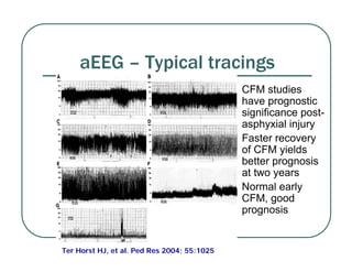
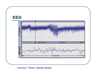
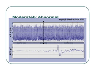
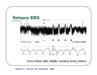
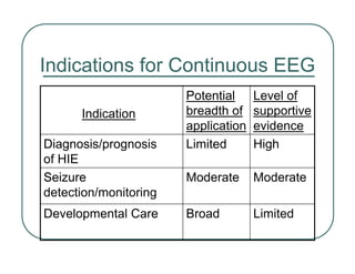
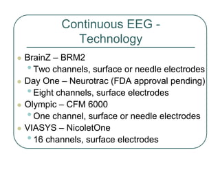
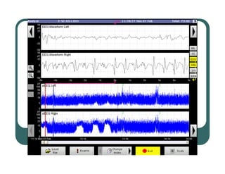
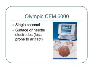
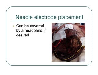
Continuous EEG Monitoring
in the Newborn
Robert White, MD
Learning Objectives
Upon completion of this session, you will be
able to:
z Discuss what current continuous EEG
monitors can and can't tell us
z Review the possible uses (and misuses) of
this technology
Presentation Outline
z What we know about continuous EEG
monitoring
z A little bit about the technique itself
z Exploring logic of aEEG use for seizure
detection and monitoring of therapy
z Suggestions for acquiring expertise
z Resources
Continuous EEG Monitoring
in the Newborn - Why?
z A real-time indicator of cerebral activity
•For diagnosis/prognosis following HIE
•For seizure detection and monitoring
•For developmental care
Why not?
•Cost
•Unconvinced of value/fear of misuse
aEEG – How is it derived?
z Background + Transients
z Filtered, rectified
z Compressed - reams of paper in one screen
EEG
Courtesy T.
Courtesy T. Weiler
Weiler, Olympic Medical
, Olympic Medical
Rectification
General Categories of aEEG
Activity
Burst-
Suppression
Discontinuous
Continuous
aEEG
Raw EEG
Burst Suppression
Courtesy T.
Courtesy T. Weiler
Weiler, Olympic Medical
, Olympic Medical
General Categories of aEEG
Activity - Continued
Seizures
Inactive
Continuous Low
Voltage
aEEG
Raw EEG
aEEG – Typical tracings
z CFM studies
have prognostic
significance post-
asphyxial injury
z Faster recovery
of CFM yields
better prognosis
at two years
z Normal early
CFM, good
prognosis
Ter Horst HJ, et al. Ped Res 2004; 55:1025
EEG
Courtesy T.
Courtesy T. Weiler
Weiler, Olympic Medical
, Olympic Medical
Moderately Abnormal
Seizure EEG
DeVries
DeVries L,
L, Toet
Toet M, Clin. Perinatol., 2006
M, Clin. Perinatol., 2006
Term infant with middle cerebral artery infarct
Premature infant seizures
25 wk. infant with Grade 4 IVH
DeVries
DeVries L, Clin. Perinatol., 2006
L, Clin. Perinatol., 2006
Seizures; Burst-suppression
and jet ventilator background
Immature aEEG
z Less mature CFM
recordings were
characterized by
discontinuous
background activity,
with depressed
electrical amplitude
and wide bandwidth
(Burdjalov, et al)
26 weeks PCA
26 weeks PCA
z With advancing age,
the CFM pattern
became increasingly
continuous, with
progressive elevation
of the minimum level of
electrical amplitude,
and narrowing of the
amplitude’s bandwidth
29 weeks PCA
29 weeks PCA
z Cyclic 20 - 30 minute
periods of wider
amplitude began to
emerge after the third
week of life, and
were recognizable by
30 - 31 weeks post-
conceptional age in
the healthy pre-term
infant
31 weeks PCA
31 weeks PCA
30 weeks
30 weeks
PCA
PCA
The effect of IVH on aEEG
z In babies with grade
III and grade IV IVH,
discontinuity of the
tracing, and the
degree of electrical
amplitude
depression were
even more marked
than normal for
gestational age
31 weeks PCA, Grade 3 IVH
31 weeks PCA, no IVH
z In all patients with IVH,
there was delay in the
appearance of
maturational changes
and emergence of the
cyclic pattern of
cerebral activity, with
persisting discontinuity
compared to infants
without IVH of
comparable
developmental age. 34 weeks PCA, IVH
34 weeks PCA, IVH
33 weeks PCA,
33 weeks PCA,
IVH
IVH
General Categories of aEEG
Activity
Rare or absent
Near-silence
(1-3 uV)
Low-voltage background with little
or no spontaneous activity
Continuous Low
Voltage
None
Artifact only
No detectable cortical activity
Inactive
High-voltage bursts
(25-100 uV)
Near-silence
(1-3 uV)
Mostly near-silence, with
occasional high-voltage bursts
Burst-Suppression
NA
NA
Rhythmic, stereotypical, high-
voltage spikes
Seizure
Significantly higher
than background
Low voltage
(2-5 uV)
Periods of activity interspersed
with quiet periods
Discontinuous
Modestly higher
than background
Modest voltage
(5-10 uV)
Always something going on, with
or without sleep-wake cycling
Continuous
Spontaneous
Activity
Background
Brief description
How Can aEEG Be Used?
HIE Evaluation,
Treatment and
Prognosis
aEEG Enhances Diagnosis and
Prognosis Following HIE
z Al Naqueeb et al (Pediatrics 1999)
•56 cases of neonatal encephalopathy
•21 infants with normal aEEG
•19 were normal at 18-24 month
follow-up
•35 infants with abnormal aEEG
•27 died or had neurological
abnormalities
Diagnosis/Prognosis
Following HIE
z Toet et al (Arch Dis Child Neo Ed 1999)
•33 infants normal or mildly abnormal aEEG
•30 were normal at follow-up, 1 died, and
2 had global delay
•35 infants burst-suppression or low voltage
•22 died; 8 major handicaps; 5 normal
Diagnosis/Prognosis
Following HIE
z Subsequent studies have shown that
combining aEEG with clinical exam and
imaging provides earlier prognostic
information, with better sensitivity and
specificity, than exam and imaging alone.
z aEEG is used as an entry criterion for
some therapeutic hypothermia protocols
How Can aEEG Be Used?
Seizure Detection
and Management
aEEG Improves Seizure
Detection/Monitoring
z Majority of seizures (~70%) in neonates are
electrographic, without clinical correlates
z Electrographic seizures are not more benign than
electroclinical seizures
z Continuous recording detects/documents seizure
frequency better than observation + conventional
EEG alone
z Anticonvulsant therapy can produce electro-clinical
dissociation, obscuring continued seizure activity
Seizure Detection/Monitoring
z Continuous EEG is a supplement to
conventional EEG, not a substitute
z No guidance yet on treatment
Neonatal Seizure Patterns
R. Clancy, Clin. Perinatol., 2006
How Can aEEG Be Used?
Developmental Care
aEEG in Developmental Care
z Allows identification of sleep/wake periods,
which might improve care practices and
provide an additional parameter in
research trials
z Normal developmental progression of
aEEg by gestational age has been
described, which might assist in providing
age-appropriate care and in identifying
high-risk infants for follow-up
Indications for Continuous EEG
Limited
Broad
Developmental Care
Moderate
Moderate
Seizure
detection/monitoring
High
Limited
Diagnosis/prognosis
of HIE
Level of
supportive
evidence
Potential
breadth of
application
Indication
Continuous EEG -
Technology
z BrainZ – BRM2
•Two channels, surface or needle electrodes
z Day One – Neurotrac (FDA approval pending)
•Eight channels, surface electrodes
z Olympic – CFM 6000
•One channel, surface or needle electrodes
z VIASYS – NicoletOne
•16 channels, surface electrodes
BrainZ BRM2 Monitor
z Two-channel
recording allows
detection of some
asymmetries
Olympic CFM 6000
z Single channel
z Surface or needle
electrodes (less
prone to artifact)
Needle electrode placement
z Can be covered
by a headband, if
desired
Putting what we know about
aEEG into clinical context…..
Key Questions
z Who should be monitored?
z What seizures to treat?
z Can additional diagnostic/prognostic info be
provided by aEEG in high-risk infants?
z Appropriate training/documentation
•Without formal training/certification, the
potential for misadventure is considerable
z Costs/charges/reimbursement
Proposed Indications for
Seizure Monitoring
z 5 minute Apgar <5, or other evidence for HIE
z Clinical suspicion of seizures
z Sarnat stage 2-3
z High-risk infants during neuromuscular
blockade
z Severe apnea
z IVH/ICH
z ELBW infants
z Infants requiring ECMO or surgery for CHD
Possible Indications for
Treatment of Seizures
z Status epilecticus
z Repetitive, prolonged seizures in a
child, especially when clinical indicators
of seizure activity (e.g., severe apnea)
might be obscured or misinterpreted as
“normal”
Treatment of Repetitive Seizure
Activity Using aEEG
z If any seizures treated (94% of neonates with seizures in
2007 Bartha survey), then aEEG probably better than clinical
evaluation and conventional EEG alone.
z If in a given situation you would treat seizures seen on
conventional EEG, it is logical to use same criteria when
seizures identified by aEEG, since clinical implications are
similar.
z If you could get a reliable conventional EEG at 0200 on a
ventilator + multiple other devices aEEG might not be
needed, but since most of us can’t…..
Why Monitor?
The ABGs Analogy
z Continuous EEG is to conventional EEG as
SaO2 monitoring is to ABGs.
z ABGs – more info, more accurately, but
SaO2 allows real-time monitoring, something
ABGs cannot do.
z Conventional EEG – more info, more precise
than aEEG, but continuous EEG allows real-
time monitoring, which conventional EEG
cannot provide.
Why Monitor?
The ABGs Analogy
z In a very stable baby, perhaps monitoring O2
sat for 30 minutes a day is enough, but in a
sick infant, it’s grossly inadequate
z If a 30 minute EEG gave a representative
picture of the full 24 hour activity, one could
logically base therapy on the conventional
EEG, but we now know that it often doesn’t
Further Context
z Use of aEEG has not increased frequency of our
infants who go home on anticonvulsants (much
lower than the 75% found in the Bartha survey)
z Concerns about side-effects of phenobarb are
largely related to its long-term use
z Everybody intervenes with some seizures!
• Example – seizure leading to severe apnea,
requiring ventilatory assistance
So, bottom line suggestion is to –
monitor aggressively, treat cautiously
More “Bottom Line”
z Continuous EEG monitoring has the potential
to make our treatment of neonatal seizures
more informed and more rational
z On the other hand, it also has the potential to
make our treatment of neonatal seizures
more irrational, more invasive, and more
expensive
So...monitor aggressively; treat cautiously
Proposed Training Sequence
z Preliminary training
•Read the Atlas and Clinics
•Review key references
•Attend pertinent conferences
•Establish contact with mentor(s)
•Attend in-service by manufacturer’s
representative
Proposed Training Sequence
z Early clinical use
•Daily “EEG review” rounds
•Review challenging tracings with
mentor(s)
•Attend a formal training course
Proposed Training Sequence
z Continuing Education
•Regular “journal club”
•Competency checks
•Conferences
The Future
z More screening/diagnostic tools – e.g., NIRS,
multiple-array EEG and others?
z New neuroprotective interventions may
require screening devices available 24/7.
z Window into the neonatal brain is opening for
real-time evaluation of well-intended but not
always benign interventions – we need better
monitoring tools!
Resources
z Dr. Whitelaw’s course: Cerebral Function Monitoring
and Neurophysiology for Neonatologists -
www.neonatalneurology.org.uk/neurophysPoster.htm
z 3rd International Conf. on Neonatal Brain Monitoring
and Neuroprotection, Feb 19-22, 2009, Disney World
www.cme.hsc.usf.edu/brain/
z Hellstrom-Westas, deVries, Rosen. An Atlas of
Amplitude-integrated EEGs in the Newborn
(new edition due this year).
z Brain Monitoring in the Neonate
Clinics in Perinatology, Sept. 2006
Thank You!
Please feel free to reach me at:
Robert_White@pediatrix.com
Financial
z Hospital billing code we use – 95950 –
“Special EEG Tests”
z Clinically apparent is not the same as
clinically significant
•But what is clinically significant
remains to be determined

eeg.newborn.classroom.pdf.99999999999999

  • 1.
    Continuous EEG Monitoring inthe Newborn Robert White, MD
  • 2.
    Learning Objectives Upon completionof this session, you will be able to: z Discuss what current continuous EEG monitors can and can't tell us z Review the possible uses (and misuses) of this technology
  • 3.
    Presentation Outline z Whatwe know about continuous EEG monitoring z A little bit about the technique itself z Exploring logic of aEEG use for seizure detection and monitoring of therapy z Suggestions for acquiring expertise z Resources
  • 4.
    Continuous EEG Monitoring inthe Newborn - Why? z A real-time indicator of cerebral activity •For diagnosis/prognosis following HIE •For seizure detection and monitoring •For developmental care Why not? •Cost •Unconvinced of value/fear of misuse
  • 5.
    aEEG – Howis it derived? z Background + Transients z Filtered, rectified z Compressed - reams of paper in one screen
  • 6.
    EEG Courtesy T. Courtesy T.Weiler Weiler, Olympic Medical , Olympic Medical
  • 7.
  • 8.
    General Categories ofaEEG Activity Burst- Suppression Discontinuous Continuous aEEG Raw EEG
  • 9.
    Burst Suppression Courtesy T. CourtesyT. Weiler Weiler, Olympic Medical , Olympic Medical
  • 10.
    General Categories ofaEEG Activity - Continued Seizures Inactive Continuous Low Voltage aEEG Raw EEG
  • 11.
    aEEG – Typicaltracings z CFM studies have prognostic significance post- asphyxial injury z Faster recovery of CFM yields better prognosis at two years z Normal early CFM, good prognosis Ter Horst HJ, et al. Ped Res 2004; 55:1025
  • 12.
    EEG Courtesy T. Courtesy T.Weiler Weiler, Olympic Medical , Olympic Medical
  • 13.
  • 14.
    Seizure EEG DeVries DeVries L, L,Toet Toet M, Clin. Perinatol., 2006 M, Clin. Perinatol., 2006 Term infant with middle cerebral artery infarct Term infant with middle cerebral artery infarct
  • 15.
    Premature infant seizures 25wk. infant with Grade 4 IVH 25 wk. infant with Grade 4 IVH DeVries DeVries L, Clin. Perinatol., 2006 L, Clin. Perinatol., 2006
  • 16.
  • 18.
    Immature aEEG z Lessmature CFM recordings were characterized by discontinuous background activity, with depressed electrical amplitude and wide bandwidth (Burdjalov, et al) 26 weeks PCA 26 weeks PCA
  • 19.
    z With advancingage, the CFM pattern became increasingly continuous, with progressive elevation of the minimum level of electrical amplitude, and narrowing of the amplitude’s bandwidth 29 weeks PCA 29 weeks PCA
  • 20.
    z Cyclic 20- 30 minute periods of wider amplitude began to emerge after the third week of life, and were recognizable by 30 - 31 weeks post- conceptional age in the healthy pre-term infant 31 weeks PCA 31 weeks PCA 30 weeks 30 weeks PCA PCA
  • 21.
    The effect ofIVH on aEEG z In babies with grade III and grade IV IVH, discontinuity of the tracing, and the degree of electrical amplitude depression were even more marked than normal for gestational age 31 weeks PCA, Grade 3 IVH 31 weeks PCA, Grade 3 IVH 31 weeks PCA, no IVH 31 weeks PCA, no IVH
  • 22.
    z In allpatients with IVH, there was delay in the appearance of maturational changes and emergence of the cyclic pattern of cerebral activity, with persisting discontinuity compared to infants without IVH of comparable developmental age. 34 weeks PCA, IVH 34 weeks PCA, IVH 33 weeks PCA, 33 weeks PCA, IVH IVH
  • 23.
    General Categories ofaEEG Activity Rare or absent Near-silence (1-3 uV) Low-voltage background with little or no spontaneous activity Continuous Low Voltage None Artifact only No detectable cortical activity Inactive High-voltage bursts (25-100 uV) Near-silence (1-3 uV) Mostly near-silence, with occasional high-voltage bursts Burst-Suppression NA NA Rhythmic, stereotypical, high- voltage spikes Seizure Significantly higher than background Low voltage (2-5 uV) Periods of activity interspersed with quiet periods Discontinuous Modestly higher than background Modest voltage (5-10 uV) Always something going on, with or without sleep-wake cycling Continuous Spontaneous Activity Background Brief description
  • 24.
    How Can aEEGBe Used? HIE Evaluation, Treatment and Prognosis
  • 25.
    aEEG Enhances Diagnosisand Prognosis Following HIE z Al Naqueeb et al (Pediatrics 1999) •56 cases of neonatal encephalopathy •21 infants with normal aEEG •19 were normal at 18-24 month follow-up •35 infants with abnormal aEEG •27 died or had neurological abnormalities
  • 26.
    Diagnosis/Prognosis Following HIE z Toetet al (Arch Dis Child Neo Ed 1999) •33 infants normal or mildly abnormal aEEG •30 were normal at follow-up, 1 died, and 2 had global delay •35 infants burst-suppression or low voltage •22 died; 8 major handicaps; 5 normal
  • 27.
    Diagnosis/Prognosis Following HIE z Subsequentstudies have shown that combining aEEG with clinical exam and imaging provides earlier prognostic information, with better sensitivity and specificity, than exam and imaging alone. z aEEG is used as an entry criterion for some therapeutic hypothermia protocols
  • 28.
    How Can aEEGBe Used? Seizure Detection and Management
  • 29.
    aEEG Improves Seizure Detection/Monitoring zMajority of seizures (~70%) in neonates are electrographic, without clinical correlates z Electrographic seizures are not more benign than electroclinical seizures z Continuous recording detects/documents seizure frequency better than observation + conventional EEG alone z Anticonvulsant therapy can produce electro-clinical dissociation, obscuring continued seizure activity
  • 30.
    Seizure Detection/Monitoring z ContinuousEEG is a supplement to conventional EEG, not a substitute z No guidance yet on treatment
  • 31.
    Neonatal Seizure Patterns R.Clancy, Clin. Perinatol., 2006 R. Clancy, Clin. Perinatol., 2006
  • 32.
    How Can aEEGBe Used? Developmental Care
  • 33.
    aEEG in DevelopmentalCare z Allows identification of sleep/wake periods, which might improve care practices and provide an additional parameter in research trials z Normal developmental progression of aEEg by gestational age has been described, which might assist in providing age-appropriate care and in identifying high-risk infants for follow-up
  • 34.
    Indications for ContinuousEEG Limited Broad Developmental Care Moderate Moderate Seizure detection/monitoring High Limited Diagnosis/prognosis of HIE Level of supportive evidence Potential breadth of application Indication
  • 35.
    Continuous EEG - Technology zBrainZ – BRM2 •Two channels, surface or needle electrodes z Day One – Neurotrac (FDA approval pending) •Eight channels, surface electrodes z Olympic – CFM 6000 •One channel, surface or needle electrodes z VIASYS – NicoletOne •16 channels, surface electrodes
  • 36.
    BrainZ BRM2 Monitor zTwo-channel recording allows detection of some asymmetries
  • 38.
    Olympic CFM 6000 zSingle channel z Surface or needle electrodes (less prone to artifact)
  • 39.
    Needle electrode placement zCan be covered by a headband, if desired
  • 40.
    Putting what weknow about aEEG into clinical context…..
  • 41.
    Key Questions z Whoshould be monitored? z What seizures to treat? z Can additional diagnostic/prognostic info be provided by aEEG in high-risk infants? z Appropriate training/documentation •Without formal training/certification, the potential for misadventure is considerable z Costs/charges/reimbursement
  • 42.
    Proposed Indications for SeizureMonitoring z 5 minute Apgar <5, or other evidence for HIE z Clinical suspicion of seizures z Sarnat stage 2-3 z High-risk infants during neuromuscular blockade z Severe apnea z IVH/ICH z ELBW infants z Infants requiring ECMO or surgery for CHD
  • 43.
    Possible Indications for Treatmentof Seizures z Status epilecticus z Repetitive, prolonged seizures in a child, especially when clinical indicators of seizure activity (e.g., severe apnea) might be obscured or misinterpreted as “normal”
  • 44.
    Treatment of RepetitiveSeizure Activity Using aEEG z If any seizures treated (94% of neonates with seizures in 2007 Bartha survey), then aEEG probably better than clinical evaluation and conventional EEG alone. z If in a given situation you would treat seizures seen on conventional EEG, it is logical to use same criteria when seizures identified by aEEG, since clinical implications are similar. z If you could get a reliable conventional EEG at 0200 on a ventilator + multiple other devices aEEG might not be needed, but since most of us can’t…..
  • 45.
    Why Monitor? The ABGsAnalogy z Continuous EEG is to conventional EEG as SaO2 monitoring is to ABGs. z ABGs – more info, more accurately, but SaO2 allows real-time monitoring, something ABGs cannot do. z Conventional EEG – more info, more precise than aEEG, but continuous EEG allows real- time monitoring, which conventional EEG cannot provide.
  • 46.
    Why Monitor? The ABGsAnalogy z In a very stable baby, perhaps monitoring O2 sat for 30 minutes a day is enough, but in a sick infant, it’s grossly inadequate z If a 30 minute EEG gave a representative picture of the full 24 hour activity, one could logically base therapy on the conventional EEG, but we now know that it often doesn’t
  • 47.
    Further Context z Useof aEEG has not increased frequency of our infants who go home on anticonvulsants (much lower than the 75% found in the Bartha survey) z Concerns about side-effects of phenobarb are largely related to its long-term use z Everybody intervenes with some seizures! • Example – seizure leading to severe apnea, requiring ventilatory assistance So, bottom line suggestion is to – monitor aggressively, treat cautiously
  • 48.
    More “Bottom Line” zContinuous EEG monitoring has the potential to make our treatment of neonatal seizures more informed and more rational z On the other hand, it also has the potential to make our treatment of neonatal seizures more irrational, more invasive, and more expensive So...monitor aggressively; treat cautiously
  • 49.
    Proposed Training Sequence zPreliminary training •Read the Atlas and Clinics •Review key references •Attend pertinent conferences •Establish contact with mentor(s) •Attend in-service by manufacturer’s representative
  • 50.
    Proposed Training Sequence zEarly clinical use •Daily “EEG review” rounds •Review challenging tracings with mentor(s) •Attend a formal training course
  • 51.
    Proposed Training Sequence zContinuing Education •Regular “journal club” •Competency checks •Conferences
  • 52.
    The Future z Morescreening/diagnostic tools – e.g., NIRS, multiple-array EEG and others? z New neuroprotective interventions may require screening devices available 24/7. z Window into the neonatal brain is opening for real-time evaluation of well-intended but not always benign interventions – we need better monitoring tools!
  • 53.
    Resources z Dr. Whitelaw’scourse: Cerebral Function Monitoring and Neurophysiology for Neonatologists - www.neonatalneurology.org.uk/neurophysPoster.htm z 3rd International Conf. on Neonatal Brain Monitoring and Neuroprotection, Feb 19-22, 2009, Disney World www.cme.hsc.usf.edu/brain/ z Hellstrom-Westas, deVries, Rosen. An Atlas of Amplitude-integrated EEGs in the Newborn (new edition due this year). z Brain Monitoring in the Neonate Clinics in Perinatology, Sept. 2006
  • 54.
    Thank You! Please feelfree to reach me at: Please feel free to reach me at: Robert_White@pediatrix.com Robert_White@pediatrix.com
  • 55.
    Financial z Hospital billingcode we use – 95950 – “Special EEG Tests”
  • 56.
    z Clinically apparentis not the same as clinically significant •But what is clinically significant remains to be determined