SlideShare a Scribd company logo
1 of 11
Download to read offline
Dr. P.W. Chaudhari, Dept of Zoology Shri Vitthal Rukhmini Mahavidyalaya Sawana 1
The Structure and Function of a Cell Wall
A cell wall is a rigid, semi-permeable protective layer in some cell types. This outer
covering is positioned next to the cell membrane (plasma membrane) in most plant
cells, fungi, bacteria, algae, and some archaea. Animal cells however, do not have a cell wall.
The cell wall has many important functions in a cell including protection, structure, and support.
Cell wall composition varies depending on the organism. In plants, the cell wall is
composed mainly of strong fibers of the carbohydrate polymer cellulose. Cellulose is the major
component of cotton fiber and wood, and it is used in paper production. Bacterial cell walls are
composed of a sugar and amino acid polymer called peptidoglycan. The main components of
fungal cell walls are chitin, glucans, and proteins.
Plant Cell Wall Structure
Dr. P.W. Chaudhari, Dept of Zoology Shri Vitthal Rukhmini Mahavidyalaya Sawana 2
The plant cell wall is multi-layered and consists of up to three sections. From the outermost layer
of the cell wall, these layers are identified as the middle lamella, primary cell wall, and
secondary cell wall. While all plant cells have a middle lamella and primary cell wall, not all
have a secondary cell wall.
• Middle lamella: This outer cell wall layer contains polysaccharides called pectins. Pectins
aid in cell adhesion by helping the cell walls of adjacent cells to bind to one another.
• Primary cell wall: This layer is formed between the middle lamella and plasma
membrane in growing plant cells. It is primarily composed of cellulose microfibrils
contained within a gel-like matrix of hemicellulose fibers and pectin polysaccharides.
The primary cell wall provides the strength and flexibility needed to allow for cell
growth.
• Secondary cell wall: This layer is formed between the primary cell wall and plasma
membrane in some plant cells. Once the primary cell wall has stopped dividing and
growing, it may thicken to form a secondary cell wall. This rigid layer strengthens and
supports the cell. In addition to cellulose and hemicellulose, some secondary cell walls
contain lignin. Lignin strengthens the cell wall and aids in water conductivity in plant
vascular tissue cells.
A major role of the cell wall is to form a framework for the cell to prevent over expansion.
Cellulose fibers, structural proteins, and other polysaccharides help to maintain the shape and
form of the cell. Additional functions of the cell wall include:
• Support: The cell wall provides mechanical strength and support. It also controls the
direction of cell growth.
• Withstand turgor pressure: Turgor pressure is the force exerted against the cell wall as the
contents of the cell push the plasma membrane against the cell wall. This pressure helps a
plant to remain rigid and erect, but can also cause a cell to rupture.
• Regulate growth: The cell wall sends signals for the cell to enter the cell cycle in order to
divide and grow.
• Regulate diffusion: The cell wall is porous allowing some substances, including proteins,
to pass into the cell while keeping other substances out.
• Communication: Cells communicate with one another via plasmodesmata (pores or
channels between plant cell walls that allow molecules and communication signals to
pass between individual plant cells).
• Protection: The cell wall provides a barrier to protect against plant viruses and other
pathogens. It also helps to prevent water loss.
• Storage: The cell wall stores carbohydrates for use in plant growth, especially in seeds.
Dr. P.W. Chaudhari, Dept of Zoology Shri Vitthal Rukhmini Mahavidyalaya Sawana 3
• Plasmodesmata is a thin channel through plant cells that allows them to
communicate.
• Plant cells differ in many ways from animal cells, both in terms of some of their
internal organelles and the fact that plant cells have cell walls, where animal cells
do not. The two cell types also differ in the way they communicate with one
another and in how they translocate molecules.
Plasmodesmata
• Plasmodesmata (singular form: plasmodesma) are intercellular organelles found
only in plant and algal cells. (The animal cell "equivalent" is called the gap
junction.)
• The plasmodesmata consist of pores, or channels, lying between individual plant
cells, and connect the symplastic space in the plant. They can also be termed as
"bridges" between two plant cells.
• The plasmodesmata separate the outer cell membranes of the plant cells. The
actual air space separating the cells is called the desmotubule.
• The desmotubule possesses a rigid membrane that runs the length of the
plasmodesma. Cytoplasm lies between the cell membrane and the desmotubule.
The entire plasmodesma is covered with the smooth endoplasmic reticulum of
the connected cells.
• Plasmodesmata form during cell division of plant development. They form when
parts of the smooth endoplasmic reticulum from the parent cells become trapped
in the newly formed plant cell wall.
• Primary plasmodesmata are formed while the cell wall and endoplasmic
reticulum are formed as well; secondary plasmodesmata are formed afterward.
Secondary plasmodesmata are more complex and may have different functional
properties in terms of the size and nature of the molecules able to pass through.
Activity and Function
• Plasmodesmata play roles in both cellular communication and in molecule
translocation. Plant cells must work together as part of a multicellular organism
(the plant); in other words, the individual cells must work to benefit the common
good.
• Therefore, communication between cells is crucial for plant survival. The
problem with the plant cells is the tough, rigid cell wall. It is difficult for larger
molecules to penetrate the cell wall, which is why plasmodesmata are necessary.
Dr. P.W. Chaudhari, Dept of Zoology Shri Vitthal Rukhmini Mahavidyalaya Sawana 4
• The plasmodesmata link tissue cells to one another, so they have functional
importance for tissue growth and development. Researchers clarified in
2009 that the development and design of major organs were dependent on the
transport of transcription factors (proteins that help convert RNA to DNA)
through the plasmodesmata.
• Plasmodesmata were previously thought to be passive pores through which
nutrients and water moved, but now it is known that there are active dynamics
involved.
• Researchers believe that the sugar molecule controlling the mechanism for
closing the plasmodesmal pore is callose. In response to a trigger such as a
pathogen invader, callose is deposited in the cell wall around the plasmodesmal
pore and the pore closes.
• The gene that gives the command for callose to be synthesized and deposited is
called CalS3. Therefore, it is likely that the plasmodesmata density may affect
the induced resistance response to pathogen attack in plants.
• This idea was clarified when it was discovered that a protein,
named PDLP5 (plasmodesmata-located protein 5), causes the production of
salicylic acid, which enhances the defense response against plant pathogenic
bacterial attack.
Dr. P.W. Chaudhari, Dept of Zoology Shri Vitthal Rukhmini Mahavidyalaya Sawana 5
Structure of the plasma membrane
Introduction
Each cell of your body is encased in a tiny bubble of membrane. This membrane has about the
consistency of...salad oil^11start superscript, 1, end superscript. The first time I read that factoid,
I didn't find it very reassuring! Salad oil seems like an awfully fragile boundary to place between
a cell and the rest of the world. Luckily, the plasma membrane turns out to be very well-suited to
its job, salad oil texture and all.
What exactly is its job? The plasma membrane not only defines the borders of the cell, but also
allows the cell to interact with its environment in a controlled way. Cells must be able to
exclude, take in, and excrete various substances, all in specific amounts. In addition, they must
able to communicate with other cells, identifying themselves and sharing information.
To perform these roles, the plasma membrane needs lipids, which make a semi-permeable barrier
between the cell and its environment. It also needs proteins, which are involved in cross-
membrane transport and cell communication, and carbohydrates (sugars and sugar chains),
which decorate both the proteins and lipids and help cells recognize each other.
Here, we’ll take a closer look at the different components of the plasma membrane, examining
their roles, their diversity, and how they work together to make a flexible, sensitive, and secure
boundary around the cell.
Dr. P.W. Chaudhari, Dept of Zoology Shri Vitthal Rukhmini Mahavidyalaya Sawana 6
Fluid mosaic model
The currently accepted model for the structure of the plasma membrane, called the fluid mosaic
model, was first proposed in 1972. This model has evolved over time, but it still provides a good
basic description of the structure and behavior of membranes in many cells.
According to the fluid mosaic model, the plasma membrane is a mosaic of components—
primarily, phospholipids, cholesterol, and proteins—that move freely and fluidly in the plane of
the membrane. In other words, a diagram of the membrane (like the one below) is just a snapshot
of a dynamic process in which phospholipids and proteins are continually sliding past one
another.
Interestingly enough, this fluidity means that if you insert a very fine needle into a cell, the
membrane will simply part to flow around the needle; once the needle is removed, the membrane
will flow back together seamlessly.
Image of the plasma membrane, showing the phospholipidbilayer with peripheral and integral
membrane proteins, glycoproteins (proteins with a carbohydrate attached), glycolipids (lipids
with a carbohydrate attached), and cholesterol molecules.
The principal components of the plasma membrane are lipids (phospholipids and cholesterol),
proteins, and carbohydrate groups that are attached to some of the lipids and proteins.
• A phospholipid is a lipid made of glycerol, two fatty acid tails, and a phosphate-linked head
group. Biological membranes usually involve two layers of phospholipids with their tails
pointing inward, an arrangement called a phospholipidbilayer.
• Cholesterol, another lipid composed of four fused carbon rings, is found alongside phospholipids
in the core of the membrane.
• Membrane proteins may extend partway into the plasma membrane, cross the membrane
entirely, or be loosely attached to its inside or outside face.
Dr. P.W. Chaudhari, Dept of Zoology Shri Vitthal Rukhmini Mahavidyalaya Sawana 7
• Carbohydrate groups are present only on the outer surface of the plasma membrane and are
attached to proteins, forming glycoproteins, or lipids, forming glycolipids.
The proportions of proteins, lipids, and carbohydrates in the plasma membrane vary between
different types of cells. For a typical human cell, however, proteins account for about 50 percent
of the composition by mass, lipids (of all types) account for about 40 percent, and the remaining
10 percent comes from carbohydrates.
Phospholipids
Phospholipids, arranged in a bilayer, make up the basic fabric of the plasma membrane. They are
well-suited for this role because they are amphipathic, meaning that they have both hydrophilic
and hydrophobic regions.
Chemical structure of a phospholipid, showing the hydrophilic head and hydrophobic tails.
The hydrophilic, or “water-loving,” portion of a phospholipid is its head, which contains a
negatively charged phosphate group as well as an additional small group (of varying identity,
“R” in the diagram at left), which may also or be charged or polar. The hydrophilic heads of
phospholipids in a membrane bilayer face outward, contacting the aqueous (watery) fluid both
inside and outside the cell. Since water is a polar molecule, it readily forms electrostatic (charge-
based) interactions with the phospholipid heads.
The hydrophobic, or “water-fearing,” part of a phospholipid consists of its long, nonpolar fatty
acid tails. The fatty acid tails can easily interact with other nonpolar molecules, but they interact
poorly with water. Because of this, it’s more energetically favorable for the phospholipids to tuck
their fatty acid tails away in the interior of the membrane, where they are shielded from the
surrounding water. The phospholipidbilayer formed by these interactions makes a good barrier
Dr. P.W. Chaudhari, Dept of Zoology Shri Vitthal Rukhmini Mahavidyalaya Sawana 8
between the interior and exterior of the cell, because water and other polar or charged substances
cannot easily cross the hydrophobic core of the membrane.
a micelle and a liposome.
Phospholipids aren’t just well-suited to form a membrane bilayer. Instead, this is something
they’ll do spontaneously under the right conditions! In water or aqueous solution, phospholipids
tend to arrange themselves with their hydrophobic tails facing each other and their hydrophilic
heads facing out. If the phospholipids have small tails, they may form a micelle (a small, single-
layered sphere), while if they have bulkier tails, they may form a liposome (a hollow droplet of
bilayer membrane)^22squared.
Proteins
Proteins are the second major component of plasma membranes. There are two main categories
of membrane proteins: integral and peripheral.
a single-pass transmembrane protein with a single membrane-spanning alpha helix and a
three-pass transmembrane protein with three membrane-spanning alpha helices.
Dr. P.W. Chaudhari, Dept of Zoology Shri Vitthal Rukhmini Mahavidyalaya Sawana 9
Integral membrane proteins are, as their name suggests, integrated into the membrane: they have
at least one hydrophobic region that anchors them to the hydrophobic core of the
phospholipidbilayer. Some stick only partway into the membrane, while others stretch from one
side of the membrane to the other and are exposed on either side^11start superscript, 1, end
superscript. Proteins that extend all the way across the membrane are called transmembrane
proteins.
The portions of an integral membrane protein found inside the membrane are hydrophobic, while
those that are exposed to the cytoplasm or extracellular fluid tend to be hydrophilic.
Transmembrane proteins may cross the membrane just once, or may have as many as twelve
different membrane-spanning sections. A typical membrane-spanning segment consists of 20-25
hydrophobic amino acids arranged in an alpha helix, although not all transmembrane proteins fit
this model. Some integral membrane proteins form a channel that allows ions or other small
molecules to pass, as shown below.
Peripheral membrane proteins are found on the outside and inside surfaces of
membranes, attached either to integral proteins or to phospholipids. Unlike integral membrane
proteins, peripheral membrane proteins do not stick into the hydrophobic core of the membrane,
and they tend to be more loosely attached.
Carbohydrates
Carbohydrates are the third major component of plasma membranes. In general, they are
found on the outside surface of cells and are bound either to proteins (forming glycoproteins) or
to lipids (forming glycolipids). These carbohydrate chains may consist of 2-60 monosaccharide
units and can be either straight or branched.
Along with membrane proteins, these carbohydrates form distinctive cellular markers,
sort of like molecular ID badges, that allow cells to recognize each other. These markers are very
Dr. P.W. Chaudhari, Dept of Zoology Shri Vitthal Rukhmini Mahavidyalaya Sawana 10
important in the immune system, allowing immune cells to differentiate between body cells,
which they shouldn’t attack, and foreign cells or tissues, which they should.
Membrane fluidity
The structure of the fatty acid tails of the phospholipids is important in determining the
properties of the membrane, and in particular, how fluid it is.
Saturated fatty acids have no double bonds (are saturated with hydrogens), so they are
relatively straight. Unsaturated fatty acids, on the other hand, contain one or more double bonds,
often resulting in a bend or kink. (You can see an example of a bent, unsaturated tail in the
diagram of phospholipid structure that appears earlier in this article.) The saturated and
unsaturated fatty acid tails of phospholipids behave differently as temperature drops:
• At cooler temperatures, the straight tails of saturated fatty acids can pack tightly together,
making a dense and fairly rigid membrane.
• Phospholipids with unsaturated fatty acid tails cannot pack together as tightly because of the bent
structure of the tails. Because of this, a membrane containing unsaturated phospholipids will stay
fluid at lower temperatures than a membrane made of saturated ones.
Most cell membranes contain a mixture of phospholipids, some with two saturated (straight) tails
and others with one saturated and one unsaturated (bent) tail. Many organisms—fish are one
example—can adjust physiologically to cold environments by changing the proportion of
unsaturated fatty acids in their membranes. For more information about saturated and
unsaturated fatty acids, see the article on lipids.
In addition to phospholipids, animals have an additional membrane component that helps to
maintain fluidity. Cholesterol, another type of lipid that is embedded among the phospholipids of
the membrane, helps to minimize the effects of temperature on fluidity.
At low temperatures, cholesterol increases fluidity by keeping phospholipids from
packing tightly together, while at high temperatures, it actually reduces fluidity^{3,4}3,4start
Dr. P.W. Chaudhari, Dept of Zoology Shri Vitthal Rukhmini Mahavidyalaya Sawana 11
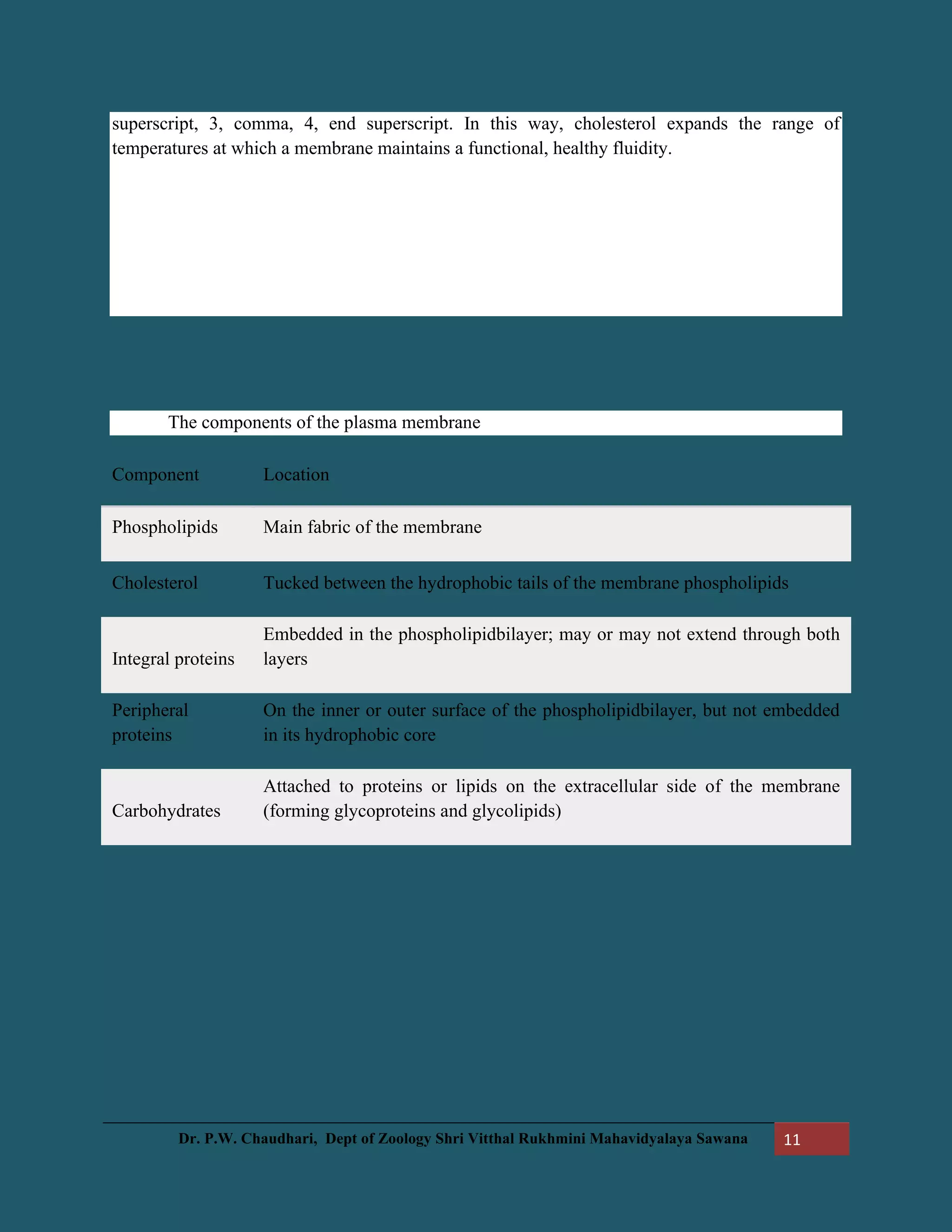
superscript, 3, comma, 4, end superscript. In this way, cholesterol expands the range of
temperatures at which a membrane maintains a functional, healthy fluidity.
The components of the plasma membrane
Component Location
Phospholipids Main fabric of the membrane
Cholesterol Tucked between the hydrophobic tails of the membrane phospholipids
Integral proteins
Embedded in the phospholipidbilayer; may or may not extend through both
layers
Peripheral
proteins
On the inner or outer surface of the phospholipidbilayer, but not embedded
in its hydrophobic core
Carbohydrates
Attached to proteins or lipids on the extracellular side of the membrane
(forming glycoproteins and glycolipids)

More Related Content

What's hot (20)

Plant organelles (Plasmodesmata, Vacuole, Plastids)
Plant organelles (Plasmodesmata, Vacuole, Plastids)Plant organelles (Plasmodesmata, Vacuole, Plastids)
Plant organelles (Plasmodesmata, Vacuole, Plastids)
 
Cell wall
Cell wallCell wall
Cell wall
 
Cell wall
Cell wallCell wall
Cell wall
 
Cell wall By KK Sahu Sir
Cell wall By KK Sahu SirCell wall By KK Sahu Sir
Cell wall By KK Sahu Sir
 
Cell Wall
Cell WallCell Wall
Cell Wall
 
plant cell wall components and Composition
plant cell wall components and Composition plant cell wall components and Composition
plant cell wall components and Composition
 
Different types of plant cells
Different types of plant cellsDifferent types of plant cells
Different types of plant cells
 
PLANT CELL WALL SMG
PLANT CELL WALL   SMGPLANT CELL WALL   SMG
PLANT CELL WALL SMG
 
Cell Wall
Cell WallCell Wall
Cell Wall
 
Plant cell wall.pptx
Plant cell wall.pptxPlant cell wall.pptx
Plant cell wall.pptx
 
Ergastic Substances
Ergastic SubstancesErgastic Substances
Ergastic Substances
 
Primary and secondary cell wall
Primary and secondary cell wallPrimary and secondary cell wall
Primary and secondary cell wall
 
E. organelles3
E. organelles3 E. organelles3
E. organelles3
 
Cellular components
Cellular componentsCellular components
Cellular components
 
Plasmodesmata
PlasmodesmataPlasmodesmata
Plasmodesmata
 
Cell Wall
Cell WallCell Wall
Cell Wall
 
Lecture 1 animal cell types and tissues
Lecture 1 animal cell types and tissuesLecture 1 animal cell types and tissues
Lecture 1 animal cell types and tissues
 
Cell Ingrowths: Adcrustation & Incrustation, Transport cells
Cell Ingrowths: Adcrustation & Incrustation, Transport cellsCell Ingrowths: Adcrustation & Incrustation, Transport cells
Cell Ingrowths: Adcrustation & Incrustation, Transport cells
 
Cell wall
Cell wallCell wall
Cell wall
 
The Fundamental Unit Of Life Class - 9
The Fundamental Unit Of Life Class - 9The Fundamental Unit Of Life Class - 9
The Fundamental Unit Of Life Class - 9
 

Similar to Eassy notes on cell wall

unit 1 cell wall , vacuole.pdf
unit 1 cell wall , vacuole.pdfunit 1 cell wall , vacuole.pdf
unit 1 cell wall , vacuole.pdfMSCW Mysore
 
Structure of plant cell
Structure of plant cellStructure of plant cell
Structure of plant cellAnhad Naad
 
PART 1 - CHAPTER 1 - CELL THE FUNDAMENTAL UNIT OF LIFE
PART 1 - CHAPTER 1 - CELL THE FUNDAMENTAL UNIT OF LIFEPART 1 - CHAPTER 1 - CELL THE FUNDAMENTAL UNIT OF LIFE
PART 1 - CHAPTER 1 - CELL THE FUNDAMENTAL UNIT OF LIFEMISSRITIMABIOLOGYEXP
 
cell wall cell membrane.pptx
cell wall cell membrane.pptxcell wall cell membrane.pptx
cell wall cell membrane.pptxAKHILRDONGA
 
Plant cell wall and plasma membrane
Plant cell wall and plasma membranePlant cell wall and plasma membrane
Plant cell wall and plasma membraneBaljinder Gill
 
Plant Anatomy and Microtechniques
Plant Anatomy and MicrotechniquesPlant Anatomy and Microtechniques
Plant Anatomy and MicrotechniquesChellappanRaju
 
Presentation on Cell Wall and Endoplasmic Recticulum
Presentation on Cell Wall and Endoplasmic RecticulumPresentation on Cell Wall and Endoplasmic Recticulum
Presentation on Cell Wall and Endoplasmic RecticulumDr. Kaushik Kumar Panigrahi
 
Introduction Biochemistry for BPT Students
Introduction Biochemistry for BPT StudentsIntroduction Biochemistry for BPT Students
Introduction Biochemistry for BPT StudentsKalaivanisathishr
 
Important Notes - NEET - Biology - Cell Structure and Function
Important Notes - NEET - Biology - Cell Structure and FunctionImportant Notes - NEET - Biology - Cell Structure and Function
Important Notes - NEET - Biology - Cell Structure and FunctionEdnexa
 
Cell_Modifications.pptx
Cell_Modifications.pptxCell_Modifications.pptx
Cell_Modifications.pptxRichmonAlaman1
 

Similar to Eassy notes on cell wall (20)

1cell wall.pptx
1cell wall.pptx1cell wall.pptx
1cell wall.pptx
 
unit 1 cell wall , vacuole.pdf
unit 1 cell wall , vacuole.pdfunit 1 cell wall , vacuole.pdf
unit 1 cell wall , vacuole.pdf
 
Structure of plant cell
Structure of plant cellStructure of plant cell
Structure of plant cell
 
Cell project
Cell projectCell project
Cell project
 
Cell As A System Analysis
Cell As A System AnalysisCell As A System Analysis
Cell As A System Analysis
 
Prokaryotic Cells Essay
Prokaryotic Cells EssayProkaryotic Cells Essay
Prokaryotic Cells Essay
 
Ppt
PptPpt
Ppt
 
PART 1 - CHAPTER 1 - CELL THE FUNDAMENTAL UNIT OF LIFE
PART 1 - CHAPTER 1 - CELL THE FUNDAMENTAL UNIT OF LIFEPART 1 - CHAPTER 1 - CELL THE FUNDAMENTAL UNIT OF LIFE
PART 1 - CHAPTER 1 - CELL THE FUNDAMENTAL UNIT OF LIFE
 
Cytology & physiology module
Cytology & physiology moduleCytology & physiology module
Cytology & physiology module
 
cell wall cell membrane.pptx
cell wall cell membrane.pptxcell wall cell membrane.pptx
cell wall cell membrane.pptx
 
Cell biology by Dr.Kshirsagar R.V
Cell biology by Dr.Kshirsagar R.VCell biology by Dr.Kshirsagar R.V
Cell biology by Dr.Kshirsagar R.V
 
Plant cell wall and plasma membrane
Plant cell wall and plasma membranePlant cell wall and plasma membrane
Plant cell wall and plasma membrane
 
Ultra structure of cell wall
Ultra structure of cell wallUltra structure of cell wall
Ultra structure of cell wall
 
Plant Anatomy and Microtechniques
Plant Anatomy and MicrotechniquesPlant Anatomy and Microtechniques
Plant Anatomy and Microtechniques
 
Biochemistry Introduction
Biochemistry IntroductionBiochemistry Introduction
Biochemistry Introduction
 
Presentation on Cell Wall and Endoplasmic Recticulum
Presentation on Cell Wall and Endoplasmic RecticulumPresentation on Cell Wall and Endoplasmic Recticulum
Presentation on Cell Wall and Endoplasmic Recticulum
 
Introduction Biochemistry for BPT Students
Introduction Biochemistry for BPT StudentsIntroduction Biochemistry for BPT Students
Introduction Biochemistry for BPT Students
 
Important Notes - NEET - Biology - Cell Structure and Function
Important Notes - NEET - Biology - Cell Structure and FunctionImportant Notes - NEET - Biology - Cell Structure and Function
Important Notes - NEET - Biology - Cell Structure and Function
 
Cell_Modifications.pptx
Cell_Modifications.pptxCell_Modifications.pptx
Cell_Modifications.pptx
 
Cell Wall-I.ppt
Cell Wall-I.pptCell Wall-I.ppt
Cell Wall-I.ppt
 

Recently uploaded

BAG TECHNIQUE Bag technique-a tool making use of public health bag through wh...
BAG TECHNIQUE Bag technique-a tool making use of public health bag through wh...BAG TECHNIQUE Bag technique-a tool making use of public health bag through wh...
BAG TECHNIQUE Bag technique-a tool making use of public health bag through wh...Sapna Thakur
 
Presentation by Andreas Schleicher Tackling the School Absenteeism Crisis 30 ...
Presentation by Andreas Schleicher Tackling the School Absenteeism Crisis 30 ...Presentation by Andreas Schleicher Tackling the School Absenteeism Crisis 30 ...
Presentation by Andreas Schleicher Tackling the School Absenteeism Crisis 30 ...EduSkills OECD
 
Class 11th Physics NEET formula sheet pdf
Class 11th Physics NEET formula sheet pdfClass 11th Physics NEET formula sheet pdf
Class 11th Physics NEET formula sheet pdfAyushMahapatra5
 
Measures of Central Tendency: Mean, Median and Mode
Measures of Central Tendency: Mean, Median and ModeMeasures of Central Tendency: Mean, Median and Mode
Measures of Central Tendency: Mean, Median and ModeThiyagu K
 
Student login on Anyboli platform.helpin
Student login on Anyboli platform.helpinStudent login on Anyboli platform.helpin
Student login on Anyboli platform.helpinRaunakKeshri1
 
Paris 2024 Olympic Geographies - an activity
Paris 2024 Olympic Geographies - an activityParis 2024 Olympic Geographies - an activity
Paris 2024 Olympic Geographies - an activityGeoBlogs
 
Grant Readiness 101 TechSoup and Remy Consulting
Grant Readiness 101 TechSoup and Remy ConsultingGrant Readiness 101 TechSoup and Remy Consulting
Grant Readiness 101 TechSoup and Remy ConsultingTechSoup
 
Software Engineering Methodologies (overview)
Software Engineering Methodologies (overview)Software Engineering Methodologies (overview)
Software Engineering Methodologies (overview)eniolaolutunde
 
The Most Excellent Way | 1 Corinthians 13
The Most Excellent Way | 1 Corinthians 13The Most Excellent Way | 1 Corinthians 13
The Most Excellent Way | 1 Corinthians 13Steve Thomason
 
SOCIAL AND HISTORICAL CONTEXT - LFTVD.pptx
SOCIAL AND HISTORICAL CONTEXT - LFTVD.pptxSOCIAL AND HISTORICAL CONTEXT - LFTVD.pptx
SOCIAL AND HISTORICAL CONTEXT - LFTVD.pptxiammrhaywood
 
Key note speaker Neum_Admir Softic_ENG.pdf
Key note speaker Neum_Admir Softic_ENG.pdfKey note speaker Neum_Admir Softic_ENG.pdf
Key note speaker Neum_Admir Softic_ENG.pdfAdmir Softic
 
social pharmacy d-pharm 1st year by Pragati K. Mahajan
social pharmacy d-pharm 1st year by Pragati K. Mahajansocial pharmacy d-pharm 1st year by Pragati K. Mahajan
social pharmacy d-pharm 1st year by Pragati K. Mahajanpragatimahajan3
 
Q4-W6-Restating Informational Text Grade 3
Q4-W6-Restating Informational Text Grade 3Q4-W6-Restating Informational Text Grade 3
Q4-W6-Restating Informational Text Grade 3JemimahLaneBuaron
 
Sanyam Choudhary Chemistry practical.pdf
Sanyam Choudhary Chemistry practical.pdfSanyam Choudhary Chemistry practical.pdf
Sanyam Choudhary Chemistry practical.pdfsanyamsingh5019
 
1029 - Danh muc Sach Giao Khoa 10 . pdf
1029 -  Danh muc Sach Giao Khoa 10 . pdf1029 -  Danh muc Sach Giao Khoa 10 . pdf
1029 - Danh muc Sach Giao Khoa 10 . pdfQucHHunhnh
 
Activity 01 - Artificial Culture (1).pdf
Activity 01 - Artificial Culture (1).pdfActivity 01 - Artificial Culture (1).pdf
Activity 01 - Artificial Culture (1).pdfciinovamais
 
Russian Escort Service in Delhi 11k Hotel Foreigner Russian Call Girls in Delhi
Russian Escort Service in Delhi 11k Hotel Foreigner Russian Call Girls in DelhiRussian Escort Service in Delhi 11k Hotel Foreigner Russian Call Girls in Delhi
Russian Escort Service in Delhi 11k Hotel Foreigner Russian Call Girls in Delhikauryashika82
 
Advanced Views - Calendar View in Odoo 17
Advanced Views - Calendar View in Odoo 17Advanced Views - Calendar View in Odoo 17
Advanced Views - Calendar View in Odoo 17Celine George
 
Explore beautiful and ugly buildings. Mathematics helps us create beautiful d...
Explore beautiful and ugly buildings. Mathematics helps us create beautiful d...Explore beautiful and ugly buildings. Mathematics helps us create beautiful d...
Explore beautiful and ugly buildings. Mathematics helps us create beautiful d...christianmathematics
 

Recently uploaded (20)

BAG TECHNIQUE Bag technique-a tool making use of public health bag through wh...
BAG TECHNIQUE Bag technique-a tool making use of public health bag through wh...BAG TECHNIQUE Bag technique-a tool making use of public health bag through wh...
BAG TECHNIQUE Bag technique-a tool making use of public health bag through wh...
 
Presentation by Andreas Schleicher Tackling the School Absenteeism Crisis 30 ...
Presentation by Andreas Schleicher Tackling the School Absenteeism Crisis 30 ...Presentation by Andreas Schleicher Tackling the School Absenteeism Crisis 30 ...
Presentation by Andreas Schleicher Tackling the School Absenteeism Crisis 30 ...
 
Class 11th Physics NEET formula sheet pdf
Class 11th Physics NEET formula sheet pdfClass 11th Physics NEET formula sheet pdf
Class 11th Physics NEET formula sheet pdf
 
Measures of Central Tendency: Mean, Median and Mode
Measures of Central Tendency: Mean, Median and ModeMeasures of Central Tendency: Mean, Median and Mode
Measures of Central Tendency: Mean, Median and Mode
 
Student login on Anyboli platform.helpin
Student login on Anyboli platform.helpinStudent login on Anyboli platform.helpin
Student login on Anyboli platform.helpin
 
Paris 2024 Olympic Geographies - an activity
Paris 2024 Olympic Geographies - an activityParis 2024 Olympic Geographies - an activity
Paris 2024 Olympic Geographies - an activity
 
Grant Readiness 101 TechSoup and Remy Consulting
Grant Readiness 101 TechSoup and Remy ConsultingGrant Readiness 101 TechSoup and Remy Consulting
Grant Readiness 101 TechSoup and Remy Consulting
 
Software Engineering Methodologies (overview)
Software Engineering Methodologies (overview)Software Engineering Methodologies (overview)
Software Engineering Methodologies (overview)
 
The Most Excellent Way | 1 Corinthians 13
The Most Excellent Way | 1 Corinthians 13The Most Excellent Way | 1 Corinthians 13
The Most Excellent Way | 1 Corinthians 13
 
SOCIAL AND HISTORICAL CONTEXT - LFTVD.pptx
SOCIAL AND HISTORICAL CONTEXT - LFTVD.pptxSOCIAL AND HISTORICAL CONTEXT - LFTVD.pptx
SOCIAL AND HISTORICAL CONTEXT - LFTVD.pptx
 
Key note speaker Neum_Admir Softic_ENG.pdf
Key note speaker Neum_Admir Softic_ENG.pdfKey note speaker Neum_Admir Softic_ENG.pdf
Key note speaker Neum_Admir Softic_ENG.pdf
 
social pharmacy d-pharm 1st year by Pragati K. Mahajan
social pharmacy d-pharm 1st year by Pragati K. Mahajansocial pharmacy d-pharm 1st year by Pragati K. Mahajan
social pharmacy d-pharm 1st year by Pragati K. Mahajan
 
Q4-W6-Restating Informational Text Grade 3
Q4-W6-Restating Informational Text Grade 3Q4-W6-Restating Informational Text Grade 3
Q4-W6-Restating Informational Text Grade 3
 
Sanyam Choudhary Chemistry practical.pdf
Sanyam Choudhary Chemistry practical.pdfSanyam Choudhary Chemistry practical.pdf
Sanyam Choudhary Chemistry practical.pdf
 
1029 - Danh muc Sach Giao Khoa 10 . pdf
1029 -  Danh muc Sach Giao Khoa 10 . pdf1029 -  Danh muc Sach Giao Khoa 10 . pdf
1029 - Danh muc Sach Giao Khoa 10 . pdf
 
Activity 01 - Artificial Culture (1).pdf
Activity 01 - Artificial Culture (1).pdfActivity 01 - Artificial Culture (1).pdf
Activity 01 - Artificial Culture (1).pdf
 
Russian Escort Service in Delhi 11k Hotel Foreigner Russian Call Girls in Delhi
Russian Escort Service in Delhi 11k Hotel Foreigner Russian Call Girls in DelhiRussian Escort Service in Delhi 11k Hotel Foreigner Russian Call Girls in Delhi
Russian Escort Service in Delhi 11k Hotel Foreigner Russian Call Girls in Delhi
 
Advanced Views - Calendar View in Odoo 17
Advanced Views - Calendar View in Odoo 17Advanced Views - Calendar View in Odoo 17
Advanced Views - Calendar View in Odoo 17
 
Advance Mobile Application Development class 07
Advance Mobile Application Development class 07Advance Mobile Application Development class 07
Advance Mobile Application Development class 07
 
Explore beautiful and ugly buildings. Mathematics helps us create beautiful d...
Explore beautiful and ugly buildings. Mathematics helps us create beautiful d...Explore beautiful and ugly buildings. Mathematics helps us create beautiful d...
Explore beautiful and ugly buildings. Mathematics helps us create beautiful d...
 

Eassy notes on cell wall

  • 1. Dr. P.W. Chaudhari, Dept of Zoology Shri Vitthal Rukhmini Mahavidyalaya Sawana 1 The Structure and Function of a Cell Wall A cell wall is a rigid, semi-permeable protective layer in some cell types. This outer covering is positioned next to the cell membrane (plasma membrane) in most plant cells, fungi, bacteria, algae, and some archaea. Animal cells however, do not have a cell wall. The cell wall has many important functions in a cell including protection, structure, and support. Cell wall composition varies depending on the organism. In plants, the cell wall is composed mainly of strong fibers of the carbohydrate polymer cellulose. Cellulose is the major component of cotton fiber and wood, and it is used in paper production. Bacterial cell walls are composed of a sugar and amino acid polymer called peptidoglycan. The main components of fungal cell walls are chitin, glucans, and proteins. Plant Cell Wall Structure
  • 2. Dr. P.W. Chaudhari, Dept of Zoology Shri Vitthal Rukhmini Mahavidyalaya Sawana 2 The plant cell wall is multi-layered and consists of up to three sections. From the outermost layer of the cell wall, these layers are identified as the middle lamella, primary cell wall, and secondary cell wall. While all plant cells have a middle lamella and primary cell wall, not all have a secondary cell wall. • Middle lamella: This outer cell wall layer contains polysaccharides called pectins. Pectins aid in cell adhesion by helping the cell walls of adjacent cells to bind to one another. • Primary cell wall: This layer is formed between the middle lamella and plasma membrane in growing plant cells. It is primarily composed of cellulose microfibrils contained within a gel-like matrix of hemicellulose fibers and pectin polysaccharides. The primary cell wall provides the strength and flexibility needed to allow for cell growth. • Secondary cell wall: This layer is formed between the primary cell wall and plasma membrane in some plant cells. Once the primary cell wall has stopped dividing and growing, it may thicken to form a secondary cell wall. This rigid layer strengthens and supports the cell. In addition to cellulose and hemicellulose, some secondary cell walls contain lignin. Lignin strengthens the cell wall and aids in water conductivity in plant vascular tissue cells. A major role of the cell wall is to form a framework for the cell to prevent over expansion. Cellulose fibers, structural proteins, and other polysaccharides help to maintain the shape and form of the cell. Additional functions of the cell wall include: • Support: The cell wall provides mechanical strength and support. It also controls the direction of cell growth. • Withstand turgor pressure: Turgor pressure is the force exerted against the cell wall as the contents of the cell push the plasma membrane against the cell wall. This pressure helps a plant to remain rigid and erect, but can also cause a cell to rupture. • Regulate growth: The cell wall sends signals for the cell to enter the cell cycle in order to divide and grow. • Regulate diffusion: The cell wall is porous allowing some substances, including proteins, to pass into the cell while keeping other substances out. • Communication: Cells communicate with one another via plasmodesmata (pores or channels between plant cell walls that allow molecules and communication signals to pass between individual plant cells). • Protection: The cell wall provides a barrier to protect against plant viruses and other pathogens. It also helps to prevent water loss. • Storage: The cell wall stores carbohydrates for use in plant growth, especially in seeds.
  • 3. Dr. P.W. Chaudhari, Dept of Zoology Shri Vitthal Rukhmini Mahavidyalaya Sawana 3 • Plasmodesmata is a thin channel through plant cells that allows them to communicate. • Plant cells differ in many ways from animal cells, both in terms of some of their internal organelles and the fact that plant cells have cell walls, where animal cells do not. The two cell types also differ in the way they communicate with one another and in how they translocate molecules. Plasmodesmata • Plasmodesmata (singular form: plasmodesma) are intercellular organelles found only in plant and algal cells. (The animal cell "equivalent" is called the gap junction.) • The plasmodesmata consist of pores, or channels, lying between individual plant cells, and connect the symplastic space in the plant. They can also be termed as "bridges" between two plant cells. • The plasmodesmata separate the outer cell membranes of the plant cells. The actual air space separating the cells is called the desmotubule. • The desmotubule possesses a rigid membrane that runs the length of the plasmodesma. Cytoplasm lies between the cell membrane and the desmotubule. The entire plasmodesma is covered with the smooth endoplasmic reticulum of the connected cells. • Plasmodesmata form during cell division of plant development. They form when parts of the smooth endoplasmic reticulum from the parent cells become trapped in the newly formed plant cell wall. • Primary plasmodesmata are formed while the cell wall and endoplasmic reticulum are formed as well; secondary plasmodesmata are formed afterward. Secondary plasmodesmata are more complex and may have different functional properties in terms of the size and nature of the molecules able to pass through. Activity and Function • Plasmodesmata play roles in both cellular communication and in molecule translocation. Plant cells must work together as part of a multicellular organism (the plant); in other words, the individual cells must work to benefit the common good. • Therefore, communication between cells is crucial for plant survival. The problem with the plant cells is the tough, rigid cell wall. It is difficult for larger molecules to penetrate the cell wall, which is why plasmodesmata are necessary.
  • 4. Dr. P.W. Chaudhari, Dept of Zoology Shri Vitthal Rukhmini Mahavidyalaya Sawana 4 • The plasmodesmata link tissue cells to one another, so they have functional importance for tissue growth and development. Researchers clarified in 2009 that the development and design of major organs were dependent on the transport of transcription factors (proteins that help convert RNA to DNA) through the plasmodesmata. • Plasmodesmata were previously thought to be passive pores through which nutrients and water moved, but now it is known that there are active dynamics involved. • Researchers believe that the sugar molecule controlling the mechanism for closing the plasmodesmal pore is callose. In response to a trigger such as a pathogen invader, callose is deposited in the cell wall around the plasmodesmal pore and the pore closes. • The gene that gives the command for callose to be synthesized and deposited is called CalS3. Therefore, it is likely that the plasmodesmata density may affect the induced resistance response to pathogen attack in plants. • This idea was clarified when it was discovered that a protein, named PDLP5 (plasmodesmata-located protein 5), causes the production of salicylic acid, which enhances the defense response against plant pathogenic bacterial attack.
  • 5. Dr. P.W. Chaudhari, Dept of Zoology Shri Vitthal Rukhmini Mahavidyalaya Sawana 5 Structure of the plasma membrane Introduction Each cell of your body is encased in a tiny bubble of membrane. This membrane has about the consistency of...salad oil^11start superscript, 1, end superscript. The first time I read that factoid, I didn't find it very reassuring! Salad oil seems like an awfully fragile boundary to place between a cell and the rest of the world. Luckily, the plasma membrane turns out to be very well-suited to its job, salad oil texture and all. What exactly is its job? The plasma membrane not only defines the borders of the cell, but also allows the cell to interact with its environment in a controlled way. Cells must be able to exclude, take in, and excrete various substances, all in specific amounts. In addition, they must able to communicate with other cells, identifying themselves and sharing information. To perform these roles, the plasma membrane needs lipids, which make a semi-permeable barrier between the cell and its environment. It also needs proteins, which are involved in cross- membrane transport and cell communication, and carbohydrates (sugars and sugar chains), which decorate both the proteins and lipids and help cells recognize each other. Here, we’ll take a closer look at the different components of the plasma membrane, examining their roles, their diversity, and how they work together to make a flexible, sensitive, and secure boundary around the cell.
  • 6. Dr. P.W. Chaudhari, Dept of Zoology Shri Vitthal Rukhmini Mahavidyalaya Sawana 6 Fluid mosaic model The currently accepted model for the structure of the plasma membrane, called the fluid mosaic model, was first proposed in 1972. This model has evolved over time, but it still provides a good basic description of the structure and behavior of membranes in many cells. According to the fluid mosaic model, the plasma membrane is a mosaic of components— primarily, phospholipids, cholesterol, and proteins—that move freely and fluidly in the plane of the membrane. In other words, a diagram of the membrane (like the one below) is just a snapshot of a dynamic process in which phospholipids and proteins are continually sliding past one another. Interestingly enough, this fluidity means that if you insert a very fine needle into a cell, the membrane will simply part to flow around the needle; once the needle is removed, the membrane will flow back together seamlessly. Image of the plasma membrane, showing the phospholipidbilayer with peripheral and integral membrane proteins, glycoproteins (proteins with a carbohydrate attached), glycolipids (lipids with a carbohydrate attached), and cholesterol molecules. The principal components of the plasma membrane are lipids (phospholipids and cholesterol), proteins, and carbohydrate groups that are attached to some of the lipids and proteins. • A phospholipid is a lipid made of glycerol, two fatty acid tails, and a phosphate-linked head group. Biological membranes usually involve two layers of phospholipids with their tails pointing inward, an arrangement called a phospholipidbilayer. • Cholesterol, another lipid composed of four fused carbon rings, is found alongside phospholipids in the core of the membrane. • Membrane proteins may extend partway into the plasma membrane, cross the membrane entirely, or be loosely attached to its inside or outside face.
  • 7. Dr. P.W. Chaudhari, Dept of Zoology Shri Vitthal Rukhmini Mahavidyalaya Sawana 7 • Carbohydrate groups are present only on the outer surface of the plasma membrane and are attached to proteins, forming glycoproteins, or lipids, forming glycolipids. The proportions of proteins, lipids, and carbohydrates in the plasma membrane vary between different types of cells. For a typical human cell, however, proteins account for about 50 percent of the composition by mass, lipids (of all types) account for about 40 percent, and the remaining 10 percent comes from carbohydrates. Phospholipids Phospholipids, arranged in a bilayer, make up the basic fabric of the plasma membrane. They are well-suited for this role because they are amphipathic, meaning that they have both hydrophilic and hydrophobic regions. Chemical structure of a phospholipid, showing the hydrophilic head and hydrophobic tails. The hydrophilic, or “water-loving,” portion of a phospholipid is its head, which contains a negatively charged phosphate group as well as an additional small group (of varying identity, “R” in the diagram at left), which may also or be charged or polar. The hydrophilic heads of phospholipids in a membrane bilayer face outward, contacting the aqueous (watery) fluid both inside and outside the cell. Since water is a polar molecule, it readily forms electrostatic (charge- based) interactions with the phospholipid heads. The hydrophobic, or “water-fearing,” part of a phospholipid consists of its long, nonpolar fatty acid tails. The fatty acid tails can easily interact with other nonpolar molecules, but they interact poorly with water. Because of this, it’s more energetically favorable for the phospholipids to tuck their fatty acid tails away in the interior of the membrane, where they are shielded from the surrounding water. The phospholipidbilayer formed by these interactions makes a good barrier
  • 8. Dr. P.W. Chaudhari, Dept of Zoology Shri Vitthal Rukhmini Mahavidyalaya Sawana 8 between the interior and exterior of the cell, because water and other polar or charged substances cannot easily cross the hydrophobic core of the membrane. a micelle and a liposome. Phospholipids aren’t just well-suited to form a membrane bilayer. Instead, this is something they’ll do spontaneously under the right conditions! In water or aqueous solution, phospholipids tend to arrange themselves with their hydrophobic tails facing each other and their hydrophilic heads facing out. If the phospholipids have small tails, they may form a micelle (a small, single- layered sphere), while if they have bulkier tails, they may form a liposome (a hollow droplet of bilayer membrane)^22squared. Proteins Proteins are the second major component of plasma membranes. There are two main categories of membrane proteins: integral and peripheral. a single-pass transmembrane protein with a single membrane-spanning alpha helix and a three-pass transmembrane protein with three membrane-spanning alpha helices.
  • 9. Dr. P.W. Chaudhari, Dept of Zoology Shri Vitthal Rukhmini Mahavidyalaya Sawana 9 Integral membrane proteins are, as their name suggests, integrated into the membrane: they have at least one hydrophobic region that anchors them to the hydrophobic core of the phospholipidbilayer. Some stick only partway into the membrane, while others stretch from one side of the membrane to the other and are exposed on either side^11start superscript, 1, end superscript. Proteins that extend all the way across the membrane are called transmembrane proteins. The portions of an integral membrane protein found inside the membrane are hydrophobic, while those that are exposed to the cytoplasm or extracellular fluid tend to be hydrophilic. Transmembrane proteins may cross the membrane just once, or may have as many as twelve different membrane-spanning sections. A typical membrane-spanning segment consists of 20-25 hydrophobic amino acids arranged in an alpha helix, although not all transmembrane proteins fit this model. Some integral membrane proteins form a channel that allows ions or other small molecules to pass, as shown below. Peripheral membrane proteins are found on the outside and inside surfaces of membranes, attached either to integral proteins or to phospholipids. Unlike integral membrane proteins, peripheral membrane proteins do not stick into the hydrophobic core of the membrane, and they tend to be more loosely attached. Carbohydrates Carbohydrates are the third major component of plasma membranes. In general, they are found on the outside surface of cells and are bound either to proteins (forming glycoproteins) or to lipids (forming glycolipids). These carbohydrate chains may consist of 2-60 monosaccharide units and can be either straight or branched. Along with membrane proteins, these carbohydrates form distinctive cellular markers, sort of like molecular ID badges, that allow cells to recognize each other. These markers are very
  • 10. Dr. P.W. Chaudhari, Dept of Zoology Shri Vitthal Rukhmini Mahavidyalaya Sawana 10 important in the immune system, allowing immune cells to differentiate between body cells, which they shouldn’t attack, and foreign cells or tissues, which they should. Membrane fluidity The structure of the fatty acid tails of the phospholipids is important in determining the properties of the membrane, and in particular, how fluid it is. Saturated fatty acids have no double bonds (are saturated with hydrogens), so they are relatively straight. Unsaturated fatty acids, on the other hand, contain one or more double bonds, often resulting in a bend or kink. (You can see an example of a bent, unsaturated tail in the diagram of phospholipid structure that appears earlier in this article.) The saturated and unsaturated fatty acid tails of phospholipids behave differently as temperature drops: • At cooler temperatures, the straight tails of saturated fatty acids can pack tightly together, making a dense and fairly rigid membrane. • Phospholipids with unsaturated fatty acid tails cannot pack together as tightly because of the bent structure of the tails. Because of this, a membrane containing unsaturated phospholipids will stay fluid at lower temperatures than a membrane made of saturated ones. Most cell membranes contain a mixture of phospholipids, some with two saturated (straight) tails and others with one saturated and one unsaturated (bent) tail. Many organisms—fish are one example—can adjust physiologically to cold environments by changing the proportion of unsaturated fatty acids in their membranes. For more information about saturated and unsaturated fatty acids, see the article on lipids. In addition to phospholipids, animals have an additional membrane component that helps to maintain fluidity. Cholesterol, another type of lipid that is embedded among the phospholipids of the membrane, helps to minimize the effects of temperature on fluidity. At low temperatures, cholesterol increases fluidity by keeping phospholipids from packing tightly together, while at high temperatures, it actually reduces fluidity^{3,4}3,4start
  • 11. Dr. P.W. Chaudhari, Dept of Zoology Shri Vitthal Rukhmini Mahavidyalaya Sawana 11 superscript, 3, comma, 4, end superscript. In this way, cholesterol expands the range of temperatures at which a membrane maintains a functional, healthy fluidity. The components of the plasma membrane Component Location Phospholipids Main fabric of the membrane Cholesterol Tucked between the hydrophobic tails of the membrane phospholipids Integral proteins Embedded in the phospholipidbilayer; may or may not extend through both layers Peripheral proteins On the inner or outer surface of the phospholipidbilayer, but not embedded in its hydrophobic core Carbohydrates Attached to proteins or lipids on the extracellular side of the membrane (forming glycoproteins and glycolipids)