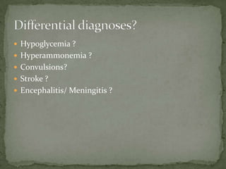
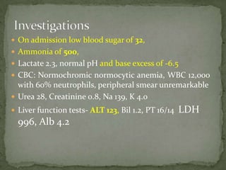
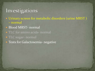
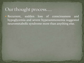
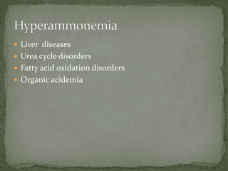
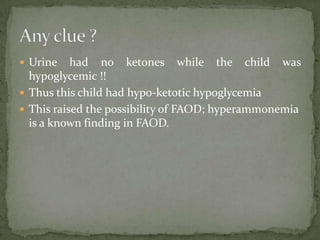
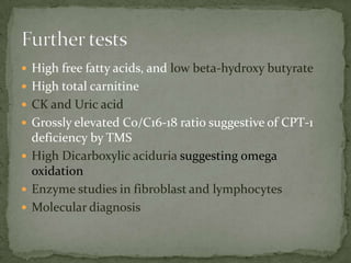
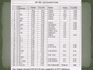
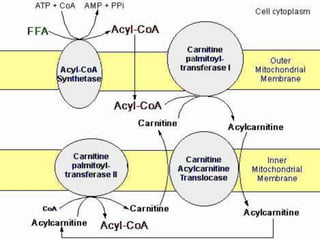
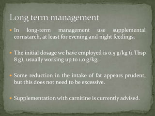
Dr. Raj Kumar
Consultant Pediatrician
  Rajasthan Hospital
 3 year old boy
 Referred for recurrent loss of consciousness
 The child would be perfectly well before these
  episodes would start
 Following trivial illness, he would start with
  vomiting and soon loose consciousness.
 Would be admitted to hospital, where he would be
  given iv fluids and recover again in no time!
 Non-consanguineous family
 Normal birth details and early development
 Family history of death in a male sibling at 2 days
  of age- unexplained
 In between episodes normal examination
 Notes from previous admissions suggested that he was
  noted to have hypoglycemia during these episodes.
 Neurological examination was unremarkable except
  hypotonia in one note; no neck rigidity and no focal
  weakness was noted
On admission:
 Unarousable,
 Febrile ; T =101F
 No bruises, petechae,
 No jaundice
 No edema feet
 No lymphnodes were palpable
 Gr III coma
 Moved limbs on deep painful stimuli
 Hypotonia +
 Pupils equal and reacting to light
 No neck rigidity
 Planters: extensor
 No distention
 No visible veins
 No umbilical hernia
 Liver enlargement of 3 cms, soft with sharp edges
 Spleen not palpable
 No ascites


 Other systems were found to be normal
 Hypoglycemia ?
 Hyperammonemia ?
 Convulsions?
 Stroke ?
 Encephalitis/ Meningitis ?
 On admission low blood sugar of 32,
 Ammonia of 500,
 Lactate 2.3, normal pH and base excess of -6.5
 CBC: Normochromic normocytic anemia, WBC 12,000
  with 60% neutrophils, peripheral smear unremarkable
 Urea 28, Creatinine 0.8, Na 139, K 4.0
 Liver function tests- ALT 123, Bil 1.2, PT 16/14   LDH
  996, Alb 4.2
 Urinary screen for metabolic disorders (urine MRST )
    – normal
   Blood MRST- normal
   TLC for amino acids- normal
   TLC sugar- normal
   Tests for Galactosemia- negative
 Biotinidase- negative
 G6PD- normal levels
 Urinary orotic acid- normal
 Plasma amino acids
 HPLC- normal
 HPLC nucleic acid- normal
 Plasma amino acids LC/MS- normal pattern
 GC-MS Organic acids- Elevated 2-oxoglutaric acid
 Recurrent,sudden loss of consciousness and
 hypoglycemia and severe hyperammonemia suggested
 neurometabolic syndrome more than anything else.
If you have a patient with :
 Neonatal progressive or recurrent encephalopathy
 Epilepsy that is refractory to treatment, myoclonic
   epilepsy
 Extrapyramidal movement disorders
 Ptosis, miosis and oculogyric crisis
 Disturbances of autonomic functions
 Ammonia
 AA including homocysteine
 OA in urine
 Purines and pyrimidines in urine and bed side sulphite
    test
   Prolactin in serum
   Whole blood serotonine
   Metabolic tests in CSF
   MRS of brain
   NMR (Nuclear Magnetic Resonance) of CSF
 Liver diseases
 Urea cycle disorders
 Fatty acid oxidation disorders
 Organic acidemia
 Urine    had no ketones while the child was
  hypoglycemic !!
 Thus this child had hypo-ketotic hypoglycemia
 This raised the possibility of FAOD; hyperammonemia
  is a known finding in FAOD.
 High free fatty acids, and low beta-hydroxy butyrate
 High total carnitine
 CK and Uric acid
 Grossly elevated C0/C16-18 ratio suggestive of CPT-1
  deficiency by TMS
 High Dicarboxylic aciduria suggesting omega
  oxidation
 Enzyme studies in fibroblast and lymphocytes
 Molecular diagnosis
 Fatty Acid Oxidation Disorder, CPT-I deficiency
 For acute episodes he was treated with oral sodium
  benzoate, IV Dextrose, IV carnitine and lactulose.
 Within 3 days ammonia decreased to 100 and
  subsequently 41- level of consciousness improved
Disorders of fatty oxidation display two general types of
 presentation.

 First, hypoketotic hypoglycemia, and clinical picture
 of Reye syndrome.

 In fact, it is now clear that most patients who appear
 to have Reye syndrome have an inborn error of
 metabolism, the most common, being MCAD
 deficiency and OTC deficiency.
 Second, reflects the chronic disruption of muscle
 function with symptoms relevant to myopathy or
 cardiomyopathy, including weakness, hypotonia,
 congestive heart failure, or arrhythmia.


 Both types of presentations may be seen in the same
 family or even in the same individual.

 Another presentation is with the sudden infant death
 syndrome (SIDS)
 Episodic illness usually occurs first between 6 months
 and 2 years, usually following fasting for 12 hours or
 more as a consequence of intercurrent infectious
 disease.

 The episode may be ushered in with vomiting or
 lethargy, or it may begin with a seizure. It is
 progressive rapidly to coma
 Hepatomegaly is usually present at the time of the
 acute illness.

 Liver biopsy at the time reveals abundant deposits of
 lipid in microvesicular pattern.

 This and hyperammonemia have often led to a
 diagnosis of Reye syndrome

 Cerebral edema and herniation have been reported in
 an acute lethal episode
 In  long-term management use supplemental
 cornstarch, at least for evening and night feedings.

 The initial dosage we have employed is 0.5 g/kg (1 Tbsp
 8 g), usually working up to 1.0 g/kg.

 Some reduction in the intake of fat appears prudent,
 but this does not need to be excessive.

 Supplementation with carnitine is currently advised.
Dr Rajkumar

Dr Rajkumar

  • 1.
    Dr. Raj Kumar ConsultantPediatrician Rajasthan Hospital
  • 2.
     3 yearold boy  Referred for recurrent loss of consciousness
  • 3.
     The childwould be perfectly well before these episodes would start  Following trivial illness, he would start with vomiting and soon loose consciousness.  Would be admitted to hospital, where he would be given iv fluids and recover again in no time!
  • 4.
     Non-consanguineous family Normal birth details and early development  Family history of death in a male sibling at 2 days of age- unexplained  In between episodes normal examination
  • 5.
     Notes fromprevious admissions suggested that he was noted to have hypoglycemia during these episodes.  Neurological examination was unremarkable except hypotonia in one note; no neck rigidity and no focal weakness was noted
  • 6.
    On admission:  Unarousable, Febrile ; T =101F  No bruises, petechae,  No jaundice  No edema feet  No lymphnodes were palpable
  • 7.
     Gr IIIcoma  Moved limbs on deep painful stimuli  Hypotonia +  Pupils equal and reacting to light  No neck rigidity  Planters: extensor
  • 8.
     No distention No visible veins  No umbilical hernia  Liver enlargement of 3 cms, soft with sharp edges  Spleen not palpable  No ascites  Other systems were found to be normal
  • 9.
     Hypoglycemia ? Hyperammonemia ?  Convulsions?  Stroke ?  Encephalitis/ Meningitis ?
  • 11.
     On admissionlow blood sugar of 32,  Ammonia of 500,  Lactate 2.3, normal pH and base excess of -6.5  CBC: Normochromic normocytic anemia, WBC 12,000 with 60% neutrophils, peripheral smear unremarkable  Urea 28, Creatinine 0.8, Na 139, K 4.0  Liver function tests- ALT 123, Bil 1.2, PT 16/14 LDH 996, Alb 4.2
  • 12.
     Urinary screenfor metabolic disorders (urine MRST ) – normal  Blood MRST- normal  TLC for amino acids- normal  TLC sugar- normal  Tests for Galactosemia- negative
  • 13.
     Biotinidase- negative G6PD- normal levels  Urinary orotic acid- normal  Plasma amino acids  HPLC- normal  HPLC nucleic acid- normal  Plasma amino acids LC/MS- normal pattern  GC-MS Organic acids- Elevated 2-oxoglutaric acid
  • 14.
     Recurrent,sudden lossof consciousness and hypoglycemia and severe hyperammonemia suggested neurometabolic syndrome more than anything else.
  • 15.
    If you havea patient with :  Neonatal progressive or recurrent encephalopathy  Epilepsy that is refractory to treatment, myoclonic epilepsy  Extrapyramidal movement disorders  Ptosis, miosis and oculogyric crisis  Disturbances of autonomic functions
  • 16.
     Ammonia  AAincluding homocysteine  OA in urine  Purines and pyrimidines in urine and bed side sulphite test  Prolactin in serum  Whole blood serotonine  Metabolic tests in CSF  MRS of brain  NMR (Nuclear Magnetic Resonance) of CSF
  • 17.
     Liver diseases Urea cycle disorders  Fatty acid oxidation disorders  Organic acidemia
  • 18.
     Urine had no ketones while the child was hypoglycemic !!  Thus this child had hypo-ketotic hypoglycemia  This raised the possibility of FAOD; hyperammonemia is a known finding in FAOD.
  • 19.
     High freefatty acids, and low beta-hydroxy butyrate  High total carnitine  CK and Uric acid  Grossly elevated C0/C16-18 ratio suggestive of CPT-1 deficiency by TMS  High Dicarboxylic aciduria suggesting omega oxidation  Enzyme studies in fibroblast and lymphocytes  Molecular diagnosis
  • 21.
     Fatty AcidOxidation Disorder, CPT-I deficiency
  • 22.
     For acuteepisodes he was treated with oral sodium benzoate, IV Dextrose, IV carnitine and lactulose.  Within 3 days ammonia decreased to 100 and subsequently 41- level of consciousness improved
  • 24.
    Disorders of fattyoxidation display two general types of presentation.  First, hypoketotic hypoglycemia, and clinical picture of Reye syndrome.  In fact, it is now clear that most patients who appear to have Reye syndrome have an inborn error of metabolism, the most common, being MCAD deficiency and OTC deficiency.
  • 25.
     Second, reflectsthe chronic disruption of muscle function with symptoms relevant to myopathy or cardiomyopathy, including weakness, hypotonia, congestive heart failure, or arrhythmia.  Both types of presentations may be seen in the same family or even in the same individual.  Another presentation is with the sudden infant death syndrome (SIDS)
  • 26.
     Episodic illnessusually occurs first between 6 months and 2 years, usually following fasting for 12 hours or more as a consequence of intercurrent infectious disease.  The episode may be ushered in with vomiting or lethargy, or it may begin with a seizure. It is progressive rapidly to coma
  • 27.
     Hepatomegaly isusually present at the time of the acute illness.  Liver biopsy at the time reveals abundant deposits of lipid in microvesicular pattern.  This and hyperammonemia have often led to a diagnosis of Reye syndrome  Cerebral edema and herniation have been reported in an acute lethal episode
  • 28.
     In long-term management use supplemental cornstarch, at least for evening and night feedings.  The initial dosage we have employed is 0.5 g/kg (1 Tbsp 8 g), usually working up to 1.0 g/kg.  Some reduction in the intake of fat appears prudent, but this does not need to be excessive.  Supplementation with carnitine is currently advised.