Downloaded 13 times


![Pathophysiology of ARDS
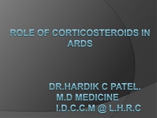
Although the cellular and molecular basis of acute lung injury and ARDS remains an area
of active investigation,
it appears that in ARDS, lung injury is caused by an imbalance of pro-inflammatory
and anti-inflammatory mediators.[4]
The most proximate signals leading to uncontrolled activation of the acute inflammatory
response are not yet understood.
However, nuclear factor κB (NF-κB), a transcription factor whose activation itself is
tightly regulated under normal conditions, has emerged as a likely candidate shifting the
balance in favor of a pro-inflammatory state.
As early as 30 minutes after an acute insult, there is increased synthesis of interleukin-8
(IL-8), a potent neutrophil chemotactic and activating agent, by pulmonary macrophages.
Release of this and similar compounds, such as IL-1 and tumor necrosis factor (TNF),
leads to endothelial activation, and pulmonary microvascular sequestration and
activation of neutrophils.
Neutrophils are thought to have an important role in the pathogenesis of ARDS.](https://image.slidesharecdn.com/drhardik-latestards-130109023135-phpapp01/85/Dr-hardik-patel-7-320.jpg)


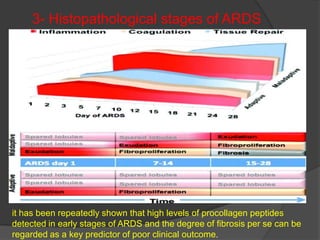


![ large trial by ARDS clinical trial network (ARDSnet),
consisting of 180 patients with ARDS (both early [73%] and late
[27%]), showed that
MP (2 mg/kg bolus in first 24 hours, followed by a dose of 0.5 mg/kg
every 6 hours for 14 days, 0.5 mg/kg every 12 hours for 7 days, and
then tapering over 4 days) has no survival benefits.
It showed that in patients with early ARDS (enrolled 7-13 days after
onset of ARDS), there were no significant differences in 60-day
mortality (36% vs 27%; P = 0.26) in the placebo and in MP group.
Similar results were seen at 180-day with mortality of 31.9 and 31.5%
in the placebo and MP group, respectively.
Other workers have also shown that corticosteroids use have no
statistically significant mortality benefits in patients with ARDS.](https://image.slidesharecdn.com/drhardik-latestards-130109023135-phpapp01/85/Dr-hardik-patel-33-320.jpg)


![ A recent meta-analysis has shown that there
was no difference in the incidence of
infection, neuromyopathy, GI bleeding, and
life-threatening complications, such as major
organ failure (heart, kidney, and liver),
between corticosteroid and placebo group.
Tang BM, Craig JC, Eslick GD, Seppelt I, McLean AS. Use of corticosteroids
in acute lung injury and acute respiratory distress syndrome: A systematic
.
review and meta-analysis. Crit Care Med 2009;37:1594–603.[PubMed]](https://image.slidesharecdn.com/drhardik-latestards-130109023135-phpapp01/85/Dr-hardik-patel-41-320.jpg)


1. The document discusses the history and pathophysiology of acute respiratory distress syndrome (ARDS), including its original description in 1821 and formal naming/definition in 1967. 2. A key development was the use of mechanical ventilation in the 1950s, which extended patient survival from hours to days/weeks and allowed more time for recovery. 3. The role of the nuclear factor κB transcription factor and associated inflammatory cytokines and neutrophils in the pathogenesis of ARDS is described. Uncontrolled inflammation can lead to endothelial and epithelial damage in the lungs. 4. Progression of ARDS is discussed as either resolving or unresolving, with the latter associated with worse outcomes. Corticosteroids are


