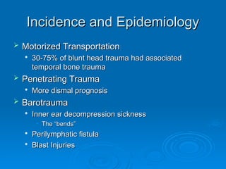
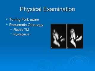
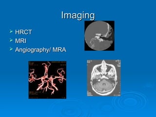
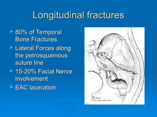
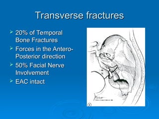
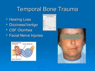
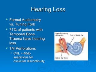
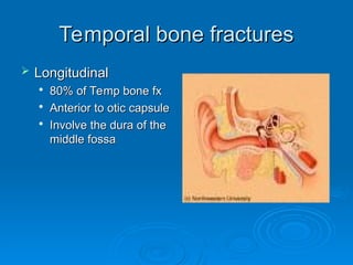
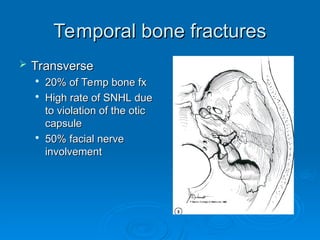
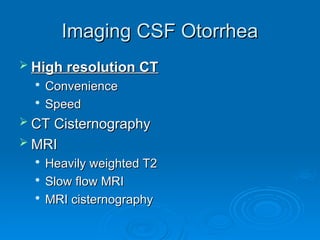
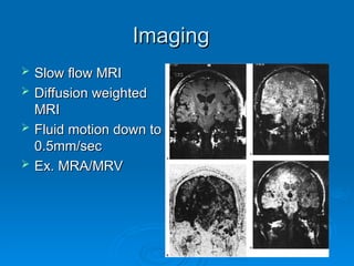
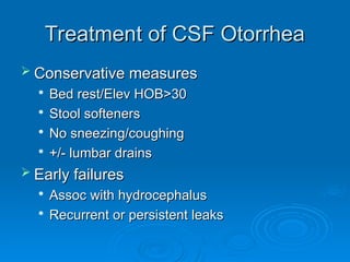
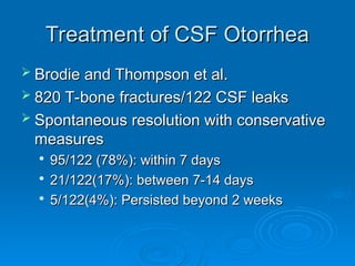
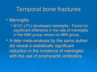
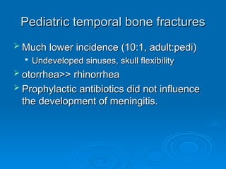
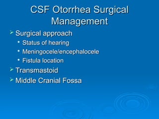
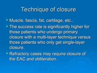
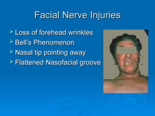
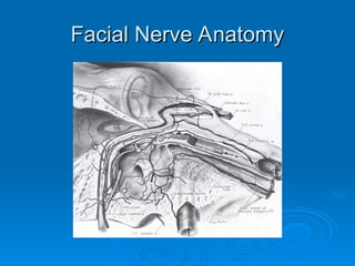
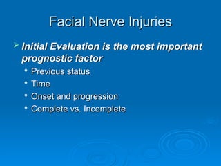
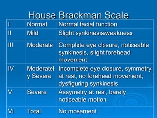
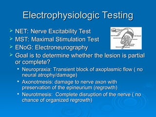
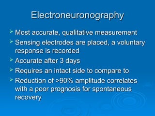
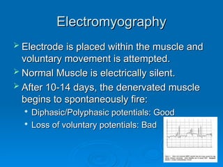
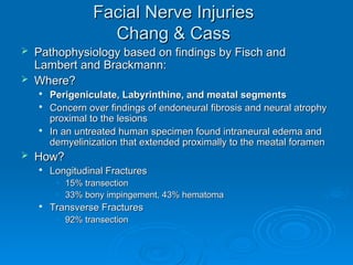
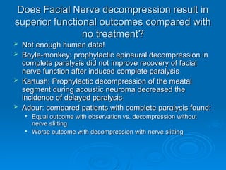
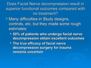
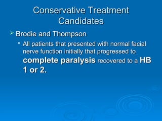
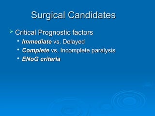
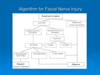
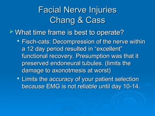
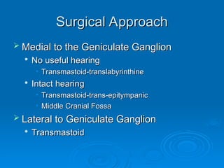
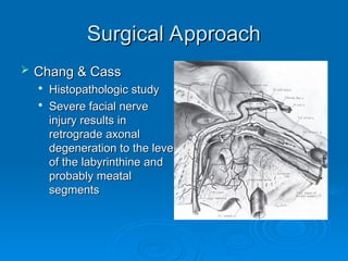
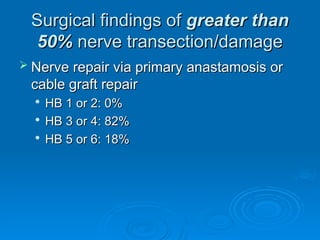
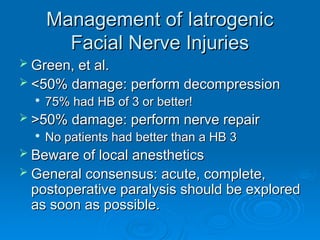
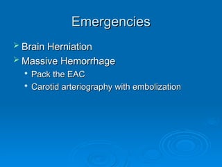
Temporal Bone Trauma
October 12, 2005
October 12, 2005
Steven T. Wright, M.D.
Matthew Ryan, M.D.
Matthew Ryan, M.D.
Temporal Bone Trauma
 Wide spectrum of
Wide spectrum of
clinical findings
clinical findings
 Knowledge of the
Knowledge of the
anatomy is vital to
anatomy is vital to
proper diagnosis and
appropriate
appropriate
management
management
Incidence and Epidemiology
 Motorized Transportation
Motorized Transportation

30-75% of blunt head trauma had associated
temporal bone trauma
 Penetrating Trauma
Penetrating Trauma

More dismal prognosis
 Barotrauma
Barotrauma

Inner ear decompression sickness
• The “bends”
The “bends”

Perilymphatic fistula

Blast Injuries
Blast Injuries
Evaluation and Management
 ATLS
ATLS

Airway
Airway

Breathing
Breathing

Circulation
Circulation
 H & P
H & P

Thorough head & neck
examination
examination
Physical Examination
 Basilar Skull
Basilar Skull
Fractures
Fractures

Periorbital Ecchymosis
(Raccoon’s Eyes)
(Raccoon’s Eyes)

Mastoid Ecchymosis
Mastoid Ecchymosis
(Battle’s Sign)
(Battle’s Sign)

Hemotympanum
Hemotympanum
Physical Examination
 Tuning Fork exam
Tuning Fork exam
 Pneumatic Otoscopy
Pneumatic Otoscopy

Flaccid TM
Flaccid TM

Nystagmus
Nystagmus
Imaging
Imaging
 HRCT
HRCT
 MRI
MRI
 Angiography/ MRA
Angiography/ MRA
Longitudinal fractures
 80% of Temporal
80% of Temporal
Bone Fractures
Bone Fractures
 Lateral Forces along
Lateral Forces along
the petrosquamous
the petrosquamous
suture line
suture line
 15-20% Facial Nerve
15-20% Facial Nerve
involvement
involvement
 EAC laceration
EAC laceration
Transverse fractures
 20% of Temporal
20% of Temporal
Bone Fractures
Bone Fractures
 Forces in the Antero-
Forces in the Antero-
Posterior direction
Posterior direction
 50% Facial Nerve
50% Facial Nerve
Involvement
Involvement
 EAC intact
EAC intact
Temporal Bone Trauma
 Hearing Loss
Hearing Loss
 Dizziness/Vertigo
Dizziness/Vertigo
 CSF Otorrhea
CSF Otorrhea
 Facial Nerve Injuries
Facial Nerve Injuries
Hearing Loss
Hearing Loss
 Formal Audiometry
Formal Audiometry
vs. Tuning Fork
vs. Tuning Fork
 71% of patients with
71% of patients with
Temporal Bone
Temporal Bone
Trauma have hearing
Trauma have hearing
loss
loss
 TM Perforations
TM Perforations

CHL > 40db
CHL > 40db
suspicious for
suspicious for
ossicular discontinuity
Hearing Loss
Hearing Loss
Longitudinal Fractures

Conductive or mixed hearing loss

80% of CHL resolve spontaneously
 Transverse Fractures
Transverse Fractures

Sensorineural hearing loss

Less likely to improve
Hearing Loss
Hearing Loss
 Tympanic Membrane Perforations
Tympanic Membrane Perforations
 Ossicular fracture or discontinuity
Ossicular fracture or discontinuity
 Hemotympanum
Hemotympanum
 Treatment:
Treatment:

Observation
Observation

Otic solutions may only mask CSF leaks
Dizziness
Dizziness
 Fracture through the otic capsule or a
Fracture through the otic capsule or a
labyrinthine concussion
 Difficult diagnosis- bed rest, obtundation,
Difficult diagnosis- bed rest, obtundation,
sedation
sedation
 Treatment: reserved for vomiting,
Treatment: reserved for vomiting,
limitation of activity

Vestibular suppressants

Allow for maximal central compensation
Dizziness
Dizziness
 Perilymphatic Fistulas
Perilymphatic Fistulas

SCUBA diver with ETD

Fluctuating dizziness and/or hearing loss

Tullio’s Phenomenon
Tullio’s Phenomenon

Management
Management
• Conservative treatment in first 10-14 days
Conservative treatment in first 10-14 days
• 40% spontaneously close
40% spontaneously close
• Surgical management for persistent vertigo or
Surgical management for persistent vertigo or
hearing loss
hearing loss
• Regardless of visualization of fistula site, the
Regardless of visualization of fistula site, the
majority of patients get better
Dizziness
Dizziness
 Inner Ear
Inner Ear
Decompression
Decompression
Sickness
Sickness

Too rapid an ascent
Too rapid an ascent
leads to percolation of
nitrogen bubbles within
the otic capsule.
the otic capsule.

Greater than 30 ft….
Decompression stages
upon ascent are
upon ascent are
needed
needed
Dizziness
Dizziness
 BPPV
BPPV

Acute, latent, and
Acute, latent, and
fatiguable vertigo
fatiguable vertigo

Can occur any time
Can occur any time
following injury
following injury

Dix Hallpike
Dix Hallpike

Epley Maneuver
Epley Maneuver
CSF Otorrhea
CSF Otorrhea
 Acquired
Acquired

Postoperative (58%)
Postoperative (58%)

Trauma (32%)
Trauma (32%)

Nontraumatic (11%)
Nontraumatic (11%)
 Spontaneous
Spontaneous

Bony defect theory
Bony defect theory

Arachnoid granulation theory
Temporal bone fractures
 Longitudinal
Longitudinal

80% of Temp bone fx
80% of Temp bone fx

Anterior to otic capsule

Involve the dura of the
middle fossa
middle fossa
Temporal bone fractures
 Transverse
Transverse

20% of Temp bone fx
20% of Temp bone fx

High rate of SNHL due
to violation of the otic
capsule
capsule

50% facial nerve
50% facial nerve
involvement
involvement
Testing of Nasal Secretions
 Beta-2-transferrin is highly sensitive and
Beta-2-transferrin is highly sensitive and
specific
specific

1/50
1/50th
th
of a drop
of a drop

Gold top tube, may need to send a sample of
the patients serum also.

Found in Vitreous Humor, Perilymph, CSF
 Electronic nose has shown early success
Electronic nose has shown early success

Faster (<24hrs)
Faster (<24hrs)

Very Accurate
Very Accurate
Imaging CSF Otorrhea
 High resolution CT
High resolution CT

Convenience
Convenience

Speed
Speed
 CT Cisternography
CT Cisternography
 MRI
MRI

Heavily weighted T2
Heavily weighted T2

Slow flow MRI
Slow flow MRI

MRI cisternography
MRI cisternography
Imaging
Imaging
 Slow flow MRI
Slow flow MRI
 Diffusion weighted
Diffusion weighted
MRI
MRI
 Fluid motion down to
Fluid motion down to
0.5mm/sec
0.5mm/sec
 Ex. MRA/MRV
Ex. MRA/MRV
Treatment of CSF Otorrhea
 Conservative measures
Conservative measures

Bed rest/Elev HOB>30

Stool softeners
Stool softeners

No sneezing/coughing

+/- lumbar drains
+/- lumbar drains
 Early failures
Early failures

Assoc with hydrocephalus

Recurrent or persistent leaks
Treatment of CSF Otorrhea
 Brodie and Thompson et al.
Brodie and Thompson et al.
 820 T-bone fractures/122 CSF leaks
820 T-bone fractures/122 CSF leaks
 Spontaneous resolution with conservative
Spontaneous resolution with conservative
measures
measures

95/122 (78%): within 7 days

21/122(17%): between 7-14 days

5/122(4%): Persisted beyond 2 weeks
Temporal bone fractures
 Meningitis
Meningitis

9/121 (7%) developed meningitis. Found no
significant difference in the rate of meningitis
in the ABX group versus no ABX group.
 A later meta-analysis by the same author
A later meta-analysis by the same author
did reveal a statistically significant
reduction in the incidence of meningitis
with the use of prophylactic antibiotics.
Pediatric temporal bone fractures
 Much lower incidence (10:1, adult:pedi)
Much lower incidence (10:1, adult:pedi)

Undeveloped sinuses, skull flexibility
 otorrhea>> rhinorrhea
otorrhea>> rhinorrhea
 Prophylactic antibiotics did not influence
Prophylactic antibiotics did not influence
the development of meningitis.
CSF Otorrhea Surgical
Management
Management
 Surgical approach
Surgical approach

Status of hearing
Status of hearing

Meningocele/encephalocele

Fistula location
Fistula location
 Transmastoid
Transmastoid
 Middle Cranial Fossa
Middle Cranial Fossa
Overlay vs Underlay
Overlay vs Underlay
technique
technique
 Meta-analysis
Meta-analysis
showed that both
showed that both
techniques have
techniques have
similar success rates
 Onlay: adjacent
Onlay: adjacent
structures at risk, or if
the underlay is not
the underlay is not
possible
possible
Technique of closure
 Muscle, fascia, fat, cartilage, etc..
Muscle, fascia, fat, cartilage, etc..
 The success rate is significantly higher for
The success rate is significantly higher for
those patients who undergo primary
closure with a multi-layer technique versus
those patients who only get single-layer
closure.
closure.
 Refractory cases may require closure of
Refractory cases may require closure of
the EAC and obliteration.
Facial Nerve Injuries
 Loss of forehead wrinkles
Loss of forehead wrinkles
 Bell’s Phenomenon
Bell’s Phenomenon
 Nasal tip pointing away
Nasal tip pointing away
 Flattened Nasofacial groove
Flattened Nasofacial groove
Facial Nerve Anatomy
Facial Nerve Injuries
 Initial Evaluation is the most important
Initial Evaluation is the most important
prognostic factor
prognostic factor

Previous status
Previous status

Time
Time

Onset and progression

Complete vs. Incomplete
House Brackman Scale
I
I Normal
Normal Normal facial function
Normal facial function
II
II Mild
Mild Slight synkinesis/weakness
Slight synkinesis/weakness
III
III Moderate
Moderate Complete eye closure, noticeable
Complete eye closure, noticeable
synkinesis, slight forehead
movement
movement
IV
IV Moderatel
Moderatel
y Severe
y Severe
Incomplete eye closure, symmetry
at rest, no forehead movement,
dysfiguring synkinesis
V
V Severe
Severe Assymetry at rest, barely
Assymetry at rest, barely
noticeable motion
noticeable motion
VI
VI Total
Total No movement
No movement
Electrophysiologic Testing
 NET: Nerve Excitability Test
NET: Nerve Excitability Test
 MST: Maximal Stimulation Test
MST: Maximal Stimulation Test
 ENoG: Electroneurography
ENoG: Electroneurography
 Goal is to determine whether the lesion is partial
Goal is to determine whether the lesion is partial
or complete?
or complete?

Neuropraxia: Transient block of axoplasmic flow ( no
neural atrophy/damage)

Axonotmesis: damage to nerve axon with
preservation of the epineurium (regrowth)

Neurotmesis: Complete disruption of the nerve ( no
chance of organized regrowth)
Nerve Excitability Test
Maximal Stimulation Test
 Stimulating electrodes are placed and a
Stimulating electrodes are placed and a
gross movement is recorded

Not as objective and reliable
 >3.5mA difference suggests a poor
>3.5mA difference suggests a poor
prognosis for return of facial function.

Correlates with >90% degeneration on ENoG
Electroneuronography
 Most accurate, qualitative measurement
Most accurate, qualitative measurement
 Sensing electrodes are placed, a voluntary
Sensing electrodes are placed, a voluntary
response is recorded
 Accurate after 3 days
Accurate after 3 days
 Requires an intact side to compare to
Requires an intact side to compare to
 Reduction of >90% amplitude correlates
Reduction of >90% amplitude correlates
with a poor prognosis for spontaneous
recovery
recovery
Electromyography
Electromyography
 Electrode is placed within the muscle and
Electrode is placed within the muscle and
voluntary movement is attempted.
 Normal Muscle is electrically silent.
Normal Muscle is electrically silent.
 After 10-14 days, the denervated muscle
After 10-14 days, the denervated muscle
begins to spontaneously fire:

Diphasic/Polyphasic potentials: Good

Loss of voluntary potentials: Bad
Facial Nerve Injuries
WHO GETS TREATMENT?
WHO GETS TREATMENT?
 Conservative treatment candidates
Conservative treatment candidates
 Surgical treatment candidates
Surgical treatment candidates
Facial Nerve Injuries
 Chang & Cass
Chang & Cass

Medline search back to 1966

Individually reviewed each article

1) Understand the pathophysiology of facial
nerve damage in temporal bone trauma.

2) What is the effect of surgical intervention
on the ultimate outcome of the facial nerve.

3) Propose a rational course for evaluation
and treatment.
and treatment.
Facial Nerve Injuries
Chang & Cass
Chang & Cass
 Pathophysiology based on findings by Fisch and
Pathophysiology based on findings by Fisch and
Lambert and Brackmann:
 Where?
Where?

Perigeniculate, Labyrinthine, and meatal segments

Concern over findings of endoneural fibrosis and neural atrophy
proximal to the lesions

In an untreated human specimen found intraneural edema and
demyelinization that extended proximally to the meatal foramen
 How?
How?

Longitudinal Fractures
• 15% transection
15% transection
• 33% bony impingement, 43% hematoma
33% bony impingement, 43% hematoma

Transverse Fractures
• 92% transection
92% transection
Does Facial Nerve decompression result in
superior functional outcomes compared with
no treatment?
no treatment?
 Not enough human data!
Not enough human data!
 Boyle-monkey: prophylactic epineural decompression in
Boyle-monkey: prophylactic epineural decompression in
complete paralysis did not improve recovery of facial
nerve function after induced complete paralysis
 Kartush: Prophylactic decompression of the meatal
Kartush: Prophylactic decompression of the meatal
segment during acoustic neuroma decreased the
incidence of delayed paralysis
 Adour: compared patients with complete paralysis found:
Adour: compared patients with complete paralysis found:

Equal outcome with observation vs. decompression without
nerve slitting
nerve slitting

Worse outcome with decompression with nerve slitting
Does Facial Nerve decompression result in
superior functional outcomes compared with
no treatment?
no treatment?
 Many difficulties in Study designs,
Many difficulties in Study designs,
controls, etc, but they made some rough
estimates:
estimates:

50% of patients who undergo facial nerve
decompression obtain excellent outcomes

The true efficacy of facial nerve
decompression surgery for trauma
remains uncertain
remains uncertain
Conservative Treatment
Candidates
Candidates
 Chang and Cass
Chang and Cass

Present with
Present with Normal Facial Function
Normal Facial Function
regardless of progression

Incomplete paralysis and no
progression
progression to complete paralysis
to complete paralysis

Less than
Less than 95%
95% degeneration by ENoG
degeneration by ENoG
• Most data comes from Bell’s palsy/tumor studies
Most data comes from Bell’s palsy/tumor studies
by Fisch.
by Fisch.
Conservative Treatment
Candidates
Candidates
 Brodie and Thompson
Brodie and Thompson

All patients that presented with normal facial
nerve function initially that progressed to
complete paralysis
complete paralysis recovered to a
recovered to a HB
HB
1 or 2.
1 or 2.
Surgical Candidates
Surgical Candidates
 Critical Prognostic factors
Critical Prognostic factors

Immediate
Immediate vs. Delayed
vs. Delayed

Complete
Complete vs. Incomplete paralysis
vs. Incomplete paralysis

ENoG criteria
ENoG criteria
Algorithm for Facial Nerve Injury
Facial Nerve Injuries
Chang & Cass
Chang & Cass
 What time frame is best to operate?
What time frame is best to operate?

Fisch-cats: Decompression of the nerve within
a 12 day period resulted in “excellent”
functional recovery. Presumption was that it
preserved endoneural tubules. (limits the
damage to axonotmesis at worst)

Limits the accuracy of your patient selection
because EMG is not reliable until day 10-14.
Surgical Approach
Surgical Approach
 Medial to the Geniculate Ganglion
Medial to the Geniculate Ganglion

No useful hearing
No useful hearing
• Transmastoid-translabyrinthine
Transmastoid-translabyrinthine

Intact hearing
Intact hearing
• Transmastoid-trans-epitympanic
Transmastoid-trans-epitympanic
• Middle Cranial Fossa
Middle Cranial Fossa
 Lateral to Geniculate Ganglion
Lateral to Geniculate Ganglion

Transmastoid
Transmastoid
Surgical Approach
Surgical Approach
 Chang & Cass
Chang & Cass

Histopathologic study

Severe facial nerve
Severe facial nerve
injury results in
injury results in
retrograde axonal
retrograde axonal
degeneration to the level
of the labyrinthine and
probably meatal
probably meatal
segments
segments
Surgical findings of
Surgical findings of greater than
greater than
50%
50% nerve transection/damage
nerve transection/damage
 Nerve repair via primary anastamosis or
Nerve repair via primary anastamosis or
cable graft repair
cable graft repair

HB 1 or 2: 0%
HB 1 or 2: 0%

HB 3 or 4: 82%
HB 3 or 4: 82%

HB 5 or 6: 18%
HB 5 or 6: 18%
Iatrogenic Facial Nerve Injuries
 Mastoidectomy (55%)
Mastoidectomy (55%)
 Tympanoplasty (14%)
Tympanoplasty (14%)
 Bony Exostoses (14%)
Bony Exostoses (14%)
 Lower tympanic segment is the most
Lower tympanic segment is the most
common location injury
 79% were not identified at the time of
79% were not identified at the time of
surgery
surgery
Management of Iatrogenic
Facial Nerve Injuries
 Green, et al.
Green, et al.
 <50% damage: perform decompression
<50% damage: perform decompression

75% had HB of 3 or better!
 >50% damage: perform nerve repair
>50% damage: perform nerve repair

No patients had better than a HB 3
 Beware of local anesthetics
Beware of local anesthetics
 General consensus: acute, complete,
General consensus: acute, complete,
postoperative paralysis should be explored
as soon as possible.
Emergencies
Emergencies
 Brain Herniation
Brain Herniation
 Massive Hemorrhage
Massive Hemorrhage

Pack the EAC
Pack the EAC

Carotid arteriography with embolization
Bibliography
Bibliography
 Bailey, Byron J., ed. Head and Neck surgery- Otolaryngology. Philadelphia, P.A. J.B. Lippincott Co., 1993.
Bailey, Byron J., ed. Head and Neck surgery- Otolaryngology. Philadelphia, P.A. J.B. Lippincott Co., 1993.
 Brodie, HA, Thompson TC. Management of Complications from 820 Temporal Bone Fractures. American
Brodie, HA, Thompson TC. Management of Complications from 820 Temporal Bone Fractures. American
Journal of Otology; 18: 188-197, 1997.
 Brodie HA, Prophylactic Antibiotic for Posttraumatic CSF Fistulas. Arch of Otolaryngology- Head and Neck
Brodie HA, Prophylactic Antibiotic for Posttraumatic CSF Fistulas. Arch of Otolaryngology- Head and Neck
Surgery; 123; 749-752, 1997.
 Black, et al. Surgical Management of Perilymphatic Fistulas: A Portland experience. American Journal of
Black, et al. Surgical Management of Perilymphatic Fistulas: A Portland experience. American Journal of
Otology; 3: 254-261, 1992.
 Chang CY, Cass SP. Management of Facial Nerve Injury Due to Temporal Bone Trauma. The American Journal
Chang CY, Cass SP. Management of Facial Nerve Injury Due to Temporal Bone Trauma. The American Journal
of Otology; 20: 96-114, 1999.
 Coker N, Traumatic Intratemporal Facial Nerve Injuries: Management Rationale for Preservation of Function.
Coker N, Traumatic Intratemporal Facial Nerve Injuries: Management Rationale for Preservation of Function.
Otolaryngology- Head and Neck Surgery; 97:262-269, 1987.
 Green, JD. Surgical Management of Iatrogenic Facial Nerve Injuries. Otolaryngolgoy- Head and Neck Surgery;
Green, JD. Surgical Management of Iatrogenic Facial Nerve Injuries. Otolaryngolgoy- Head and Neck Surgery;
111; 606-610, 1994.
111; 606-610, 1994.
 Lambert PR, Brackman DE.
Lambert PR, Brackman DE. Facial Paralysis in Longitudinal Temporal Bone Fractures : A Review of 26 cases.
Facial Paralysis in Longitudinal Temporal Bone Fractures : A Review of 26 cases.
Laryngoscope; 94:1022-1026, 1984.
 Lee D, Honrado C, Har-El G. Pediatric Temporal Bone Fractures. Laryngoscope: vol 108(6). June 1998, p816-
Lee D, Honrado C, Har-El G. Pediatric Temporal Bone Fractures. Laryngoscope: vol 108(6). June 1998, p816-
821.
821.
 Mckennan KX, Chole RA. Facial Paralysis in Temporal Bone Trauma. American Journal of Otology; 13: 354-
Mckennan KX, Chole RA. Facial Paralysis in Temporal Bone Trauma. American Journal of Otology; 13: 354-
261, 1982.
261, 1982.
 Savva A, Taylor M, Beatty C. Management of Cerebrospinal Fluid Leaks involving the Temporal Bone: Report on
Savva A, Taylor M, Beatty C. Management of Cerebrospinal Fluid Leaks involving the Temporal Bone: Report on
92 Patients. Laryngoscope: vol 113(1). January 2003, p50-56
 Thaler E, Bruney F, Kennedy D, et al. Use of an Electronic Nose to Distinguish Cerebrospinal Fluid from Serum.
Thaler E, Bruney F, Kennedy D, et al. Use of an Electronic Nose to Distinguish Cerebrospinal Fluid from Serum.
Archives of Otolaryngology; vol 126(1). Jan 2000, p71-74.

Temporal bone trauma slides, Diagnosis and Therapy

  • 1.
    Temporal Bone Trauma TemporalBone Trauma October 12, 2005 October 12, 2005 Steven T. Wright, M.D. Steven T. Wright, M.D. Matthew Ryan, M.D. Matthew Ryan, M.D.
  • 2.
    Temporal Bone Trauma TemporalBone Trauma  Wide spectrum of Wide spectrum of clinical findings clinical findings  Knowledge of the Knowledge of the anatomy is vital to anatomy is vital to proper diagnosis and proper diagnosis and appropriate appropriate management management
  • 3.
    Incidence and Epidemiology Incidenceand Epidemiology  Motorized Transportation Motorized Transportation  30-75% of blunt head trauma had associated 30-75% of blunt head trauma had associated temporal bone trauma temporal bone trauma  Penetrating Trauma Penetrating Trauma  More dismal prognosis More dismal prognosis  Barotrauma Barotrauma  Inner ear decompression sickness Inner ear decompression sickness • The “bends” The “bends”  Perilymphatic fistula Perilymphatic fistula  Blast Injuries Blast Injuries
  • 4.
    Evaluation and Management Evaluationand Management  ATLS ATLS  Airway Airway  Breathing Breathing  Circulation Circulation  H & P H & P  Thorough head & neck Thorough head & neck examination examination
  • 5.
    Physical Examination Physical Examination Basilar Skull Basilar Skull Fractures Fractures  Periorbital Ecchymosis Periorbital Ecchymosis (Raccoon’s Eyes) (Raccoon’s Eyes)  Mastoid Ecchymosis Mastoid Ecchymosis (Battle’s Sign) (Battle’s Sign)  Hemotympanum Hemotympanum
  • 6.
    Physical Examination Physical Examination Tuning Fork exam Tuning Fork exam  Pneumatic Otoscopy Pneumatic Otoscopy  Flaccid TM Flaccid TM  Nystagmus Nystagmus
  • 7.
    Imaging Imaging  HRCT HRCT  MRI MRI Angiography/ MRA Angiography/ MRA
  • 8.
    Longitudinal fractures Longitudinal fractures 80% of Temporal 80% of Temporal Bone Fractures Bone Fractures  Lateral Forces along Lateral Forces along the petrosquamous the petrosquamous suture line suture line  15-20% Facial Nerve 15-20% Facial Nerve involvement involvement  EAC laceration EAC laceration
  • 9.
    Transverse fractures Transverse fractures 20% of Temporal 20% of Temporal Bone Fractures Bone Fractures  Forces in the Antero- Forces in the Antero- Posterior direction Posterior direction  50% Facial Nerve 50% Facial Nerve Involvement Involvement  EAC intact EAC intact
  • 10.
    Temporal Bone Trauma TemporalBone Trauma  Hearing Loss Hearing Loss  Dizziness/Vertigo Dizziness/Vertigo  CSF Otorrhea CSF Otorrhea  Facial Nerve Injuries Facial Nerve Injuries
  • 11.
    Hearing Loss Hearing Loss Formal Audiometry Formal Audiometry vs. Tuning Fork vs. Tuning Fork  71% of patients with 71% of patients with Temporal Bone Temporal Bone Trauma have hearing Trauma have hearing loss loss  TM Perforations TM Perforations  CHL > 40db CHL > 40db suspicious for suspicious for ossicular discontinuity ossicular discontinuity
  • 12.
    Hearing Loss Hearing Loss LongitudinalFractures Longitudinal Fractures  Conductive or mixed hearing loss Conductive or mixed hearing loss  80% of CHL resolve spontaneously 80% of CHL resolve spontaneously  Transverse Fractures Transverse Fractures  Sensorineural hearing loss Sensorineural hearing loss  Less likely to improve Less likely to improve
  • 13.
    Hearing Loss Hearing Loss Tympanic Membrane Perforations Tympanic Membrane Perforations  Ossicular fracture or discontinuity Ossicular fracture or discontinuity  Hemotympanum Hemotympanum  Treatment: Treatment:  Observation Observation  Otic solutions may only mask CSF leaks Otic solutions may only mask CSF leaks
  • 14.
    Dizziness Dizziness  Fracture throughthe otic capsule or a Fracture through the otic capsule or a labyrinthine concussion labyrinthine concussion  Difficult diagnosis- bed rest, obtundation, Difficult diagnosis- bed rest, obtundation, sedation sedation  Treatment: reserved for vomiting, Treatment: reserved for vomiting, limitation of activity limitation of activity  Vestibular suppressants Vestibular suppressants  Allow for maximal central compensation Allow for maximal central compensation
  • 15.
    Dizziness Dizziness  Perilymphatic Fistulas PerilymphaticFistulas  SCUBA diver with ETD SCUBA diver with ETD  Fluctuating dizziness and/or hearing loss Fluctuating dizziness and/or hearing loss  Tullio’s Phenomenon Tullio’s Phenomenon  Management Management • Conservative treatment in first 10-14 days Conservative treatment in first 10-14 days • 40% spontaneously close 40% spontaneously close • Surgical management for persistent vertigo or Surgical management for persistent vertigo or hearing loss hearing loss • Regardless of visualization of fistula site, the Regardless of visualization of fistula site, the majority of patients get better majority of patients get better
  • 16.
    Dizziness Dizziness  Inner Ear InnerEar Decompression Decompression Sickness Sickness  Too rapid an ascent Too rapid an ascent leads to percolation of leads to percolation of nitrogen bubbles within nitrogen bubbles within the otic capsule. the otic capsule.  Greater than 30 ft…. Greater than 30 ft…. Decompression stages Decompression stages upon ascent are upon ascent are needed needed
  • 17.
    Dizziness Dizziness  BPPV BPPV  Acute, latent,and Acute, latent, and fatiguable vertigo fatiguable vertigo  Can occur any time Can occur any time following injury following injury  Dix Hallpike Dix Hallpike  Epley Maneuver Epley Maneuver
  • 18.
    CSF Otorrhea CSF Otorrhea Acquired Acquired  Postoperative (58%) Postoperative (58%)  Trauma (32%) Trauma (32%)  Nontraumatic (11%) Nontraumatic (11%)  Spontaneous Spontaneous  Bony defect theory Bony defect theory  Arachnoid granulation theory Arachnoid granulation theory
  • 19.
    Temporal bone fractures Temporalbone fractures  Longitudinal Longitudinal  80% of Temp bone fx 80% of Temp bone fx  Anterior to otic capsule Anterior to otic capsule  Involve the dura of the Involve the dura of the middle fossa middle fossa
  • 20.
    Temporal bone fractures Temporalbone fractures  Transverse Transverse  20% of Temp bone fx 20% of Temp bone fx  High rate of SNHL due High rate of SNHL due to violation of the otic to violation of the otic capsule capsule  50% facial nerve 50% facial nerve involvement involvement
  • 21.
    Testing of NasalSecretions Testing of Nasal Secretions  Beta-2-transferrin is highly sensitive and Beta-2-transferrin is highly sensitive and specific specific  1/50 1/50th th of a drop of a drop  Gold top tube, may need to send a sample of Gold top tube, may need to send a sample of the patients serum also. the patients serum also.  Found in Vitreous Humor, Perilymph, CSF Found in Vitreous Humor, Perilymph, CSF  Electronic nose has shown early success Electronic nose has shown early success  Faster (<24hrs) Faster (<24hrs)  Very Accurate Very Accurate
  • 22.
    Imaging CSF Otorrhea ImagingCSF Otorrhea  High resolution CT High resolution CT  Convenience Convenience  Speed Speed  CT Cisternography CT Cisternography  MRI MRI  Heavily weighted T2 Heavily weighted T2  Slow flow MRI Slow flow MRI  MRI cisternography MRI cisternography
  • 23.
    Imaging Imaging  Slow flowMRI Slow flow MRI  Diffusion weighted Diffusion weighted MRI MRI  Fluid motion down to Fluid motion down to 0.5mm/sec 0.5mm/sec  Ex. MRA/MRV Ex. MRA/MRV
  • 24.
    Treatment of CSFOtorrhea Treatment of CSF Otorrhea  Conservative measures Conservative measures  Bed rest/Elev HOB>30 Bed rest/Elev HOB>30  Stool softeners Stool softeners  No sneezing/coughing No sneezing/coughing  +/- lumbar drains +/- lumbar drains  Early failures Early failures  Assoc with hydrocephalus Assoc with hydrocephalus  Recurrent or persistent leaks Recurrent or persistent leaks
  • 25.
    Treatment of CSFOtorrhea Treatment of CSF Otorrhea  Brodie and Thompson et al. Brodie and Thompson et al.  820 T-bone fractures/122 CSF leaks 820 T-bone fractures/122 CSF leaks  Spontaneous resolution with conservative Spontaneous resolution with conservative measures measures  95/122 (78%): within 7 days 95/122 (78%): within 7 days  21/122(17%): between 7-14 days 21/122(17%): between 7-14 days  5/122(4%): Persisted beyond 2 weeks 5/122(4%): Persisted beyond 2 weeks
  • 26.
    Temporal bone fractures Temporalbone fractures  Meningitis Meningitis  9/121 (7%) developed meningitis. Found no 9/121 (7%) developed meningitis. Found no significant difference in the rate of meningitis significant difference in the rate of meningitis in the ABX group versus no ABX group. in the ABX group versus no ABX group.  A later meta-analysis by the same author A later meta-analysis by the same author did reveal a statistically significant did reveal a statistically significant reduction in the incidence of meningitis reduction in the incidence of meningitis with the use of prophylactic antibiotics. with the use of prophylactic antibiotics.
  • 27.
    Pediatric temporal bonefractures Pediatric temporal bone fractures  Much lower incidence (10:1, adult:pedi) Much lower incidence (10:1, adult:pedi)  Undeveloped sinuses, skull flexibility Undeveloped sinuses, skull flexibility  otorrhea>> rhinorrhea otorrhea>> rhinorrhea  Prophylactic antibiotics did not influence Prophylactic antibiotics did not influence the development of meningitis. the development of meningitis.
  • 28.
    CSF Otorrhea Surgical CSFOtorrhea Surgical Management Management  Surgical approach Surgical approach  Status of hearing Status of hearing  Meningocele/encephalocele Meningocele/encephalocele  Fistula location Fistula location  Transmastoid Transmastoid  Middle Cranial Fossa Middle Cranial Fossa
  • 29.
    Overlay vs Underlay Overlayvs Underlay technique technique  Meta-analysis Meta-analysis showed that both showed that both techniques have techniques have similar success rates similar success rates  Onlay: adjacent Onlay: adjacent structures at risk, or if structures at risk, or if the underlay is not the underlay is not possible possible
  • 30.
    Technique of closure Techniqueof closure  Muscle, fascia, fat, cartilage, etc.. Muscle, fascia, fat, cartilage, etc..  The success rate is significantly higher for The success rate is significantly higher for those patients who undergo primary those patients who undergo primary closure with a multi-layer technique versus closure with a multi-layer technique versus those patients who only get single-layer those patients who only get single-layer closure. closure.  Refractory cases may require closure of Refractory cases may require closure of the EAC and obliteration. the EAC and obliteration.
  • 31.
    Facial Nerve Injuries FacialNerve Injuries  Loss of forehead wrinkles Loss of forehead wrinkles  Bell’s Phenomenon Bell’s Phenomenon  Nasal tip pointing away Nasal tip pointing away  Flattened Nasofacial groove Flattened Nasofacial groove
  • 32.
  • 33.
    Facial Nerve Injuries FacialNerve Injuries  Initial Evaluation is the most important Initial Evaluation is the most important prognostic factor prognostic factor  Previous status Previous status  Time Time  Onset and progression Onset and progression  Complete vs. Incomplete Complete vs. Incomplete
  • 34.
    House Brackman Scale HouseBrackman Scale I I Normal Normal Normal facial function Normal facial function II II Mild Mild Slight synkinesis/weakness Slight synkinesis/weakness III III Moderate Moderate Complete eye closure, noticeable Complete eye closure, noticeable synkinesis, slight forehead synkinesis, slight forehead movement movement IV IV Moderatel Moderatel y Severe y Severe Incomplete eye closure, symmetry Incomplete eye closure, symmetry at rest, no forehead movement, at rest, no forehead movement, dysfiguring synkinesis dysfiguring synkinesis V V Severe Severe Assymetry at rest, barely Assymetry at rest, barely noticeable motion noticeable motion VI VI Total Total No movement No movement
  • 35.
    Electrophysiologic Testing Electrophysiologic Testing NET: Nerve Excitability Test NET: Nerve Excitability Test  MST: Maximal Stimulation Test MST: Maximal Stimulation Test  ENoG: Electroneurography ENoG: Electroneurography  Goal is to determine whether the lesion is partial Goal is to determine whether the lesion is partial or complete? or complete?  Neuropraxia: Transient block of axoplasmic flow ( no Neuropraxia: Transient block of axoplasmic flow ( no neural atrophy/damage) neural atrophy/damage)  Axonotmesis: damage to nerve axon with Axonotmesis: damage to nerve axon with preservation of the epineurium (regrowth) preservation of the epineurium (regrowth)  Neurotmesis: Complete disruption of the nerve ( no Neurotmesis: Complete disruption of the nerve ( no chance of organized regrowth) chance of organized regrowth)
  • 36.
    Nerve Excitability Test NerveExcitability Test Maximal Stimulation Test Maximal Stimulation Test  Stimulating electrodes are placed and a Stimulating electrodes are placed and a gross movement is recorded gross movement is recorded  Not as objective and reliable Not as objective and reliable  >3.5mA difference suggests a poor >3.5mA difference suggests a poor prognosis for return of facial function. prognosis for return of facial function.  Correlates with >90% degeneration on ENoG Correlates with >90% degeneration on ENoG
  • 37.
    Electroneuronography Electroneuronography  Most accurate,qualitative measurement Most accurate, qualitative measurement  Sensing electrodes are placed, a voluntary Sensing electrodes are placed, a voluntary response is recorded response is recorded  Accurate after 3 days Accurate after 3 days  Requires an intact side to compare to Requires an intact side to compare to  Reduction of >90% amplitude correlates Reduction of >90% amplitude correlates with a poor prognosis for spontaneous with a poor prognosis for spontaneous recovery recovery
  • 38.
    Electromyography Electromyography  Electrode isplaced within the muscle and Electrode is placed within the muscle and voluntary movement is attempted. voluntary movement is attempted.  Normal Muscle is electrically silent. Normal Muscle is electrically silent.  After 10-14 days, the denervated muscle After 10-14 days, the denervated muscle begins to spontaneously fire: begins to spontaneously fire:  Diphasic/Polyphasic potentials: Good Diphasic/Polyphasic potentials: Good  Loss of voluntary potentials: Bad Loss of voluntary potentials: Bad
  • 39.
    Facial Nerve Injuries FacialNerve Injuries WHO GETS TREATMENT? WHO GETS TREATMENT?  Conservative treatment candidates Conservative treatment candidates  Surgical treatment candidates Surgical treatment candidates
  • 40.
    Facial Nerve Injuries FacialNerve Injuries  Chang & Cass Chang & Cass  Medline search back to 1966 Medline search back to 1966  Individually reviewed each article Individually reviewed each article  1) Understand the pathophysiology of facial 1) Understand the pathophysiology of facial nerve damage in temporal bone trauma. nerve damage in temporal bone trauma.  2) What is the effect of surgical intervention 2) What is the effect of surgical intervention on the ultimate outcome of the facial nerve. on the ultimate outcome of the facial nerve.  3) Propose a rational course for evaluation 3) Propose a rational course for evaluation and treatment. and treatment.
  • 41.
    Facial Nerve Injuries FacialNerve Injuries Chang & Cass Chang & Cass  Pathophysiology based on findings by Fisch and Pathophysiology based on findings by Fisch and Lambert and Brackmann: Lambert and Brackmann:  Where? Where?  Perigeniculate, Labyrinthine, and meatal segments Perigeniculate, Labyrinthine, and meatal segments  Concern over findings of endoneural fibrosis and neural atrophy Concern over findings of endoneural fibrosis and neural atrophy proximal to the lesions proximal to the lesions  In an untreated human specimen found intraneural edema and In an untreated human specimen found intraneural edema and demyelinization that extended proximally to the meatal foramen demyelinization that extended proximally to the meatal foramen  How? How?  Longitudinal Fractures Longitudinal Fractures • 15% transection 15% transection • 33% bony impingement, 43% hematoma 33% bony impingement, 43% hematoma  Transverse Fractures Transverse Fractures • 92% transection 92% transection
  • 42.
    Does Facial Nervedecompression result in Does Facial Nerve decompression result in superior functional outcomes compared with superior functional outcomes compared with no treatment? no treatment?  Not enough human data! Not enough human data!  Boyle-monkey: prophylactic epineural decompression in Boyle-monkey: prophylactic epineural decompression in complete paralysis did not improve recovery of facial complete paralysis did not improve recovery of facial nerve function after induced complete paralysis nerve function after induced complete paralysis  Kartush: Prophylactic decompression of the meatal Kartush: Prophylactic decompression of the meatal segment during acoustic neuroma decreased the segment during acoustic neuroma decreased the incidence of delayed paralysis incidence of delayed paralysis  Adour: compared patients with complete paralysis found: Adour: compared patients with complete paralysis found:  Equal outcome with observation vs. decompression without Equal outcome with observation vs. decompression without nerve slitting nerve slitting  Worse outcome with decompression with nerve slitting Worse outcome with decompression with nerve slitting
  • 43.
    Does Facial Nervedecompression result in Does Facial Nerve decompression result in superior functional outcomes compared with superior functional outcomes compared with no treatment? no treatment?  Many difficulties in Study designs, Many difficulties in Study designs, controls, etc, but they made some rough controls, etc, but they made some rough estimates: estimates:  50% of patients who undergo facial nerve 50% of patients who undergo facial nerve decompression obtain excellent outcomes decompression obtain excellent outcomes  The true efficacy of facial nerve The true efficacy of facial nerve decompression surgery for trauma decompression surgery for trauma remains uncertain remains uncertain
  • 44.
    Conservative Treatment Conservative Treatment Candidates Candidates Chang and Cass Chang and Cass  Present with Present with Normal Facial Function Normal Facial Function regardless of progression regardless of progression  Incomplete paralysis and no Incomplete paralysis and no progression progression to complete paralysis to complete paralysis  Less than Less than 95% 95% degeneration by ENoG degeneration by ENoG • Most data comes from Bell’s palsy/tumor studies Most data comes from Bell’s palsy/tumor studies by Fisch. by Fisch.
  • 45.
    Conservative Treatment Conservative Treatment Candidates Candidates Brodie and Thompson Brodie and Thompson  All patients that presented with normal facial All patients that presented with normal facial nerve function initially that progressed to nerve function initially that progressed to complete paralysis complete paralysis recovered to a recovered to a HB HB 1 or 2. 1 or 2.
  • 46.
    Surgical Candidates Surgical Candidates Critical Prognostic factors Critical Prognostic factors  Immediate Immediate vs. Delayed vs. Delayed  Complete Complete vs. Incomplete paralysis vs. Incomplete paralysis  ENoG criteria ENoG criteria
  • 47.
    Algorithm for FacialNerve Injury Algorithm for Facial Nerve Injury
  • 48.
    Facial Nerve Injuries FacialNerve Injuries Chang & Cass Chang & Cass  What time frame is best to operate? What time frame is best to operate?  Fisch-cats: Decompression of the nerve within Fisch-cats: Decompression of the nerve within a 12 day period resulted in “excellent” a 12 day period resulted in “excellent” functional recovery. Presumption was that it functional recovery. Presumption was that it preserved endoneural tubules. (limits the preserved endoneural tubules. (limits the damage to axonotmesis at worst) damage to axonotmesis at worst)  Limits the accuracy of your patient selection Limits the accuracy of your patient selection because EMG is not reliable until day 10-14. because EMG is not reliable until day 10-14.
  • 49.
    Surgical Approach Surgical Approach Medial to the Geniculate Ganglion Medial to the Geniculate Ganglion  No useful hearing No useful hearing • Transmastoid-translabyrinthine Transmastoid-translabyrinthine  Intact hearing Intact hearing • Transmastoid-trans-epitympanic Transmastoid-trans-epitympanic • Middle Cranial Fossa Middle Cranial Fossa  Lateral to Geniculate Ganglion Lateral to Geniculate Ganglion  Transmastoid Transmastoid
  • 50.
    Surgical Approach Surgical Approach Chang & Cass Chang & Cass  Histopathologic study Histopathologic study  Severe facial nerve Severe facial nerve injury results in injury results in retrograde axonal retrograde axonal degeneration to the level degeneration to the level of the labyrinthine and of the labyrinthine and probably meatal probably meatal segments segments
  • 51.
    Surgical findings of Surgicalfindings of greater than greater than 50% 50% nerve transection/damage nerve transection/damage  Nerve repair via primary anastamosis or Nerve repair via primary anastamosis or cable graft repair cable graft repair  HB 1 or 2: 0% HB 1 or 2: 0%  HB 3 or 4: 82% HB 3 or 4: 82%  HB 5 or 6: 18% HB 5 or 6: 18%
  • 52.
    Iatrogenic Facial NerveInjuries Iatrogenic Facial Nerve Injuries  Mastoidectomy (55%) Mastoidectomy (55%)  Tympanoplasty (14%) Tympanoplasty (14%)  Bony Exostoses (14%) Bony Exostoses (14%)  Lower tympanic segment is the most Lower tympanic segment is the most common location injury common location injury  79% were not identified at the time of 79% were not identified at the time of surgery surgery
  • 53.
    Management of Iatrogenic Managementof Iatrogenic Facial Nerve Injuries Facial Nerve Injuries  Green, et al. Green, et al.  <50% damage: perform decompression <50% damage: perform decompression  75% had HB of 3 or better! 75% had HB of 3 or better!  >50% damage: perform nerve repair >50% damage: perform nerve repair  No patients had better than a HB 3 No patients had better than a HB 3  Beware of local anesthetics Beware of local anesthetics  General consensus: acute, complete, General consensus: acute, complete, postoperative paralysis should be explored postoperative paralysis should be explored as soon as possible. as soon as possible.
  • 54.
    Emergencies Emergencies  Brain Herniation BrainHerniation  Massive Hemorrhage Massive Hemorrhage  Pack the EAC Pack the EAC  Carotid arteriography with embolization Carotid arteriography with embolization
  • 55.
    Bibliography Bibliography  Bailey, ByronJ., ed. Head and Neck surgery- Otolaryngology. Philadelphia, P.A. J.B. Lippincott Co., 1993. Bailey, Byron J., ed. Head and Neck surgery- Otolaryngology. Philadelphia, P.A. J.B. Lippincott Co., 1993.  Brodie, HA, Thompson TC. Management of Complications from 820 Temporal Bone Fractures. American Brodie, HA, Thompson TC. Management of Complications from 820 Temporal Bone Fractures. American Journal of Otology; 18: 188-197, 1997. Journal of Otology; 18: 188-197, 1997.  Brodie HA, Prophylactic Antibiotic for Posttraumatic CSF Fistulas. Arch of Otolaryngology- Head and Neck Brodie HA, Prophylactic Antibiotic for Posttraumatic CSF Fistulas. Arch of Otolaryngology- Head and Neck Surgery; 123; 749-752, 1997. Surgery; 123; 749-752, 1997.  Black, et al. Surgical Management of Perilymphatic Fistulas: A Portland experience. American Journal of Black, et al. Surgical Management of Perilymphatic Fistulas: A Portland experience. American Journal of Otology; 3: 254-261, 1992. Otology; 3: 254-261, 1992.  Chang CY, Cass SP. Management of Facial Nerve Injury Due to Temporal Bone Trauma. The American Journal Chang CY, Cass SP. Management of Facial Nerve Injury Due to Temporal Bone Trauma. The American Journal of Otology; 20: 96-114, 1999. of Otology; 20: 96-114, 1999.  Coker N, Traumatic Intratemporal Facial Nerve Injuries: Management Rationale for Preservation of Function. Coker N, Traumatic Intratemporal Facial Nerve Injuries: Management Rationale for Preservation of Function. Otolaryngology- Head and Neck Surgery; 97:262-269, 1987. Otolaryngology- Head and Neck Surgery; 97:262-269, 1987.  Green, JD. Surgical Management of Iatrogenic Facial Nerve Injuries. Otolaryngolgoy- Head and Neck Surgery; Green, JD. Surgical Management of Iatrogenic Facial Nerve Injuries. Otolaryngolgoy- Head and Neck Surgery; 111; 606-610, 1994. 111; 606-610, 1994.  Lambert PR, Brackman DE. Lambert PR, Brackman DE. Facial Paralysis in Longitudinal Temporal Bone Fractures : A Review of 26 cases. Facial Paralysis in Longitudinal Temporal Bone Fractures : A Review of 26 cases. Laryngoscope; 94:1022-1026, 1984. Laryngoscope; 94:1022-1026, 1984.  Lee D, Honrado C, Har-El G. Pediatric Temporal Bone Fractures. Laryngoscope: vol 108(6). June 1998, p816- Lee D, Honrado C, Har-El G. Pediatric Temporal Bone Fractures. Laryngoscope: vol 108(6). June 1998, p816- 821. 821.  Mckennan KX, Chole RA. Facial Paralysis in Temporal Bone Trauma. American Journal of Otology; 13: 354- Mckennan KX, Chole RA. Facial Paralysis in Temporal Bone Trauma. American Journal of Otology; 13: 354- 261, 1982. 261, 1982.  Savva A, Taylor M, Beatty C. Management of Cerebrospinal Fluid Leaks involving the Temporal Bone: Report on Savva A, Taylor M, Beatty C. Management of Cerebrospinal Fluid Leaks involving the Temporal Bone: Report on 92 Patients. Laryngoscope: vol 113(1). January 2003, p50-56 92 Patients. Laryngoscope: vol 113(1). January 2003, p50-56  Thaler E, Bruney F, Kennedy D, et al. Use of an Electronic Nose to Distinguish Cerebrospinal Fluid from Serum. Thaler E, Bruney F, Kennedy D, et al. Use of an Electronic Nose to Distinguish Cerebrospinal Fluid from Serum. Archives of Otolaryngology; vol 126(1). Jan 2000, p71-74. Archives of Otolaryngology; vol 126(1). Jan 2000, p71-74.

Editor's Notes

  • #4 Patients with multiple systems trauma must proceed according to the ATLS protocol and often the otolaryngologist is notified after its been completed, but should not be overlooked in the fundamental approach to evaluation of a trauma patient. Of course a thorough H&P consisting of a thorough and well document Head and Neck exam is necessary. Foreign bodies are more accountable for injuries to the TM, Ossicles and Facial Nerve without temporal bone fractures.
  • #5 A complete physical examination will often reveal the type of injury even before radiographic evidence can confirm it. The classic PE findings of a Basilar skull fracture are Periorbital Ecchymosis (raccoon’s eyes), Mastoid ecchymosis (battle’s sign) and Hemotympanum. The external canal and TM is more commonly lacerated in Longitudinal fractures which allows for blood or CSF otorrhea Whereas the intact external canal assoc w/ transverse fractures may lead to a buildup of blood or CSF behind and intact TM
  • #6 The tuning fork exam is a quick way to evaluate hearing in the emergency room setting. Likewise, Pneumatic otoscopy may initiate the nystagmus and vertiginous symptoms of a PLF, or even reveal a subtle fracture of the malleus.
  • #7 High resolution CT scanning with bone windows are the standard in diagnosis for identifying and classifying lesions of the middle and inner ear. MRI is becoming more prominent in identification of subclinical CNS injuries, especially in patients that are considered for surgical intervention for facial nerve lesions by documenting preexisting CNS injury and determining the risk of potential morbidity in patients undergoing middle cranial fossa approach for facial nerve injury. MRI evaluation of the facial nerve has been of interest in Bell’s Palsy, and is gaining some interest in evaluation of nerve edema/compression in trauma evaluation. In a study by Haberkamp, gadolinium enhanced MRI was found to be helpful in accurately predicting the site of facial nerve injury as a result of trauma with focal enhancement of the lesion. In the case of penetrating injuries, if there is any concern of risk to the internal Carotid artery, angiography or MRA is necessary.
  • #8 The most common temp bone fracture (80%) Results from forces applied in the lateral projection, then traveling along the path of least resistance with the petrosquamous suture line anterior to the otic capsule. Sometimes involving the TMJ. 15-20% have Facial nerve injuries, and are sometimes often delayed in onset attributed to edema rather than laceration. Vestibular and sensorineural deficits are relatively uncommon, and are attributed to concussive effects rather than direct trauma on the labyrinth or cochlea. The External Canal is often disrupted.
  • #9 A much less frequent is the transverse fracture which is generated by forces in the Antero-Posterior axis. These fractures require much more energy and are more commonly associated with more serious or even fatal head injuries. The facial nerve is involved in 50% of the cases The External canal is usually intact
  • #11 Hearing loss is a common compliant of middle and inner ear trauma. 71% of temporal bone trauma relate hearing loss. Eval of hearing should be done w/ formal audiometry, however in the ER setting tuning fork tests can give preliminary data. In general, the larger the TM perforation, the greater the hearing impairment, but this relationship is not constant, seemingly identical perforations in size and location produce different degrees of hearing loss. In CHL greater than 40Db, be suspicious for ossicular discontinuity w/ or without TM perforation
  • #12 Longitudinal fractures are more likely to cause conductive or mixed hearing loss. Transverse fractures, on the other hand, are more likely to involve the otic capsule and inner ear canal and led to Sensorineural hearing loss. In a review by Tos, 80% of CHL from Temporal bone fractures would spontaneously resolve, whereas none of the sensorineural loss resolved.
  • #15 Perilymphatic fistulas may present as fluctuating episodes of dizziness/vertigo with or without hearing loss, lasting a few seconds. Tullio’s phenomenon may be present 40% should close spontaneously by conservative treatment of light activity, elevating the HOB. In certain cases such as progressive hearing loss or worsening vertigo beyond an observation period of 10-14 days, surgical options may be considered. Usually requiring elevation of a tympanomeatal flap, and suspected defects are plugged with fascia, muscle, or fat. Regardless of visualization of a specific leak site, the majority of patients achieve resolution of their symptoms.
  • #17 Acute, latent, fatigable vertigo such as BPPV can occur ANY time following trauma. Diagnosis is by history and Dix-Hallpike . >90% success has been achieved by Epley maneuvers.
  • #28 The surgical approach depends mainly on the status of hearing , the presence of meningocele or encephalocele, and location of the fistula. Defects of the mastoid tegmen can be fixed w/ a transmastoid approach and plugging with fascia/bone… larger defects or with brain tissue may require a middle cranial fossa approach as well. Tegmen tympani defects in an intact hearing ear should be repaired through a middle fossa approach to preserve hearing.
  • #32 The facial nerve begins its course throughout the middle and inner ear as it enter s the IAC Runs 8-10mm w/in the Antero-Superior quadrant To its narrowest labyrinthine portion 2-4mm to the geniculate ganglion The tympanic segment begins just distal to this and turns 40 to 80 degrees at the first Genu Then posteroinferiorly across the tympanic cavity to the second genu, Turns 90 degrees inferiorly where the mastoid segment travels for 12-14mm in the anterior mastoid to exit the stylomastoid foramen.
  • #33 Early evaluation and a careful, thorough history are crucial when evaluating the status of the facial nerve. Particular importance to characteristics of onset (sudden vs. delayed) and complete vs. incomplete. Care must be taken not to misdiagnose a facial nerve paralysis as a paresis by attributing movement of the levator palpebrae superioris muscle innervated by CNIII. Temporal bone fractures are the most common cause of traumatic injury to the facial nerve. Fortunately the nerve is fairly robust and has a good regenerative response to mechanical injury.
  • #35 Electrophysiologic Testing of Facial Nerve Function includes the Nerve Excitability Test, Max Stimulation Test, and ENoG. Can only be used for unilateral paralysis, results are not valid until >72hrs post injury.
  • #36 NET measures the lowest current necessary to elicit a twitch when stimulating the stylomastoid foramen. Then comparing to the normal side. MST is basically a modified NET, by depolarizing all facial nerve branches.
  • #37 ENoG is considered to be the most accurate, prognostic test because it provides qualitative, objective measurements of neural degeneration. The peak to peak amplitude is measured between an electrode at the stylomastoid foramen and usually the nasolabial groove, then compared to the normal side. >90% degeneration correlates with a poor prognosis for spontaneous recovery. It should be noted that ENoG data is well known for Bell’s Palsy, however there is limited definitive ENoG data assoc w/ facial nerve injuries due to trauma.
  • #38 Development of muscular degeneration fibrillations does not develop for 10-14 days, therefore making EMG of limited value in early detection. Diphasic or Triphasic potential indicate normal voluntary contraction, Polyphasic potentials indicate reinervation, which develop 6-12 weeks before clinical return of function, which is useful in the evaluation of patients seen in the late post-traumatic period.
  • #44 There is general consensus in the conservative treatment of a patient with an incomplete paralysis. In a large overview by Chang & Cass, It was concluded that surgical treatment was not required in patients who had 1) documented normal facial nerve function after injury regardless of progression. 2)incomplete paralysis as long as there was no progression to complete paralysis 3)less than 95% ENoG
  • #46 There are still many unanswered questions and much controversy re: which patients outcome is affected by surgical intervention. According to nerve transection cases, Fisch suggested that patients with >95% degeneration by ENoG within 6 days of injury are expected to have poor functional outcome and should be treated. Chang and Cass suggest that if decompression surgery is surgery is anticipated, based on animal models by Yamamoto and Fisch, it should be done within a 14 day period. In general, patients with acute onset paralysis w/ >95% degeneration and patients whom develop a complete paralysis w/>95% degeneration within a 14 day period may benefit from surgery.
  • #49 Injuries distal to the Geniculate ganglion can be approached via the transmastoid procedure. A facial recess approach will help provide examination of the nerve from the geniculate ganglion to the second genu. Histopathologic case reports of patients with severe facial nerve injuries has shown axonal degeneration takes place to the the level of the labyrinthine segment and probably to the meatal segment. Therefore, lesions that are distal to the geniculate ganglion may not adequately be addressed by a transmastoid approach alone. Even though clinically, this is the common route that is chosen.
  • #51 If the nerve is found to be intact, decompression proceeds proximally and distally until normal nerve is encountered. In Chang and Cass’s review, 50% of decompressed nerves had excellent functional outcomes. For transected nerves >50%, nerve repair via direct end to end anastomosis or nerve graft is done. 82% will recover to a HB of 3-4, none have shown to recover to HB 1-2. It has not been shown that early vs. delayed repair leads to better functional outcome.
  • #52 Iatrogenic FN injuries are rare but devastating complications of otologic surgery. Most common Mastoidectomy (55%) Tplasty (14%), Bony exostoses (14%) Green found that 79% of injuries were not identified at the time of surgery.
  • #53 In Green’s review, <50% transection got decompression 75% had a HB of 3 or better >50% transection got nerve repair No patients did better than a HB 3 As with facial nerve injuries from trauma, there is still much controversy. However, it is generally agreed upon by otologic surgeons that an acute postoperative facial nerve paralysis should be surgically explored as soon as possible. For postoperative delayed onset injuries, serial EPS testing should be performed, and >90% degeneration within one week warrants surgical exploration.