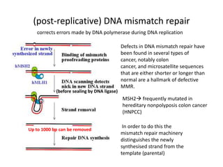
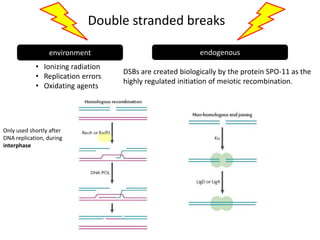
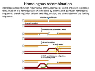
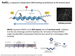
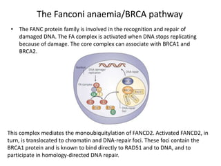
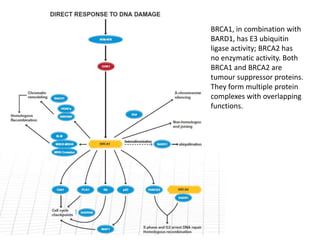
1. Genetic stability of organisms
Accurate DNA replication
+
DNA repair mechanisms
Present in prokaryotes and eukaryotes
2. 1 million individual lesions/cell/day
• Heat
• Metabolic accident
• Radiation
• Environment
• depurination
But only 1/1000 accidental base
change results in a permanent
mutation
Molecular Biology of the Cell
3. Danger of DNA damage
• Structural damage -> prevent replication/transcription
– Stalling of replication fork
• Harmful mutations -> impair survival of organism
– Mutation in tumour suppressor genes for examples
• If unrepaired:
– Senescence
– Apoptosis
– Aberrant cell division -> cancer
4. Genetic diseases associated with defects in DNA repair
Transcription-coupled repair
HNPCC
ATM
Breast, ovarian and Defect in homologous
colon cancer recombination, BRCA2
2005 W. H. Freeman Pierce, Benjamin. Genetics: A Conceptual Approach, 2nd ed.
5. the DNA molecule can be repaired fairly
easily because it carries 2 separate copies
of all the genetic information
Only viruses, that have a tiny genome (therefore tiny target for
DNA damage) can afford to encode their genetic information in
any molecule other than double stranded DNA
single-stranded DNA or RNA.
6. Multiple DNA repair pathways
• Base excision repair (BER)
• Nucleotide excision repair (NER)
• Mismatch repair (MR)
Level of damage
• DNA strand cross link repair
• Homologous recombination (HR)
• Non-homologous end joining (NHEJ)
+ transcription coupled repair
7. After T. Lindahl, Nature 362:709–715, 1993
Spontaneous DNA alteration
Nucleotides known to be modified by:
• Oxidative damage
• hydrolytic attack
• uncontrolled methylation
Purines (guanine and adenine) are more affected by those spontaneous reactions.
5000 purine bases are lost every day: DEPURINATION
Spontaneous DEAMINATION of cytosine uracil occurs at a rate of 100 bases per cell per day.
UV can covalently link two adjacent pyrimidine bases to form THYMINE DIMERS.
10. “Risky Business”
1. Replicative polymerases
2. Translesion DNA synthesis (TLS) polymerases
‘backup’ polymerases not as accurate as the normal
replicative polymerase
lack exonucleotic proofreading activity
3. DNA repair and recombination
4. Reverse transcriptase
Restrained to telomerase in eukaryotes, using a
RNA template for DNA synthesis
11. 15 different mammalian DNA polymerases
Example of specialised polymerase: Terminal
deoxynucleotidyl transferase (TDT) is expressed
only in lymphoid tissue, and adds random
nucleotides to double-strand breaks formed
during somatic recombination to promote
immunological diversity.
12. (post-replicative) DNA mismatch repair
corrects errors made by DNA polymerase during DNA replication
Defects in DNA mismatch repair have
been found in several types of
cancer, notably colon
cancer, and microsatellite sequences
that are either shorter or longer than
normal are a hallmark of defective
MMR.
(before sealing by DNA ligase)
MSH2 frequently mutated in
hereditary nonpolyposis colon cancer
(HNPCC)
In order to do this the
Up to 1000 bp can be removed
mismatch repair machinery
distinguishes the newly
synthesised strand from the
template (parental)
13. Double stranded breaks
environment endogenous
• Ionizing radiation
DSBs are created biologically by the protein SPO-11 as the
• Replication errors
highly regulated initiation of meiotic recombination.
• Oxidating agents
Only used shortly after
DNA replication, during
interphase
15. Homologous recombination
Homologous recombination requires DSB of DNA (damage or stalled or broken replication
fork), invasion of a homologous dsDNA molecule by a ssDNA end, pairing of homologous
sequences, branch migration to form a Holliday junction, and isomerisation of the flanking
sequences.
18. Rad51, a sequence-independent DNA binding recombinase at the branch point
Holliday junction
BRCA2 sequesters RAD51 via its BCR repeats and its C-terminal motif, mobilises
it to the site of damage and then facilitates the formation of helical RAD51–
single stranded DNA nucleoprotein filaments that search for a homologous DNA
template.
Owen Richard Davies & Luca Pellegrini, Nature Structural & Molecular Biology 14, 475 - 483 (2007)
19. ATM protein kinase
• DSBs create changes in
chromatin structure which
activate ATM by
autophosphorylation, which
then induces many cellular
responses by
phosphorylating a vast
number of target proteins.
• Associate with the BRCA1-
associated genome
surveillance complex
(BASC)
• Can phosphorylate p53 also
involved in DNA repair and
cell cycle arrest.
20. The Fanconi anaemia/BRCA pathway
• The FANC protein family is involved in the recognition and repair of
damaged DNA. The FA complex is activated when DNA stops replicating
because of damage. The core complex can associate with BRCA1 and
BRCA2.
This complex mediates the monoubiquitylation of FANCD2. Activated FANCD2, in
turn, is translocated to chromatin and DNA-repair foci. These foci contain the
BRCA1 protein and is known to bind directly to RAD51 and to DNA, and to
participate in homology-directed DNA repair.
21.
22. BRCA1, in combination with
BARD1, has E3 ubiquitin
ligase activity; BRCA2 has
no enzymatic activity. Both
BRCA1 and BRCA2 are
tumour suppressor proteins.
They form multiple protein
complexes with overlapping
functions.
Editor's Notes
The basic processes of DNA repair are highly conserved among both prokaryotes and eukaryotes and even among bacteriophage (virusesthat infect bacteria); however, more complex organisms with more complex genomes have correspondingly more complex repair mechanisms.[47]
A key step is an enzyme-mediated “flipping-out” of the altered nucleotide from the helix, which allows the enzyme to probe all faces of the base for damage (Figure 5-51). It is thought that DNA glycosylases travel along DNA using base-flipping to evaluate the status of each base pair. Once a damagedbase is recognized, the DNA glycosylase reaction creates a deoxyribose sugar that lacks its base. This “missing tooth” is recognized by an enzyme called AP endonuclease, which cuts the phosphodiester backbone, and the damage is then removed and repaired (see Figure 5-50A). Depurination, which is by far the most frequent type of damage suffered by DNA, also leaves a deoxyribose sugar with a missing base. Depurinations are directly repaired beginning with AP endonuclease, following the bottom half of the pathway in Figure 5-50A. he second major repair pathway is called nucleotide excision repair. This mechanism can repair the damage caused by almost any large change in the structure of the DNA double helix. Such “bulky lesions” include those created by the covalent reaction of DNA bases with large hydrocarbons (such as the carcinogen benzopyrene), as well as the various pyrimidine dimers (T-T, T-C, and C-C) caused by sunlight. In this pathway, a large multienzyme complex scans the DNA for a distortion in the double helix, rather than for a specific base change. Once a bulky lesion has been found, the phosphodiester backbone of the abnormal strand is cleaved on both sides of the distortion, and an oligonucleotide containing the lesion is peeled away from the DNA double helix by a DNA helicase enzyme. The large gap produced in the DNA helix is then repaired by DNA polymerase and DNA ligase (Figure 5-50B).
HR: single-strand invasion, branch migration, limited DNA synthesisIn mycobacteria, NHEJ is much more error prone than in yeast, with bases often added to and deleted from the ends of double-strand breaks during repair.[9] Many of the bacteria that possess NHEJ proteins spend a significant portion of their life cycle in a stationary haploid phase, in which a template for recombination is not available.[8] NHEJ may have evolved to help these organisms survive DSBs induced during dessication