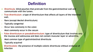
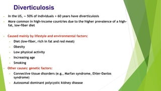
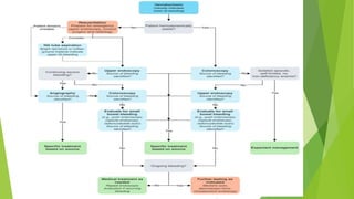
Diverticular Disease
DR Muteb ALShammari
GS Resident
Definition
❖ Diverticula: blind pouches that protrude from the gastrointestinal wall and
communicate with the lumen.
➢ True diverticulum: a type of diverticulum that affects all layers of the intestinal
wall.
• Rare (except Meckel diverticulum)
• Typically congenital
• Occur less commonly in the colon
• Most commonly occur in the cecum
➢ False diverticulum or pseudodiverticulum: type of diverticulum that involves only
the mucosa and submucosa and does not contain muscular layer or adventitia.
• Most common type of gastrointestinal diverticula
• Typically acquired
❖ Diverticulosis: the presence of multiple colonic diverticula without evidence of
infection
Diverticulosis
• In the US, ∼ 50% of individuals > 60 years have diverticulosis
• More common in high-income countries due to the higher prevalence of a high-
fat, low-fiber diet
• Caused mainly by lifestyle and environmental factors:
• Diet (low-fiber, rich in fat and red meat)
• Obesity
• Low physical activity
• Increasing age
• Smoking
• Other causes: genetic factors:
• Connective tissue disorders (e.g., Marfan syndrome, Ehler-Danlos
syndrome)
• Autosomal dominant polycystic kidney disease
Pathophysiology
• The formation of diverticula is considered multifactorial.
• Increased intraluminal pressure, e.g., due to chronic constipation.
• Weakness of the intestinal wall
• Age-related loss of elasticity of the connective tissue
• Physiological gaps in the intestinal wall, which occur where blood
vessels penetrate, predispose to protrusion and herniation of
intestinal mucosa and submucosa.
• Localized particularly in the sigmoid colon
Due to the narrow passage, intraluminal pressure is highest in
the sigmoid colon, which promotes the formation of diverticula.
Clinical features
❑ Usually asymptomatic
❑ May manifest with abdominal discomfort or pain,
especially if associated with chronic constipation
❑ Diverticular bleeding
❑ Tenderness over the affected area.
❑ Mild abdominal cramps.
❑ Swelling or bloating
Diagnostics
Asymptomatic diverticulosis:
- Typically an incidental diagnosis
E.g., during a screening colonoscopy
-No workup required
Symptomatic diverticulosis:
Colonoscopy: diagnostic modality of choice for suspected symptomatic diverticulosis
Indications:
• Lower GI bleed.
• Recurrent abdominal pain and/or diarrhea.
• Concern for underlying malignancy.
Findings: well-defined outpouching from the colonic wall
Avoid if acute diverticulitis is suspected.
Biopsy and histological analysis can be performed, if necessary
Imaging
Double-contrast barium enema: highly sensitive test to detect
diverticulosis but not commonly performed
Consider in the workup of the following:
• Recurrent LLQ pain without signs of acute inflammation .
• Altered bowel habits .
• Lower GI bleed in a hemodynamically stable patient if colonoscopy cannot
be performed.
Contraindications: suspected diverticulitis or perforated diverticulum
Findings: outpouching of the colonic wall of variable size
Imaging
Abdominal ultrasound
Indications: may be performed as part of the workup for nonspecific
LLQ pain
Findings: outpouching from the colonic wall
Colonoscopy:is the diagnostic modality of choice for symptomatic
diverticulosis
Treatment
Asymptomatic diverticulosis:
No treatment can reverse the growth of existing diverticula.
The goal is the prevention of progression
Symptomatic uncomplicated diverticular disease:
Proposed therapies include antibiotics and probiotics, however,
supportive evidence is lacking.
It is possible that symptoms attributed to diverticular disease may be
caused by irritable bowel syndrome.
Complications
• Diverticular bleeding
➢ Diverticulosis is the most common cause of lower GI bleeding in adults.
➢ Occurs in ∼ 5% of individuals with diverticulosis
• Etiology: erosions around the edge of diverticula
• Clinical findings:
➢ Painless hematochezia
➢ Signs of anemia may be present if recurrent
➢ Severe or ongoing bleeding: significant drop in hemoglobin → hemodynamic
instability (hypotension, tachycardia, dizziness, reduced level of consciousness)
• In 70–80% of cases, bleeding ceases spontaneously
• Differential diagnosis: other causes of lower gastrointestinal bleeding (e.g.
hemorrhoidal bleeding)
Treatment:
• Initial management of overt GI bleeding :
❑ Ensure patient is NPO.
❑ Insert two large-bore peripheral IVs (for possible fluid resuscitation and blood
transfusion) and obtain blood samples for laboratory studies (e.g., CBC, type and
screen).
❑ Conduct a focused history and examination (including DRE)
Risk stratify to guide further management.
❖ Prior to hemostatic procedures:
❑ Administer pretreatment (e.g., IV PPI) as needed.
✓ IV PPIs can reduce the risk of mortality and rebleeding, however, their
administration should not delay definitive hemostatic interventions or be
prioritized over resuscitation measures for unstable patients.
✓ Administer anticoagulant reversal if INR > 2.5.
✓ Consider withholding antithrombotic agents.
Stable patients:
• Restrictive transfusion strategy (transfuse pRBCs if Hb ≤ 7–8 g/dL).
• Refer for endoscopy (e.g., EGD or colonoscopy) according to risk
stratification and source of bleeding
Unstable patients:
❑ Follow an ABCDE approach.
❑ Consider intubation to protect the airway (e.g., in patients with altered
mental state and/or severe ongoing hematemesis).
❑ Urgent volume resuscitation for hemodynamic instability
✓ IV fluid resuscitation
✓ Liberal transfusion strategy: for hemorrhagic shock or massive bleeding
✓ Target normal vital signs prior to diagnostic testing if possible.
After stabilizations
• Endoscopic hemostasis during colonoscopy (e.g., epinephrine
injection, thermal coagulation, ligation)
• Angiography with vessel embolization
• Performed if bleeding cannot be localized or treated during
endoscopy
Diverticulitis
• Occurs in ∼ 4–20% of individuals with diverticulosis
• most commonly in the sigmoid colon
Inflammation:
❖ Most commonly: chronic inflammation and increased intraluminal
pressure → erosion of diverticula wall → inflammation and
bacterial translocation
❖ Rarely: stool becomes lodged in diverticula → obstruction of
intestinal lumen → inflammation
Clinical features
• Sigmoid colon most commonly affected → left lower quadrant pain
• Possibly tender, palpable mass (pericolonic inflammation)
• Fever
• Change in bowel habits (constipation in ∼ 50% of cases and diarrhea
in 25–35% of cases)
• Acute abdomen: indicates possible perforation and peritonitis
• ↑ Urinary urgency and frequency (in ∼ 15% of cases)
• Rarely: hematochezia
Diagnostics
• Suspect acute diverticulitis in adult patients presenting
with LLQ pain, fever, and leukocytosis
• Laboratory studies
• CBC: leukocytosis, possible anemia
• BMP: electrolyte abnormalities, ↑ BUN, ↑ creatinine
• CRP: ↑ CRP
• FOBT: positive in patients with diverticular bleeding
• Diverticulitis is highly likely in patients with LLQ pain and
tenderness, no vomiting, and CRP > 50 mg/L.
Imaging
CT abdomen and pelvis with IV contrast
Indications
• Preferred initial imaging modality for suspected diverticulitis
• Diagnostic confirmation in patients with no prior imaging studies
• Staging the severity of diverticulitis
Supportive findings
Colonic outpouching
Signs of inflammation
• Bowel wall thickening > 3 mm
• Peridiverticular mesenteric fat stranding
Complications may also be identified
• Peridiverticular abscess: hypodense collections with peripheral contrast enhancement
• Diverticular perforation: pneumoperitoneum
• Intestinal obstruction: dilated intestinal loops with multiple air-fluid levels
Imaging
MRI abdomen and pelvis (without and with IV contrast)Indications:
suspected diverticulitis in patients with contraindications to CT
Ultrasound abdomen
Indications
• Formal ultrasound is typically considered as an alternative to MRI in patients
with contraindications to CT
• Point-of-care ultrasound may be considered as an initial imaging modality and
can show findings of complicated diverticulitis (e.g., pneumoperitoneum, free
fluid, abscess formation).
Supportive findings:
diverticula with surrounding inflammation, abscess formation (detectable fluid),
bowel wall thickening
Imaging
• Abdominal x-ray
• Not useful in diagnosing uncomplicated diverticulitis
• Indications
• Suspected perforation or bowel obstruction
• May be performed as part of the routine workup for acute abdominal pain
• Findings that may be seen in complicated diverticulitis include
• Bowel perforation: pneumoperitoneum
• Bowel obstruction: dilated bowel loops and multiple air-fluid levels
• Screening colonoscopy
• Recommended 6–8 weeks after the resolution of the acute episode to assess the
extent of diverticulitis and rule out malignancy
• Colonoscopy is contraindicated during an acute episode because of the increased
risk of perforation.
• Not required if a recent evaluation of the colon has been performed
❖ Avoid colonoscopy during the acute phase of diverticulitis because of the risk of
perforation!
CLASSIFICATIONS
Differential diagnoses
• Crohn’s disease, ulcerative colitis
• Colorectal cancer
• Intestinal ischemia (ischemic colitis)
• Acute appendicitis
• Ileus , colonic obstruction
• Ectopic pregnancy
• Ovarian torsion
• Ovarian cancer
• Inguinal hernia
• Renal colic
• Urinary tract infection
Treatment
• Uncomplicated diverticulitis
➢ Conservative management
➢ Consider broad-spectrum oral antibiotics
➢ Complicated diverticulitis
➢ Antibiotic therapy: broad-spectrum IV antibiotics are routinely recommended
• Management of complications abscess:
• Size < 4 cm: trial of conservative management with IV antibiotics
• Size ≥ 4 cm
• Ultrasound- or CT-guided percutaneous drainage
• Consider laparoscopic or open surgical drainage if percutaneous drainage is not feasible.
• Continue IV antibiotic therapy.
• Send aspirate or pus for cultures and tailor antibiotic treatment accordingly.
Treatment
• Perforation with generalized peritonitis: emergency surgery
• Hemodynamically stable patients: laparoscopic or
open colectomy and primary anastomosis with/without a
temporary diverting stoma
• Critically ill patients: Hartmann procedure
Complications
Perforation
Locally-contained perforation: can lead to the formation of an abscess or phlegmon
Intraperitoneal perforation
Caused by:
Rupture of an inflamed diverticulum → free communication with the peritoneum → generalized fecal peritonitis
Rupture of a diverticular abscess → generalized purulent peritonitis
Can present with symptoms of acute abdomen and widespread intraperitoneal free air on imaging
Abscess
Peridiverticular localization
Causes symptoms similar to those of acute diverticulitis
Suspect an abscess in patients with persistent fever and abdominal pain despite antibiotic treatment.
Intestinal obstruction (rare)
Etiology
Narrowing due to inflammatory swelling
Compression through abscesses
Ileus caused by localized irritation
Complications
• Clinical findings
• Abdominal pain and distention
• Constipation
• Nausea, vomiting
• Acute abdomen
• Fistulas
• Epidemiology
• Most commonly colovesical
• Other forms: colovaginal, coloenteric, colocutaneous
• Clinical findings
• Pneumaturia and fecaluria
• May cause recurring urinary tract infections, including urosepsis
• Diagnosis: CT with oral contrast
• Localized thickening of the colon and bladder
• Air or contrast material in the bladder
• Treatment
• Resection and primary anastomosis
• Antibiotics if surgery is not possible
Complications
• Recurrent diverticulitis
• 13–23% of patients with uncomplicated diverticulitis
• Up to 40% of patients with complicated diverticulitis
diverticular disease [تم حفظه تلقائيا] 3.pptx

diverticular disease [تم حفظه تلقائيا] 3.pptx

  • 1.
    Diverticular Disease DR MutebALShammari GS Resident
  • 2.
    Definition ❖ Diverticula: blindpouches that protrude from the gastrointestinal wall and communicate with the lumen. ➢ True diverticulum: a type of diverticulum that affects all layers of the intestinal wall. • Rare (except Meckel diverticulum) • Typically congenital • Occur less commonly in the colon • Most commonly occur in the cecum ➢ False diverticulum or pseudodiverticulum: type of diverticulum that involves only the mucosa and submucosa and does not contain muscular layer or adventitia. • Most common type of gastrointestinal diverticula • Typically acquired ❖ Diverticulosis: the presence of multiple colonic diverticula without evidence of infection
  • 3.
    Diverticulosis • In theUS, ∼ 50% of individuals > 60 years have diverticulosis • More common in high-income countries due to the higher prevalence of a high- fat, low-fiber diet • Caused mainly by lifestyle and environmental factors: • Diet (low-fiber, rich in fat and red meat) • Obesity • Low physical activity • Increasing age • Smoking • Other causes: genetic factors: • Connective tissue disorders (e.g., Marfan syndrome, Ehler-Danlos syndrome) • Autosomal dominant polycystic kidney disease
  • 4.
    Pathophysiology • The formationof diverticula is considered multifactorial. • Increased intraluminal pressure, e.g., due to chronic constipation. • Weakness of the intestinal wall • Age-related loss of elasticity of the connective tissue • Physiological gaps in the intestinal wall, which occur where blood vessels penetrate, predispose to protrusion and herniation of intestinal mucosa and submucosa. • Localized particularly in the sigmoid colon Due to the narrow passage, intraluminal pressure is highest in the sigmoid colon, which promotes the formation of diverticula.
  • 6.
    Clinical features ❑ Usuallyasymptomatic ❑ May manifest with abdominal discomfort or pain, especially if associated with chronic constipation ❑ Diverticular bleeding ❑ Tenderness over the affected area. ❑ Mild abdominal cramps. ❑ Swelling or bloating
  • 8.
    Diagnostics Asymptomatic diverticulosis: - Typicallyan incidental diagnosis E.g., during a screening colonoscopy -No workup required Symptomatic diverticulosis: Colonoscopy: diagnostic modality of choice for suspected symptomatic diverticulosis Indications: • Lower GI bleed. • Recurrent abdominal pain and/or diarrhea. • Concern for underlying malignancy. Findings: well-defined outpouching from the colonic wall Avoid if acute diverticulitis is suspected. Biopsy and histological analysis can be performed, if necessary
  • 11.
    Imaging Double-contrast barium enema:highly sensitive test to detect diverticulosis but not commonly performed Consider in the workup of the following: • Recurrent LLQ pain without signs of acute inflammation . • Altered bowel habits . • Lower GI bleed in a hemodynamically stable patient if colonoscopy cannot be performed. Contraindications: suspected diverticulitis or perforated diverticulum Findings: outpouching of the colonic wall of variable size
  • 13.
    Imaging Abdominal ultrasound Indications: maybe performed as part of the workup for nonspecific LLQ pain Findings: outpouching from the colonic wall Colonoscopy:is the diagnostic modality of choice for symptomatic diverticulosis
  • 15.
    Treatment Asymptomatic diverticulosis: No treatmentcan reverse the growth of existing diverticula. The goal is the prevention of progression Symptomatic uncomplicated diverticular disease: Proposed therapies include antibiotics and probiotics, however, supportive evidence is lacking. It is possible that symptoms attributed to diverticular disease may be caused by irritable bowel syndrome.
  • 16.
    Complications • Diverticular bleeding ➢Diverticulosis is the most common cause of lower GI bleeding in adults. ➢ Occurs in ∼ 5% of individuals with diverticulosis • Etiology: erosions around the edge of diverticula • Clinical findings: ➢ Painless hematochezia ➢ Signs of anemia may be present if recurrent ➢ Severe or ongoing bleeding: significant drop in hemoglobin → hemodynamic instability (hypotension, tachycardia, dizziness, reduced level of consciousness) • In 70–80% of cases, bleeding ceases spontaneously • Differential diagnosis: other causes of lower gastrointestinal bleeding (e.g. hemorrhoidal bleeding)
  • 17.
    Treatment: • Initial managementof overt GI bleeding : ❑ Ensure patient is NPO. ❑ Insert two large-bore peripheral IVs (for possible fluid resuscitation and blood transfusion) and obtain blood samples for laboratory studies (e.g., CBC, type and screen). ❑ Conduct a focused history and examination (including DRE) Risk stratify to guide further management. ❖ Prior to hemostatic procedures: ❑ Administer pretreatment (e.g., IV PPI) as needed. ✓ IV PPIs can reduce the risk of mortality and rebleeding, however, their administration should not delay definitive hemostatic interventions or be prioritized over resuscitation measures for unstable patients. ✓ Administer anticoagulant reversal if INR > 2.5. ✓ Consider withholding antithrombotic agents.
  • 18.
    Stable patients: • Restrictivetransfusion strategy (transfuse pRBCs if Hb ≤ 7–8 g/dL). • Refer for endoscopy (e.g., EGD or colonoscopy) according to risk stratification and source of bleeding Unstable patients: ❑ Follow an ABCDE approach. ❑ Consider intubation to protect the airway (e.g., in patients with altered mental state and/or severe ongoing hematemesis). ❑ Urgent volume resuscitation for hemodynamic instability ✓ IV fluid resuscitation ✓ Liberal transfusion strategy: for hemorrhagic shock or massive bleeding ✓ Target normal vital signs prior to diagnostic testing if possible.
  • 19.
    After stabilizations • Endoscopichemostasis during colonoscopy (e.g., epinephrine injection, thermal coagulation, ligation) • Angiography with vessel embolization • Performed if bleeding cannot be localized or treated during endoscopy
  • 21.
    Diverticulitis • Occurs in∼ 4–20% of individuals with diverticulosis • most commonly in the sigmoid colon Inflammation: ❖ Most commonly: chronic inflammation and increased intraluminal pressure → erosion of diverticula wall → inflammation and bacterial translocation ❖ Rarely: stool becomes lodged in diverticula → obstruction of intestinal lumen → inflammation
  • 22.
    Clinical features • Sigmoidcolon most commonly affected → left lower quadrant pain • Possibly tender, palpable mass (pericolonic inflammation) • Fever • Change in bowel habits (constipation in ∼ 50% of cases and diarrhea in 25–35% of cases) • Acute abdomen: indicates possible perforation and peritonitis • ↑ Urinary urgency and frequency (in ∼ 15% of cases) • Rarely: hematochezia
  • 23.
    Diagnostics • Suspect acutediverticulitis in adult patients presenting with LLQ pain, fever, and leukocytosis • Laboratory studies • CBC: leukocytosis, possible anemia • BMP: electrolyte abnormalities, ↑ BUN, ↑ creatinine • CRP: ↑ CRP • FOBT: positive in patients with diverticular bleeding • Diverticulitis is highly likely in patients with LLQ pain and tenderness, no vomiting, and CRP > 50 mg/L.
  • 24.
    Imaging CT abdomen andpelvis with IV contrast Indications • Preferred initial imaging modality for suspected diverticulitis • Diagnostic confirmation in patients with no prior imaging studies • Staging the severity of diverticulitis Supportive findings Colonic outpouching Signs of inflammation • Bowel wall thickening > 3 mm • Peridiverticular mesenteric fat stranding Complications may also be identified • Peridiverticular abscess: hypodense collections with peripheral contrast enhancement • Diverticular perforation: pneumoperitoneum • Intestinal obstruction: dilated intestinal loops with multiple air-fluid levels
  • 25.
    Imaging MRI abdomen andpelvis (without and with IV contrast)Indications: suspected diverticulitis in patients with contraindications to CT Ultrasound abdomen Indications • Formal ultrasound is typically considered as an alternative to MRI in patients with contraindications to CT • Point-of-care ultrasound may be considered as an initial imaging modality and can show findings of complicated diverticulitis (e.g., pneumoperitoneum, free fluid, abscess formation). Supportive findings: diverticula with surrounding inflammation, abscess formation (detectable fluid), bowel wall thickening
  • 26.
    Imaging • Abdominal x-ray •Not useful in diagnosing uncomplicated diverticulitis • Indications • Suspected perforation or bowel obstruction • May be performed as part of the routine workup for acute abdominal pain • Findings that may be seen in complicated diverticulitis include • Bowel perforation: pneumoperitoneum • Bowel obstruction: dilated bowel loops and multiple air-fluid levels • Screening colonoscopy • Recommended 6–8 weeks after the resolution of the acute episode to assess the extent of diverticulitis and rule out malignancy • Colonoscopy is contraindicated during an acute episode because of the increased risk of perforation. • Not required if a recent evaluation of the colon has been performed ❖ Avoid colonoscopy during the acute phase of diverticulitis because of the risk of perforation!
  • 28.
  • 30.
    Differential diagnoses • Crohn’sdisease, ulcerative colitis • Colorectal cancer • Intestinal ischemia (ischemic colitis) • Acute appendicitis • Ileus , colonic obstruction • Ectopic pregnancy • Ovarian torsion • Ovarian cancer • Inguinal hernia • Renal colic • Urinary tract infection
  • 31.
    Treatment • Uncomplicated diverticulitis ➢Conservative management ➢ Consider broad-spectrum oral antibiotics ➢ Complicated diverticulitis ➢ Antibiotic therapy: broad-spectrum IV antibiotics are routinely recommended • Management of complications abscess: • Size < 4 cm: trial of conservative management with IV antibiotics • Size ≥ 4 cm • Ultrasound- or CT-guided percutaneous drainage • Consider laparoscopic or open surgical drainage if percutaneous drainage is not feasible. • Continue IV antibiotic therapy. • Send aspirate or pus for cultures and tailor antibiotic treatment accordingly.
  • 32.
    Treatment • Perforation withgeneralized peritonitis: emergency surgery • Hemodynamically stable patients: laparoscopic or open colectomy and primary anastomosis with/without a temporary diverting stoma • Critically ill patients: Hartmann procedure
  • 34.
    Complications Perforation Locally-contained perforation: canlead to the formation of an abscess or phlegmon Intraperitoneal perforation Caused by: Rupture of an inflamed diverticulum → free communication with the peritoneum → generalized fecal peritonitis Rupture of a diverticular abscess → generalized purulent peritonitis Can present with symptoms of acute abdomen and widespread intraperitoneal free air on imaging Abscess Peridiverticular localization Causes symptoms similar to those of acute diverticulitis Suspect an abscess in patients with persistent fever and abdominal pain despite antibiotic treatment. Intestinal obstruction (rare) Etiology Narrowing due to inflammatory swelling Compression through abscesses Ileus caused by localized irritation
  • 36.
    Complications • Clinical findings •Abdominal pain and distention • Constipation • Nausea, vomiting • Acute abdomen • Fistulas • Epidemiology • Most commonly colovesical • Other forms: colovaginal, coloenteric, colocutaneous • Clinical findings • Pneumaturia and fecaluria • May cause recurring urinary tract infections, including urosepsis • Diagnosis: CT with oral contrast • Localized thickening of the colon and bladder • Air or contrast material in the bladder • Treatment • Resection and primary anastomosis • Antibiotics if surgery is not possible
  • 38.
    Complications • Recurrent diverticulitis •13–23% of patients with uncomplicated diverticulitis • Up to 40% of patients with complicated diverticulitis