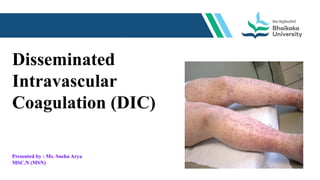
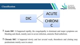
Disseminated
Intravascular
Coagulation (DIC)
Presented by : Ms. Sneha Arya
MSC.N (MSN)
GENERAL OBJECTIVE
At the end of this lecture, students will able to get in depth knowledge
about DIC (Disseminated intravascular coagulation).
SPECIFIC OBJECTIVES
Student will be able to,
 To introduce the DIC.
 To define the DIC.
 To enlist the cause & risk factors of DIC.
 To describe the pathophysiology.
 To enlist the sign and symptoms of DIC.
 To explain the diagnostic evaluation.
 To explain the management of DIC.
 To summarize the topic.
Introduction
Disseminated intravascular coagulation (DIC) is a rare and serious condition that disrupts
blood flow. It is a blood clotting disorder that can turn into uncontrollable bleeding. DIC can
affect people who have cancer or sepsis. It can also affect people recovering from
complications from pregnancy and delivery or who have been injured.
Definition
Disseminated intravascular coagulation (DIC) is a state /syndrome which is characterized by
accelerated intravascular coagulation associated with increased consumption of platelet and
plasma clotting factors.
Incidence
DIC may occur in 30-50% of patients with sepsis, and it develops in an estimated 1% of all hospitalized patients.
DIC occurs at all ages and in all races, and no particular sex predisposition has been noted.
Causes & risk factors
1. Infectious diseases: (Bacterial, viral, rickettsia, parasitic diseases and so on) in particular septicemia.
systemic infections with other microorganisms( viruses and parasites)
2. Sepsis
3. Major damage to organs or tissues: cirrhosis of the liver, pancreatitis, severe injury, burns, or major surgery
4. Severe immune reactions: failed blood transfusion, rejection of an organ transplant, toxin such as snake
venom.
5. Serious pregnancy-related problems: placenta separating from the uterus before delivery, amniotic fluid
entering the bloodstream, or serious bleeding during or after delivery.
6. Cancer (Acute promyelocytic leukemia, acute myelomonocytic monocytic leukemia, or disseminated
prostatic carcinoma, Lung, breast, gastrointestinal malignancy)
Classification
DIC
ACUTE
CHRONI
C
 Acute DIC: It happened rapidly, the coagulopathy is dominant and major symptoms are
bleeding and shock, mainly seen in severe infection, amniotic fluid embolism.
 Chronic DIC: it happened slowly and last several week, thrombosis and clotting may
predominate mainly seen in cancer
Pathophysiology
Sign & symptoms
 Malena , which may appear tarry and black from bleeding in your stomach or intestines
 hematuria
 Unusually heavy menstrual bleeding
 Bleeding from urinary catheter use
 Bleeding easily from wounds or after surgery
 Hematomas, which are larger bruises that also cause swelling and pain
 Frequent nosebleeds
 Bleeding from your gums, especially after brushing your teeth or flossing
 Yellowing of the skin or eyes (jaundice), which could indicate liver problems
 Shortness of breath
 Chest pain
 Headaches
 Dizziness or confusion seizures
Diagnostic evaluation
 Medical history and physical exam
 ADAMTS13 testing to check blood levels and activity of this protein, which can be low in
a condition called thrombotic thrombocytopenic purpura .
 Liver biopsy and liver function tests
 Simple screening
 Platelet count
 Extended screening
 Fibrin D-dimer fragment
 Fibrinogen
Types of Coagulation Tests
I. Prothrombin Time (PT): Evaluates ability to clot
II. International Normalized Ratio (INR): Ensures that results from a PT test are
the same from one lab to another
III. Partial Thromboplastin Time (PTT): Determines if blood-thinning therapy is
effective
Management
Treatment of the underlying disease
• It is the mainstay of management of either acute or chronic DIC. Avoid delay treat
vigorously (eg, shock, sepsis, obstetrical problems).
Replacement therapy
• Coagulation factor deficiency require replacement with FFP (fresh frozen plasma). Platelet
transfusion should be used to maintain a platelet count greater than 30000/µl, and 50000/pl.
• Fibrinogen is replaced with cryoprecipitate. One unit of cryoprecipitate usually raises the
fibrinogen level by 6~8mg/dl, so that 15 units of cryoprecipitate will raise the level from 50
to 150mg/dl.
Heparin therapy
In some cases, heparin therapy is contraindicated, but when DIC is producing serious clinical
consequences and the underlying cause is not rapidly reversible, heparin may be necessary.
Dose:500~750u/h is necessary.
Other Treatment
 Aminocaproic acid, 1g/h iv
 Tranexamic acid, 10mg / kg iv, q8h
Those two drugs should be added to decrease the rate of fibrinolysis, raise the fibrinogen level,
and control bleeding. Aminocaproic acid can never be used without heparin in DIC because of
the risk of thrombosis.
Nursing management:
Diagnosis:
1. Impaired Gas Exchange
2. Ineffective Tissue Perfusion
3. Deficient Knowledge
4. Risk for Bleeding
 Assess for changes in the level of consciousness.
 Assess the respiratory depth, rate, and rhythm.
 Assess the client’s breath sounds. Assess cough for signs of bloody sputum.
 Assess for tachycardia, shortness of breath, and use of accessory muscles.
 Monitor oxygen saturation and assess arterial blood gases.
Interventions:
1. Provide reassurance and allay anxiety by staying with the client during the acute episodes of respiratory
distress.
Anxiety increases dyspnea, the work of breathing, and the respiratory rate.
2. Change the client’s positioning every 2 hours, and perform chest physiotherapy.
These maneuvers facilitate the movement and drainage of secretions.
3. Position the client in a high-Fowler’s position as indicated.
An upright position allows for adequate diaphragmatic and lung excursion and promotes optimal lung expansion.
4. Assist with coughing or suction as indicated.
Productive coughing is the most effective way to remove moist secretions. If the client is unable to perform
independently, suctioning may be needed to promote airway patency and reduce the work of breathing.
5. Maintain an oxygen administration device as ordered.
The appropriate amount of oxygen must be delivered continuously so that the client maintains an oxygen
saturation of 90% or greater.
6. Anticipate the need for intubation and mechanical ventilation.
Early intubation and mechanical ventilation are recommended to prevent full decompensation of the client.
Mechanical ventilation provides supportive care to maintain adequate oxygenation and ventilation for the client.
Summary
 Introduction the DIC.
 Definition of DIC.
 Cause & risk factors of DIC.
 Pathophysiology.
 Sign and symptoms of DIC.
 Diagnostic evaluation.
 Management of DIC.
Assignment:
 Write a care plan on any one nursing diagnosis on a patient with DIC.
Questions
 What is the definition of DIC?
 Enlist the causes and risk factors of DIC.
 Enlist the medicines which is used in treatment of DIC.
Assignment:
 Write a care plan on any one nursing diagnosis on a patient
with DIC.
Bibliography and references
 Brunner and Siddharth’s Textbook of Medical-Surgical Nursing. (2018). (n.p.): Wolters kluwer india Pvt Ltd.
Page no 750 to 752.
 Hawks, J. H., Black, J. M. (n.d.). Medical Surgical Nursing: Clinical Management for Positive Outcomes, 8e
(2 Vol Set) Without CD. India: Elsevier India.
 Lippincott Manual of Medical - Surgical Nursing Adaptation of Nettina: Lippincott Manual of
Nursing. (2016). (n.p.): Wolters Kluwer India Pvt Ltd. page no 445
 C. (n.d.). Disseminated Intravascular Coagulation (DIC): What Is It, Risk Factors, Symptoms & Treatment.
Cleveland Clinic.
https://my.clevelandclinic.org/health/diseases/21836-disseminated-intravascular-coagulation-dic
 BSN, R.N., P. M. (2017, July 2). 4 Disseminated Intravascular Coagulation Nursing Care Plans. Nurseslabs.
https://nurseslabs.com/disseminated-intravascular-coagulation-nursing-care-plans/
THANK YOU

Disseminated intravascular coagulation (DIC)

  • 1.
  • 2.
    GENERAL OBJECTIVE At theend of this lecture, students will able to get in depth knowledge about DIC (Disseminated intravascular coagulation).
  • 3.
    SPECIFIC OBJECTIVES Student willbe able to,  To introduce the DIC.  To define the DIC.  To enlist the cause & risk factors of DIC.  To describe the pathophysiology.  To enlist the sign and symptoms of DIC.  To explain the diagnostic evaluation.  To explain the management of DIC.  To summarize the topic.
  • 4.
    Introduction Disseminated intravascular coagulation(DIC) is a rare and serious condition that disrupts blood flow. It is a blood clotting disorder that can turn into uncontrollable bleeding. DIC can affect people who have cancer or sepsis. It can also affect people recovering from complications from pregnancy and delivery or who have been injured. Definition Disseminated intravascular coagulation (DIC) is a state /syndrome which is characterized by accelerated intravascular coagulation associated with increased consumption of platelet and plasma clotting factors.
  • 5.
    Incidence DIC may occurin 30-50% of patients with sepsis, and it develops in an estimated 1% of all hospitalized patients. DIC occurs at all ages and in all races, and no particular sex predisposition has been noted.
  • 6.
    Causes & riskfactors 1. Infectious diseases: (Bacterial, viral, rickettsia, parasitic diseases and so on) in particular septicemia. systemic infections with other microorganisms( viruses and parasites) 2. Sepsis 3. Major damage to organs or tissues: cirrhosis of the liver, pancreatitis, severe injury, burns, or major surgery 4. Severe immune reactions: failed blood transfusion, rejection of an organ transplant, toxin such as snake venom. 5. Serious pregnancy-related problems: placenta separating from the uterus before delivery, amniotic fluid entering the bloodstream, or serious bleeding during or after delivery. 6. Cancer (Acute promyelocytic leukemia, acute myelomonocytic monocytic leukemia, or disseminated prostatic carcinoma, Lung, breast, gastrointestinal malignancy)
  • 7.
    Classification DIC ACUTE CHRONI C  Acute DIC:It happened rapidly, the coagulopathy is dominant and major symptoms are bleeding and shock, mainly seen in severe infection, amniotic fluid embolism.  Chronic DIC: it happened slowly and last several week, thrombosis and clotting may predominate mainly seen in cancer
  • 8.
  • 9.
    Sign & symptoms Malena , which may appear tarry and black from bleeding in your stomach or intestines  hematuria  Unusually heavy menstrual bleeding  Bleeding from urinary catheter use  Bleeding easily from wounds or after surgery  Hematomas, which are larger bruises that also cause swelling and pain  Frequent nosebleeds  Bleeding from your gums, especially after brushing your teeth or flossing
  • 10.
     Yellowing ofthe skin or eyes (jaundice), which could indicate liver problems  Shortness of breath  Chest pain  Headaches  Dizziness or confusion seizures
  • 11.
    Diagnostic evaluation  Medicalhistory and physical exam  ADAMTS13 testing to check blood levels and activity of this protein, which can be low in a condition called thrombotic thrombocytopenic purpura .  Liver biopsy and liver function tests  Simple screening  Platelet count  Extended screening  Fibrin D-dimer fragment  Fibrinogen
  • 12.
    Types of CoagulationTests I. Prothrombin Time (PT): Evaluates ability to clot II. International Normalized Ratio (INR): Ensures that results from a PT test are the same from one lab to another III. Partial Thromboplastin Time (PTT): Determines if blood-thinning therapy is effective
  • 13.
    Management Treatment of theunderlying disease • It is the mainstay of management of either acute or chronic DIC. Avoid delay treat vigorously (eg, shock, sepsis, obstetrical problems). Replacement therapy • Coagulation factor deficiency require replacement with FFP (fresh frozen plasma). Platelet transfusion should be used to maintain a platelet count greater than 30000/µl, and 50000/pl. • Fibrinogen is replaced with cryoprecipitate. One unit of cryoprecipitate usually raises the fibrinogen level by 6~8mg/dl, so that 15 units of cryoprecipitate will raise the level from 50 to 150mg/dl.
  • 14.
    Heparin therapy In somecases, heparin therapy is contraindicated, but when DIC is producing serious clinical consequences and the underlying cause is not rapidly reversible, heparin may be necessary. Dose:500~750u/h is necessary. Other Treatment  Aminocaproic acid, 1g/h iv  Tranexamic acid, 10mg / kg iv, q8h Those two drugs should be added to decrease the rate of fibrinolysis, raise the fibrinogen level, and control bleeding. Aminocaproic acid can never be used without heparin in DIC because of the risk of thrombosis.
  • 15.
    Nursing management: Diagnosis: 1. ImpairedGas Exchange 2. Ineffective Tissue Perfusion 3. Deficient Knowledge 4. Risk for Bleeding  Assess for changes in the level of consciousness.  Assess the respiratory depth, rate, and rhythm.  Assess the client’s breath sounds. Assess cough for signs of bloody sputum.  Assess for tachycardia, shortness of breath, and use of accessory muscles.  Monitor oxygen saturation and assess arterial blood gases.
  • 16.
    Interventions: 1. Provide reassuranceand allay anxiety by staying with the client during the acute episodes of respiratory distress. Anxiety increases dyspnea, the work of breathing, and the respiratory rate. 2. Change the client’s positioning every 2 hours, and perform chest physiotherapy. These maneuvers facilitate the movement and drainage of secretions. 3. Position the client in a high-Fowler’s position as indicated. An upright position allows for adequate diaphragmatic and lung excursion and promotes optimal lung expansion.
  • 17.
    4. Assist withcoughing or suction as indicated. Productive coughing is the most effective way to remove moist secretions. If the client is unable to perform independently, suctioning may be needed to promote airway patency and reduce the work of breathing. 5. Maintain an oxygen administration device as ordered. The appropriate amount of oxygen must be delivered continuously so that the client maintains an oxygen saturation of 90% or greater. 6. Anticipate the need for intubation and mechanical ventilation. Early intubation and mechanical ventilation are recommended to prevent full decompensation of the client. Mechanical ventilation provides supportive care to maintain adequate oxygenation and ventilation for the client.
  • 18.
    Summary  Introduction theDIC.  Definition of DIC.  Cause & risk factors of DIC.  Pathophysiology.  Sign and symptoms of DIC.  Diagnostic evaluation.  Management of DIC. Assignment:  Write a care plan on any one nursing diagnosis on a patient with DIC.
  • 19.
    Questions  What isthe definition of DIC?  Enlist the causes and risk factors of DIC.  Enlist the medicines which is used in treatment of DIC.
  • 20.
    Assignment:  Write acare plan on any one nursing diagnosis on a patient with DIC.
  • 21.
    Bibliography and references Brunner and Siddharth’s Textbook of Medical-Surgical Nursing. (2018). (n.p.): Wolters kluwer india Pvt Ltd. Page no 750 to 752.  Hawks, J. H., Black, J. M. (n.d.). Medical Surgical Nursing: Clinical Management for Positive Outcomes, 8e (2 Vol Set) Without CD. India: Elsevier India.  Lippincott Manual of Medical - Surgical Nursing Adaptation of Nettina: Lippincott Manual of Nursing. (2016). (n.p.): Wolters Kluwer India Pvt Ltd. page no 445  C. (n.d.). Disseminated Intravascular Coagulation (DIC): What Is It, Risk Factors, Symptoms & Treatment. Cleveland Clinic. https://my.clevelandclinic.org/health/diseases/21836-disseminated-intravascular-coagulation-dic  BSN, R.N., P. M. (2017, July 2). 4 Disseminated Intravascular Coagulation Nursing Care Plans. Nurseslabs. https://nurseslabs.com/disseminated-intravascular-coagulation-nursing-care-plans/
  • 22.